Datasheet
Year, pagecount:2005, 6 page(s)
Language:English
Downloads:9
Uploaded:December 18, 2011
Size:132 KB
Institution:
-
Comments:
Attachment:-
Download in PDF:Please log in!
Comments
No comments yet. You can be the first!Most popular documents in this category
Content extract
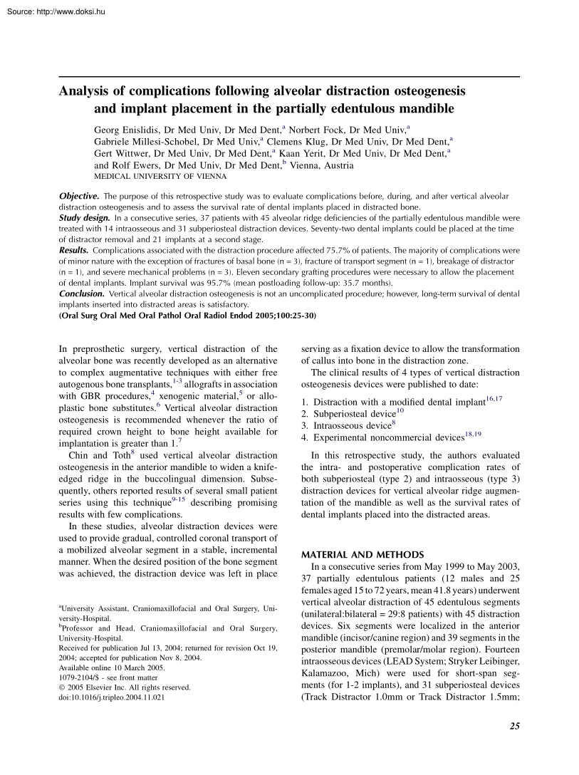
Analysis of complications following alveolar distraction osteogenesis and implant placement in the partially edentulous mandible Georg Enislidis, Dr Med Univ, Dr Med Dent,a Norbert Fock, Dr Med Univ,a Gabriele Millesi-Schobel, Dr Med Univ,a Clemens Klug, Dr Med Univ, Dr Med Dent,a Gert Wittwer, Dr Med Univ, Dr Med Dent,a Kaan Yerit, Dr Med Univ, Dr Med Dent,a and Rolf Ewers, Dr Med Univ, Dr Med Dent,b Vienna, Austria MEDICAL UNIVERSITY OF VIENNA Objective. The purpose of this retrospective study was to evaluate complications before, during, and after vertical alveolar distraction osteogenesis and to assess the survival rate of dental implants placed in distracted bone. Study design. In a consecutive series, 37 patients with 45 alveolar ridge deficiencies of the partially edentulous mandible were treated with 14 intraosseous and 31 subperiosteal distraction devices. Seventy-two dental implants could be placed at the time of distractor removal and 21 implants at a second stage. Results.
Complications associated with the distraction procedure affected 757% of patients The majority of complications were of minor nature with the exception of fractures of basal bone (n = 3), fracture of transport segment (n = 1), breakage of distractor (n = 1), and severe mechanical problems (n = 3). Eleven secondary grafting procedures were necessary to allow the placement of dental implants. Implant survival was 957% (mean postloading follow-up: 357 months) Conclusion. Vertical alveolar distraction osteogenesis is not an uncomplicated procedure; however, long-term survival of dental implants inserted into distracted areas is satisfactory. (Oral Surg Oral Med Oral Pathol Oral Radiol Endod 2005;100:25-30) In preprosthetic surgery, vertical distraction of the alveolar bone was recently developed as an alternative to complex augmentative techniques with either free autogenous bone transplants,1-3 allografts in association with GBR procedures,4 xenogenic material,5 or alloplastic bone
substitutes.6 Vertical alveolar distraction osteogenesis is recommended whenever the ratio of required crown height to bone height available for implantation is greater than 1.7 Chin and Toth8 used vertical alveolar distraction osteogenesis in the anterior mandible to widen a knifeedged ridge in the buccolingual dimension. Subsequently, others reported results of several small patient series using this technique9-15 describing promising results with few complications. In these studies, alveolar distraction devices were used to provide gradual, controlled coronal transport of a mobilized alveolar segment in a stable, incremental manner. When the desired position of the bone segment was achieved, the distraction device was left in place a University Assistant, Craniomaxillofacial and Oral Surgery, University-Hospital. b Professor and Head, Craniomaxillofacial and Oral Surgery, University-Hospital. Received for publication Jul 13, 2004; returned for revision Oct 19, 2004; accepted for
publication Nov 8, 2004. Available online 10 March 2005. 1079-2104/$ - see front matter Ó 2005 Elsevier Inc. All rights reserved doi:10.1016/jtripleo200411021 serving as a fixation device to allow the transformation of callus into bone in the distraction zone. The clinical results of 4 types of vertical distraction osteogenesis devices were published to date: 1. 2. 3. 4. Distraction with a modified dental implant16,17 Subperiosteal device10 Intraosseous device8 Experimental noncommercial devices18,19 In this retrospective study, the authors evaluated the intra- and postoperative complication rates of both subperiosteal (type 2) and intraosseous (type 3) distraction devices for vertical alveolar ridge augmentation of the mandible as well as the survival rates of dental implants placed into the distracted areas. MATERIAL AND METHODS In a consecutive series from May 1999 to May 2003, 37 partially edentulous patients (12 males and 25 females aged 15 to 72 years, mean 41.8 years)
underwent vertical alveolar distraction of 45 edentulous segments (unilateral:bilateral = 29:8 patients) with 45 distraction devices. Six segments were localized in the anterior mandible (incisor/canine region) and 39 segments in the posterior mandible (premolar/molar region). Fourteen intraosseous devices (LEAD System; Stryker Leibinger, Kalamazoo, Mich) were used for short-span segments (for 1-2 implants), and 31 subperiosteal devices (Track Distractor 1.0mm or Track Distractor 15mm; 25 OOOOE July 2005 26 Enislidis et al Table I. Treatment phases and complications Incidence of complication Treatment Phase 1 - Insertion of distractor Phase 2 - Latency period Dehiscence Occlusal interference 2 1 Conservative (1), flap/secondary wound closure (1) Orthodontic uprighting of distractor rod 3 4 3 2 2 2 1 1 1 Conservative Orthodontic uprighting of distractor rod Conservative Conservative Conservative Reuptake of correct movement IMF Replacement of distractor device Removal of
distractor, abortion of treatment 1 1 Removal of distractor, osteosynthesis of fragment, abortion of treatment Re-insertion of distractor rod into basal plate 7 3 2 2 2 2 1 Conservative (3), trimming of crestal bone (1), shortening of distractor rod (3) Conservative ORIF Conservative Conservative Conservative Conservative 1 1 Trimming of crestal bone (1) Vertical augmentation Phase 6 - Implant healing Dehiscence Infection 4 1 Conservative (1), flap/secondary wound closure (3) Conservative Phase 7 - Post implant loading Hypesthesia Pain 2 1 Conservative Conservative Phase 3 - Active Distraction Dehiscence Tilting of segment Pain Swelling Hypaesthesia Wrong direction Fracture of basal bone* Breakage of distractor* Mechanical problem (mechanical block preventing distractor activation)* Mechanical problem (instability of distractor)* Mechanical problem (disengagement of threaded rod from basal stabilizing plate)* Phase 4 - Retention Dehiscence Hypaesthesia Fracture of basal
bone* Pain Swelling Infection Inflammation Phase 5 - Between distractor explantation and implant insertion Dehiscence Fracture of transport segment (during distractor removal)* Total 53 *Major complications. Gebrueder Martin, Tuttlingen, Germany) were applied in long-span segments (for 2-3 implants). Inadequate vertical height was the primary indication for vertical distraction osteogenesis in 3 situations: when esthetic improvement prior to implantologic/ prosthetic rehabilitation was required,20 when crown to implant ratio would be unfavorable,7 and when the height above the mandibular canal would be too low for insertion of dental implants of at least 7 mm length.21 Surgical procedure All patients were operated under general anesthesia by a total of 7 experienced senior oral and maxillofacial surgeons. For subperiosteal devices, a full thickness vestibular incision was placed in the lower vestibule. After subperiosteal dissection and exposure of the buccal surface of the
mandible, L-shaped or trapezoid osteotomies were outlined but not completed. The distractor was mounted by fixing the transport and basal plates with titanium screws in the optimal position. Subsequently, the distractor was temporarily removed and the vertical and horizontal osteotomies were completed. Care was taken to preserve the lingual periosteum. In some cases of L-shaped osteotomy, a 4-hole miniplate was adapted to the dorsal end of the segment in the posterior mandible.22 The distractor was OOOOE Volume 100, Number 1 Enislidis et al 27 Table II. Intraosseous vs subperiosteal distractor type and complications Distractor type Total number of complications Major complications Intraosseous Subperiosteal Total 14 (31%) 20 (38%) 31 (69%) 33 (62%) 45 (100%) 53 (100%) 4 (50%) 4 (50%) 8 (100%) remounted and vertical transport tested to identify obstacles in the distraction path. Finally, the transport segment was returned to the most basal position and the mucoperiosteal
flap was closed. For intraosseous devices, the surgical procedure was identical with the exception that the osteotomized segment had to be mobilized before mounting the distractor. After a latency period of approximately 1 week following surgery (mean 8.2 days, range 4-18 days), bone distraction was started at a daily rate of 0.9 mm (3 activations of 0.3 mm) according to the protocol by Hidding et al.10 After vertical transportation to the desired level, the distraction rod was left in place for an average of 2.6 months (range 08-55 months) to allow initial bony healing in the regeneration chamber. Thereafter, distractors were removed and dental implants inserted. Evaluation of treatment results was routinely performed by panoramic radiographs before and after insertion of distractor, at the end of the active distraction phase, during the retention period, before and after removal of distractor rod, and after insertion of dental implants. RESULTS In all 37 patients (with 45 distraction
sites), distractors could be implanted successfully. 43 distraction procedures could be accomplished as planned with an average gain of 8.2 mm (range 5-15 mm) in alveolar bone height as measured on the pre- and postdistraction panoramic radiographs. In 2 patients (with 2 distraction sites), distraction was discontinued and distractors had to be removed owing to mechanical problems (Table I). Three patients (with 3 distraction sites) were lost to follow-up after distractor removal. The remaining 32 patients received a total of 94 dental implants (93 original implants and 1 supplementary implant) in 40 distraction sites. Complications For analysis, treatment was divided into 7 phases (Table I) and a total of 53 complications in 75.7% of patients (n = 28) were attributed accordingly. Major complications were recorded in 21.6% of patients (n = 8) and occurred more frequently in the Table III. Anterior vs posterior distraction sites and complications Distraction sites Total number of
complications Major complications Anterior Posterior Total 6 (13%) 6 (11%) 39 (87%) 47 (89%) 45 (100%) 53 (100%) 0 (0%) 8 (100%) 8 (100%) intraosseous distraction group (Table II) and in the posterior site (Table III). Fractures of basal bone (n = 3) (Fig 1) could be treated by intermaxillary fixation (IMF) in 1 case, but required open reduction and internal fixation from an intraoral approach in 2 cases. Fracture of transport segment (n = 1) required vertical augmentation with bone and alloplastic material (Algipore; Friadent, Mannheim, Germany) covered by a GBR membrane from an intraoral approach. Breakage of distractor (n = 1) (Fig 2) required removal and replacement by a new distraction device. Severe mechanical problems (n = 3) lead to preterm distractor removal and abortion of treatment in 2 cases and rescue operation for reinsertion of a disengaged distractor part in 1 case. Minor complications were easy to handle though they generally required additional controls and
intensive attention to the patients. Five patients suffered from temporary postoperative hypoesthesia in 6 locations; in another patient with fracture of basal bone and open reduction/internal fixation, hypoesthesia with occasional dysesthesia/pain had still not totally resolved after 4 years. Secondary grafting Local osseous deficiencies usually were noted during distractor removal and/or insertion of dental implants and were classified as shortcoming but not complication of the distraction procedure. Secondary grafting procedures were performed in 11 of 45 distraction sites: During insertion of dental implants when a buccal fenestration along the implants (category II)23 occurred. Horizontal augmentations with bone chips (n = 1), bone blocks (n = 2) or alloplastic bone substitutes in association with GBR (n = 3) were performed. During insertion of dental implants when a bony defect in the regenerated zone (category IV)23 was found. Local augmentation with bone chips was performed in
2 sites. When primary insertion of dental implants was not possible because of inadequate buccolingual dimension (category III).23 Additional grafting prior to insertion of dental implants had to be performed in 3 sites. OOOOE July 2005 28 Enislidis et al Fig 1. Fracture of basal bone in the left mandible after removal of distractor due to infection in the retention phase (average postloading follow-up 35.7 months, range 9.9-549 months) Three dental implants were lost before and 1 after prosthetic loading (n = 4 patients). In replacement of a lost implant, 1 supplementary implant had to be inserted for adequate distribution of prosthetic load (Table IV). The cumulative implant survival rate was 95.7% (Table V); a surviving implant had to be clinically stable and free of associated persistent pain or infection as suggested by Albrektsson et al.24 Fig 2. Breakage of subperiosteal distractor transport plate Dental implants Thirty-two patients received a total of 94 implants in 40
distraction sites. Seventy implants in 22 patients could be placed at the time of distractor removal (Table IV). In some instances, dental implants were inserted at a second stage (19 implants in 8 patients) after a mean of 4.8 months (range 1-12 months) after distractor removal. Two patients had dental implants inserted both primarily during distractor removal and at a second stage (4 implants in 2 patients). After 5.9 months (range 22-80 months) of initial osseointegration, prosthetic rehabilitation was performed. Sixty-six dental implants were loaded prosthodontically with fixed partial bridges in 22 patients DISCUSSION Distraction osteogenesis is known for its ability to produce a gain in alveolar bone height from 5 to15 mm in edentulous segments of the mandible7,9,10,13,25 with mean values from 5 mm7 to 9.9 mm10,25 In our series, a gain of 8.2mm was measured on panoramic radiographs Other techniques for vertical alveolar ridge augmentation include grafting with autogenous
bone,2,26,27 allografts,4 and xenogenic or alloplastic materials5,6 with or without GBR procedures. However, these techniques cannot be described as alternatives to alveolar distraction osteogenesis, mainly because the gain in vertical bone height is generally minor. Furthermore, an unpredictable amount of bone loss even in the uncomplicated patient of approximately 25%3 to 42%27 for onlay grafts is to be expected. Realistically, the mean vertical bone gain is limited to less than 5 mm in the OOOOE Volume 100, Number 1 Enislidis et al 29 Table IV. Timing of implant placement Number of patients Number of implants Primary placement Secondary placement Primary and secondary placement Supplementary implant Post-distraction implant placement not possible Lost to follow-up after distractor removal 22 8 2 2 70 19 4 1 0 3 0 Total 37 94 Timing of placement partially edentulous lower jaw.2,27 These figures are more or less equally valid for vertical augmentation with GBR.4 This
may be caused by the fact that adequate coverage of the grafts with soft tissue is difficult to achieve in the first instance for greater augmentation heights. In contrast, distraction osteogenesis is able to overcome this critical issue by expanding the soft tissues during the procedure of bone augmentation. Chiapasco et al20 compared the results of vertical GBR with distraction osteogenesis. In his series, bone resorption values before and after implant placement were significantly higher in the GBR group. Additionally, the success rate of implants placed in distraction osteogenesis patients was higher than that obtained in vertical GBR patients. Unfortunately, there are only few data from the literature concerning the long-term survival rate of implants in the mandible after distraction osteogenesis in partially edentulous patients. Our survival rate of 95.7% after a mean follow-up of 394 months (range 4.8-583 months) postimplantation is comparable to the survival rate of 100%
reported by Chiapasco et al.25 For the maxilla, Jensen et al18 reported a 90.4% survival rate with at least 3 years follow-up postrestoration after a vertical distraction of 3-15 mm (mean 6.5 mm) Despite the fact that alveolar distraction osteogenesis was able to produce adequate bone for implant placement and ensured a high long-term implant survival rate in our series, 75.7% of patients suffered complications. This is consistent with what others have reported. In the literature, the total percentage of complications ranges from 0%11 to 100%.28 The majority of complications in our series were of a minor nature. Tilting of segments and occlusal interference of distraction rods were rare compared to other studies13,28 and were easily corrected. Tensionrelated pain required no additional measures apart from cessation of distractor activation for a short period and/or analgesic medication. Soft tissue dehiscence was the most common minor complication (37.8% of distraction sites) with 6.7%
of these becoming infected Table V. Life table analysis: Cumulative survival rate of implants Interval Placement to loading Loading to 1y 1 to 2y 2 to 3y 3 to 4y 4 to 5y Implants Implants at Cumulative at start risk at end Implant survival of interval of interval failures rate (%) 94 68 61 50 42 10 68 61 50 42 10 0 3 1 0 0 0 0 96.8 95.7 95.7 95.7 95.7 95.7 One of the causes for this may well be the distraction rate of 0.9 mm per day; a recent study with 05 mm daily distraction rate describes uneventful recovery of the surgical sites after distraction procedure in all cases.25 Major complications arising from distraction osteogenesis were seen in every fifth patient. Three fractures of basal bone occurred in patients treated with subperiosteal distraction devices. Whether this is due to optimized tension distribution in the intraosseous device or due to unfavorable stress with subperiosteal devices cannot be answered owing to other influential parameters such as residual height
of basal bone and local bone quality. Fractures of basal bone and transport segment, breakage of distractor, and severe mechanical problems leading to abortion of treatment have been described before.8,12,13,18,28-30 Defects in bone formation after distraction osteogenesis23 were usually discovered at the time of distractor explantation and required supplementary corrective augmentation procedures in 11 of 45 distraction sites. The scope of defects ranged from minor buccal fenestrations along the dental implants to severe bony defects in the regeneration zone; supplementary operative measures therefore ranged from minor corrective interventions to true rescue procedures. As a practical consequence, placement of implants had to be delayed in 8 patients; in another 2 patients, the insertion of implants was impossible. Instability of tissue regenerated by distraction osteogenesis is blamed for decreased ossification and increased fibrous connective tissue in the distraction zone.31
Another theory attributes buccal saucerization to the surgical approach and the disruption of periosteum on the lateral aspect of the distraction site.32 CONCLUSION In conclusion, our study showed that three-quarters of our patients suffered complications which required additional treatment measures. Four-fifths of complications occurred from distractor implantation to distractor removal. These figures undermine the theory that dis- OOOOE July 2005 30 Enislidis et al traction osteogenesis is not an uncomplicated procedure. However, dental implants can be safely inserted into distracted areas in most instances and long-term survival of loaded implants is satisfactory. The authors would like to thank Dr. Frommlet, Institute for Statistics and Decision Support Systems, University of Vienna, for performing statistical evaluations. The authors also wish to thank all staff members who contributed to patient treatment and data collection. REFERENCES 1. Lekholm U, Wannfors K, Isaksson S,
Adielsson B Oral implants in combination with bone grafts. A 3-year retrospective study using the Branemark implant system. Int J Oral Maxillofac Surg 1999;28:181-7. 2. Roccuzzo M, Ramieri G, Spada M, Bianchi S, Berrone S Vertical ridge augmentation by means of a titanium mesh and autogenous bone grafts. Clin Oral Impl Res 2004;15:73-81 3. Verhoeven J, Ruijter J, Cune M, Terlou M, Zoon M Onlay grafts in combination with endosseous implants in severe mandibular atrophy: one year results of a prospective, quantitative radiological study. Clin Oral Impl Res 2000;11:583-94 4. Simion M, Jovanovic S, Tinti C, Parma Benfenati S Long-term evaluation of osseointegrated implant inserted at time or after vertical ridge augmentation: a retrospective study on 123 implants with 1-5 year follow-up. Clin Oral Impl Res 2001;12:35-45 5. Artzi Z, Dayan D, Alpern Y, Nemcovsky C Vertical ridge augmentation using xenogenic material supported by a configured titanium mesh: clinicohistopathologic and
histochemical study. Int J Oral Maxillofac Impl 2003;18:440-6 6. Block M, Kent J Long-term radiographic evaluation of hydroxyapatite-augmented mandibular alveolar ridges. J Oral Maxillofac Surg 1984;42:793-6. 7. Garcia-Garcia A, Somoza-Martin M, Gandara-Vila P, Saulacic N, Gandara-Rey J. Alveolar distraction before insertion of dental implants in the posterior mandible. Br J Oral Maxillofac Surg 2003;41:376-9. 8. Chin M, Toth B Distraction osteogenesis in maxillofacial surgery using internal devices: review of five cases. J Oral Maxillofac Surg 1996;54:45-53. 9. Chiapasco M, Romeo E, Vogel G Vertical distraction osteogenesis of edentulous ridges for improvement of oral implant positioning: a clinical report of preliminary results. Int J Oral Maxillofac Impl 2001;16:43-51. 10. Hidding J, Lazar F, Zöller J Erste Ergebnisse bei der vertikalen Distraktionsosteogenese des atrophischen Alveolarkamms. Mund Kiefer Gesichts Chir 1999;3(Suppl 1):79-83. German 11. McAllister B Histologic and
radiographic evidence of vertical ridge augmentation utilizing distraction osteogenesis: 10 consecutively placed distractors. J Periodontol 2001;72:1767-79 12. Rachmiel A, Srouji S, Peled M Alveolar ridge augmentation by distraction osteogenesis. Int J Oral Maxillofac Surg 2001;30:510-7 13. Uckan S, Haydar S, Dolanmaz D Alveolar distraction: Analysis of 10 cases. Oral Surg Oral Med Oral Pathol Oral Radiol Endod 2002;94:561-5. 14. Uckan S, Dolanmaz D, Kalayci A, Cilasun U Distraction osteogenesis of basal mandibular bone for reconstruction of the alveolar ridge. Brit J Oral Maxillofac Surg 2002;40:393-6 15. Urbani G Alveolar distraction before implantation: a report of five cases and a review of literature. Int J Periodontics Restorative Dent 2001;21:569-79. 16. Gaggl A, Schultes G, Karcher H Distraction implants: a new operative technique for alveolar ridge augmentation. J Craniomaxillofac Surg 1999;27:214-21 17. Oda T, Sawaki Y, Ueda M Alveolar ridge augmentation by distraction
osteogenesis using titanium implants: an experimental study. Int J Oral Maxillofac Surg 1999;28: 151-6. 18. Jensen O, Cockrell R, Kuhlke L, Reed C Anterior maxillary alveolar distraction osteogenesis: a prospective 5-year clincal study. Int J Oral Maxillofac Implants 2002;17:52-68 19. Zechner W, Bernhart T, Zauza K, Celar A, Watzek G Multidimensional osteodistraction for correction of implant malposition edentulous segments. Clin Oral Impl Res 2001;12: 531-8. 20. Chiapasco M, Romeo E, Casentini P, Rimondini L Alveolar distraction osteogenesis vs. vertical guided bone regeneration for the correction of vertically deficient edentulous ridges: a 1-3eyear prospective study on humans. Clin Oral Impl Res 2004;15:82-95. 21. Deporter D, Pilliar R, Todescan R, Watson P, Pharoah M Managing the posterior mandible of partially edentulous patients with short, porous-surfaced dental implants: early data from a clinical trial. Int J Oral Maxillofac Impl 2001;16: 653-8. 22. Millesi-Schobel G, Millesi
W, Glaser C, Watzinger F, Klug C, Ewers R. The L-shaped osteotomy for vertical callus distraction in the molar region of the mandible: a technical note. J Craniomaxillofac Surg 2000;28:176-80. 23. Garcia-Garcia A, Somoza-Martin M, Gandara-Vila P, GandaraRey J A preliminary morphologic classification of the alveolar ridge after distraction osteogenesis. J Oral Maxillofac Surg 2004; 62:562-6. 24. Albrektsson T, Zarb G, Worthington P, Eriksson A The longterm efficacy of currently used dental implants: a review and proposed criteria of success. Int J Oral Maxillofac Implants 1986;1:1-25. 25. Chiapasco M, Consolo U, Bianchi A, Ronchi P Alveolar distraction osteogenesis for the correction of vertically deficient edentulous ridges: a multicenter prospective study on humans. Int J Oral Maxillofac Impl 2004;19:399-407. 26. Choi B, Lee S, Huh J, Han S Use of the sandwich osteotomy plus an interpositional allograft for vertical augmentation of the alveolar ridge. J Craniomaxillofac Surg
2004;32:51-4. 27. Cordaro L, Amade D, Cordaro M Clinical results of alveolar ridge augmentation with mandibular block bone grafts in partially edentulous patients prior to implant placement. Clin Oral Impl Res 2002;13:103-11. 28. Garcia-Garcia A Minor complications arising in alveolar distraction osteogenesis. J Oral Maxillofac Surg 2002;60: 496-501. 29. Fukuda M, Iino M, Ohnuki T, Nagai H, Takahashi T Vertical alveolar distraction osteogenesis with complications in a reconstructed mandible. J Oral Impl 2003;29:185-8 30. van Strijen P, Breuning K, Becking A, Tuinzing D Complications in bilateral mandibular distraction osteogenesis using internal devices. Oral Surg Oral Med Oral Pathol Oral Radiol Endod 2003;96:392-7. 31. Ilizarov G The tension-stress effect on the genesis and growth of tissue. 1 The influence of stability of fixation and soft tissue preservation. Clin Orthop 1989;238:248-62 32. Block M, Chang A, Crawford C Mandibular alveolar ridge augmentation in the dog using
distraction osteogenesis. J Oral Maxillofac Surg 1996;54:309-14. Reprint requests: DDr Georg Enislidis Gustav-Tschermakgasse 27 1190 Vienna, Austria georg.enislidis@meduniwienacat


 Just like you draw up a plan when you’re going to war, building a house, or even going on vacation, you need to draw up a plan for your business. This tutorial will help you to clearly see where you are and make it possible to understand where you’re going.