A doksi online olvasásához kérlek jelentkezz be!
A doksi online olvasásához kérlek jelentkezz be!
Nincs még értékelés. Legyél Te az első!
Legnépszerűbb doksik ebben a kategóriában


Tartalmi kivonat
P R P R O S T H O D O N T O I CS ST H O D O N T I C S Correction of the Ptotic Chin HUGH DEVLIN AND RICHARD E. LLOYD Abstract: Where there is extensive stripping of the mentalis muscle from the mandible during vestibuloplasty procedures the chin tissues often become lax and droop, giving an unsightly ptotic defect. If this soft-tissue defect is combined with excessive chin tissue an excisional surgical procedure is required. This article describes a surgical procedure to produce an acceptable chin profile. Dent Update 2003; 30: 132-134 Clinical Relevance: A surgical procedure to improve lip contour following vestibuloplasty is described. A geing involves a loss of skin texture, wrinkles, an increasing frequency of skin blemishes and sagging of the facial tissues. Skin exposed for a long time to the damaging effects of ultraviolet radiation takes on a leathery, wrinkled appearance (photoageing). The use of sunblock to prevent sun damage, giving up smoking and reducing alcohol
consumption can all soften the savage effects of ageing. Genetic factors, lifestyle, tooth extraction and mandibular atrophy shape the maturing facial tissues and, in particular, can cause ptosis or drooping of the chin. Patients may also complain of a poor chin profile with an associated deep submental fold. Assessment of the patient’s profile includes an analysis of whether the deficiency is caused by bone or soft tissue. Ptosis of the chin soft tissues can be a complication of vestibuloplasty procedures where there is extensive reflection of the mentalis muscle from the periosteum and poor reattachment. Many surgical techniques for correcting the ptotic chin have been described.1 For patients with a good lower lip posture, excess sagging chin fat, muscle and skin can be removed. This case report describes the correction of a deformed, ptotic chin following vestibuloplasty. CASE REPORT History Hugh Devlin, PhD, MSc, BSc, BDS, Senior Lecturer in Restorative Dentistry, and Richard
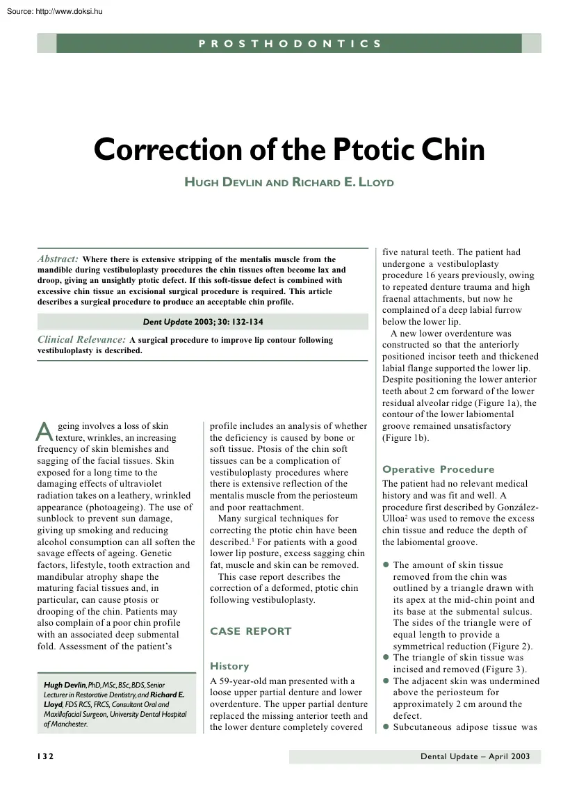
E. Lloyd, FDS RCS, FRCS, Consultant Oral and Maxillofacial Surgeon, University Dental Hospital of Manchester. 132 A 59-year-old man presented with a loose upper partial denture and lower overdenture. The upper partial denture replaced the missing anterior teeth and the lower denture completely covered five natural teeth. The patient had undergone a vestibuloplasty procedure 16 years previously, owing to repeated denture trauma and high fraenal attachments, but now he complained of a deep labial furrow below the lower lip. A new lower overdenture was constructed so that the anteriorly positioned incisor teeth and thickened labial flange supported the lower lip. Despite positioning the lower anterior teeth about 2 cm forward of the lower residual alveolar ridge (Figure 1a), the contour of the lower labiomental groove remained unsatisfactory (Figure 1b). Operative Procedure The patient had no relevant medical history and was fit and well. A procedure first described by GonzálezUlloa2
was used to remove the excess chin tissue and reduce the depth of the labiomental groove. l The amount of skin tissue removed from the chin was outlined by a triangle drawn with its apex at the mid-chin point and its base at the submental sulcus. The sides of the triangle were of equal length to provide a symmetrical reduction (Figure 2). l The triangle of skin tissue was incised and removed (Figure 3). l The adjacent skin was undermined above the periosteum for approximately 2 cm around the defect. l Subcutaneous adipose tissue was Dental Update – April 2003 P R O S T H O D O N T I C S a b Figure 1. (a) It was not possible to reduce the labiomental groove satisfactorily despite forward positioning of the lower anterior teeth of the denture. (b) The chin profile before operation (with the patient wearing the denture). removed from the base of the wound and the surrounding tissue. The wound edges were approximated using a Prolene suture that passed from the midlateral side of
the triangle through the periosteum to the same position on the other side (Figure 3). By bringing the sides of the wound together, it can be assessed whether sufficient tissue has been removed by assessing the effect on the chin profile. l Haemostasis was achieved and the Figure 2. Careful preoperative planning was followed by marking of the incision lines at operation. Figure 3. Diagrammatic representation of the surgical site. The area over and below the chin (outlined in Figure 2) was excised. The tissue surrounding the wound was undermined and closed. Dental Update – April 2003 wound closed with sutures. l The nylon skin hook was tied over a pressure gauze dressing to achieve attachment of the skin with the underlying periosteum. Further pressure dressings were applied to protect the area. Postoperative Care The sutures were removed 3 days after surgery to avoid punctate scarring. The surgery much reduced the labiomental groove and considerably improved the patient’s
lateral profile (Figure 4), and the patient was delighted with the cosmetic result. Healing of the incision wound was very satisfactory, with minimal scarring in the submental groove (Figure 5). Twelve months after surgery there has been no recurrence of ptosis. The patient has coped well with the denture, which did not require modifying, and is satisfied with the result. scar contraction resulting in lip incompetence. However, the patient described here had none of these complications. When constructing replacement dentures for a patient, obtaining an acceptable soft-tissue contour can be a daunting task for the general dental practitioner. Providing sufficient lip support to eliminate facial grooves can severely compromise the retention of the denture. Preprosthetic surgical techniques should be considered, especially as they can contribute to the success of denture treatment; however, they can also contribute to a deterioration in the facial profile – a fact that should be borne
in mind when discussing this treatment option with patients. In the atrophic edentulous mandible, vestibuloplasty procedures are often used to provide better denture stability by deepening the anterior mandibular vestibule. The denture gains stability by being better able to resist occlusal and lip forces. However, extensive stripping or ‘degloving’ of the mentalis muscle DISCUSSION Figure 4. Profile of the chin 2 months after the excision procedure (patient wearing the unmodified dentures). This technique may be useful in treating the cosmetic deformities following osseous reduction genioplasty, vestibuloplasty, or any procedure that causes extensive stripping of the mentalis muscle from the mandible. Possible short-term complications associated with the González-Ulloa procedure include wound dehiscence, infection, and haematoma formation. In the longterm, complications may include recurrence of ptosis and excessive Figure 5. The submental view 2 months after the chin
excision procedure: scarring in the neck crease is minimal. 133 P R O S T H O D O N T I C S from the mandible often causes drooping or ptosis of the lower lip. With the placement of osseointegrated implants in the anterior mandible, extensive vestibular extension for denture support and retention is not required and 10 mm of mentalis muscle attachment can remain on the mandible. This is usually sufficient to prevent lip ptosis. Prospective trials have randomly allocated patients treated with complete dentures to either a nonsurgical control group or denture provision involving preprosthetic BOOK REVIEW Oxford Handbook of Applied Dental Sciences. C Scully, ed Oxford University Press Inc., New York, 2002 (642 pp., £1995 flexicover) ISBN 0-19851096-9 The purpose of this book is ‘to outline the preclinical sciences as applied to dentistry’ and ‘to demonstrate why modern medical science is so relevant to clinical dental practice’. It is intended for candidates taking primary
dental qualifications (BDS, DDS and perhaps the IQE) and postgraduate diplomas such as MFDS. The book has 58 chapters, written by 27 authors, and the content is arranged in eight sections: Anatomy and Development, Physiology, Biochemistry, Genomics, General Immunology, Pathology, Microbiology and Behavioural Sciences. (Regrettably, Pharmacology was omitted because of lack of space.) The book provides a good, concise summary of many of the traditional preclinical topics. Information is presented in a condensed and fairly didactic manner, which I fear may encourage rote-learning, rather than deeper understanding. In several chapters, the text is augmented with diagrams, but no references are included. The text appearance is rather daunting, and I feel this could be improved by a larger typeface. The index is good, and listed most of my 134 surgery or implant abutments.3,4 Patients expressed general satisfaction with the short-term effects of preprosthetic surgery, which can be
attributed to the increase in the denture-bearing area.3 However, in this group the favourable short-term satisfaction rate with their dentures was considerably diminished after 5 years.4 It is the poor long-term success of denture treatment combined with preprosthetic surgery (and the success of implant therapy) that has reduced the need for preprosthetic surgery. sample of test words. As promised, the book does state the clinical relevance of many topics. In some chapters, the clinical application is included in the body of the text (e.g aetiology of dental developmental abnormalities) while, in the Anatomy chapters, ‘Clinical Relevance’ boxes are used. Most of these are helpful, and this format could have been adopted throughout the book. However, the box stressing the importance of lymphatic drainage (p.58) is displaced too far away from the anatomical description of the regional lymphatics (p.25) The information is up-to-date, and there is a useful section on the dental
implications of prions. The decision to divide the content into the traditional academic disciplines may be a convenient way of packaging the information. However, it does not help readers to integrate the interdisciplinary aspects of many topics. For example, the excellent account of regulation of tooth development (in Genomics) is separated from the equally good description of odontogenesis (in Anatomy & Development). These two topics were cross-referenced in the text but I feel these intimately related aspects should have been integrated. The format also creates some unnecessary duplication of material, especially between the Physiology and Biochemistry sections. There are very few errors, but I did R EFERENCES 1. 2. 3. 4. Feldman JJ. The ptotic (witch’s) chin deformity: an excisional approach. Plast Reconstr Surg 1992; 90: 207–217. González-Ulloa M. Ptosis of the chin; the witch’s chin. Plast Reconstr Surg 1972; 50: 54– 57. Boerrigter EM, Stegenga B, Raghoebar GM,
Boering G. Patient satisfaction and chewing ability with implant-retained mandibular overdentures: a comparison with new complete dentures with or without preprosthetic surgery. J Oral Maxillofac Surg 1995; 53: 1167–1173. Raghoeber GM, Meijer HJA, Stegenga B, van’t Hof MA, van Oort RP, Vissink A. Effectiveness of three treatment modalities for the edentulous mandible. A five year randomised clinical trial Clin Oral Implant Res 2000; 11: 195–201. notice that nerve axon diameters were (twice) given in millimetres rather than micrometres. The coverage is quite comprehensive but, inevitably, there are some omissions (e.g pulse oximetry; junctional epithelium; effects of acid etching on enamel; positive and negative reinforcement). Handbooks of Clinical Dentistry have proved very popular with students (and some staff). I am not sure if this Handbook of Applied Dental Sciences will prove as popular, but it does provide a portable information source and revision aid that is likely to
appeal to many students. R. Orchardson University of Glasgow CPD REMINDER Achieve all 15 hours of verifiable CPD by simply completing the questions in each issue of Dental Update or on our website: www.dental-updatecouk To gain your verifiable CPD you must submit your answers before receiving the next issue where the answers are published. Dental Update – April 2003
consumption can all soften the savage effects of ageing. Genetic factors, lifestyle, tooth extraction and mandibular atrophy shape the maturing facial tissues and, in particular, can cause ptosis or drooping of the chin. Patients may also complain of a poor chin profile with an associated deep submental fold. Assessment of the patient’s profile includes an analysis of whether the deficiency is caused by bone or soft tissue. Ptosis of the chin soft tissues can be a complication of vestibuloplasty procedures where there is extensive reflection of the mentalis muscle from the periosteum and poor reattachment. Many surgical techniques for correcting the ptotic chin have been described.1 For patients with a good lower lip posture, excess sagging chin fat, muscle and skin can be removed. This case report describes the correction of a deformed, ptotic chin following vestibuloplasty. CASE REPORT History Hugh Devlin, PhD, MSc, BSc, BDS, Senior Lecturer in Restorative Dentistry, and Richard
E. Lloyd, FDS RCS, FRCS, Consultant Oral and Maxillofacial Surgeon, University Dental Hospital of Manchester. 132 A 59-year-old man presented with a loose upper partial denture and lower overdenture. The upper partial denture replaced the missing anterior teeth and the lower denture completely covered five natural teeth. The patient had undergone a vestibuloplasty procedure 16 years previously, owing to repeated denture trauma and high fraenal attachments, but now he complained of a deep labial furrow below the lower lip. A new lower overdenture was constructed so that the anteriorly positioned incisor teeth and thickened labial flange supported the lower lip. Despite positioning the lower anterior teeth about 2 cm forward of the lower residual alveolar ridge (Figure 1a), the contour of the lower labiomental groove remained unsatisfactory (Figure 1b). Operative Procedure The patient had no relevant medical history and was fit and well. A procedure first described by GonzálezUlloa2
was used to remove the excess chin tissue and reduce the depth of the labiomental groove. l The amount of skin tissue removed from the chin was outlined by a triangle drawn with its apex at the mid-chin point and its base at the submental sulcus. The sides of the triangle were of equal length to provide a symmetrical reduction (Figure 2). l The triangle of skin tissue was incised and removed (Figure 3). l The adjacent skin was undermined above the periosteum for approximately 2 cm around the defect. l Subcutaneous adipose tissue was Dental Update – April 2003 P R O S T H O D O N T I C S a b Figure 1. (a) It was not possible to reduce the labiomental groove satisfactorily despite forward positioning of the lower anterior teeth of the denture. (b) The chin profile before operation (with the patient wearing the denture). removed from the base of the wound and the surrounding tissue. The wound edges were approximated using a Prolene suture that passed from the midlateral side of
the triangle through the periosteum to the same position on the other side (Figure 3). By bringing the sides of the wound together, it can be assessed whether sufficient tissue has been removed by assessing the effect on the chin profile. l Haemostasis was achieved and the Figure 2. Careful preoperative planning was followed by marking of the incision lines at operation. Figure 3. Diagrammatic representation of the surgical site. The area over and below the chin (outlined in Figure 2) was excised. The tissue surrounding the wound was undermined and closed. Dental Update – April 2003 wound closed with sutures. l The nylon skin hook was tied over a pressure gauze dressing to achieve attachment of the skin with the underlying periosteum. Further pressure dressings were applied to protect the area. Postoperative Care The sutures were removed 3 days after surgery to avoid punctate scarring. The surgery much reduced the labiomental groove and considerably improved the patient’s
lateral profile (Figure 4), and the patient was delighted with the cosmetic result. Healing of the incision wound was very satisfactory, with minimal scarring in the submental groove (Figure 5). Twelve months after surgery there has been no recurrence of ptosis. The patient has coped well with the denture, which did not require modifying, and is satisfied with the result. scar contraction resulting in lip incompetence. However, the patient described here had none of these complications. When constructing replacement dentures for a patient, obtaining an acceptable soft-tissue contour can be a daunting task for the general dental practitioner. Providing sufficient lip support to eliminate facial grooves can severely compromise the retention of the denture. Preprosthetic surgical techniques should be considered, especially as they can contribute to the success of denture treatment; however, they can also contribute to a deterioration in the facial profile – a fact that should be borne
in mind when discussing this treatment option with patients. In the atrophic edentulous mandible, vestibuloplasty procedures are often used to provide better denture stability by deepening the anterior mandibular vestibule. The denture gains stability by being better able to resist occlusal and lip forces. However, extensive stripping or ‘degloving’ of the mentalis muscle DISCUSSION Figure 4. Profile of the chin 2 months after the excision procedure (patient wearing the unmodified dentures). This technique may be useful in treating the cosmetic deformities following osseous reduction genioplasty, vestibuloplasty, or any procedure that causes extensive stripping of the mentalis muscle from the mandible. Possible short-term complications associated with the González-Ulloa procedure include wound dehiscence, infection, and haematoma formation. In the longterm, complications may include recurrence of ptosis and excessive Figure 5. The submental view 2 months after the chin
excision procedure: scarring in the neck crease is minimal. 133 P R O S T H O D O N T I C S from the mandible often causes drooping or ptosis of the lower lip. With the placement of osseointegrated implants in the anterior mandible, extensive vestibular extension for denture support and retention is not required and 10 mm of mentalis muscle attachment can remain on the mandible. This is usually sufficient to prevent lip ptosis. Prospective trials have randomly allocated patients treated with complete dentures to either a nonsurgical control group or denture provision involving preprosthetic BOOK REVIEW Oxford Handbook of Applied Dental Sciences. C Scully, ed Oxford University Press Inc., New York, 2002 (642 pp., £1995 flexicover) ISBN 0-19851096-9 The purpose of this book is ‘to outline the preclinical sciences as applied to dentistry’ and ‘to demonstrate why modern medical science is so relevant to clinical dental practice’. It is intended for candidates taking primary
dental qualifications (BDS, DDS and perhaps the IQE) and postgraduate diplomas such as MFDS. The book has 58 chapters, written by 27 authors, and the content is arranged in eight sections: Anatomy and Development, Physiology, Biochemistry, Genomics, General Immunology, Pathology, Microbiology and Behavioural Sciences. (Regrettably, Pharmacology was omitted because of lack of space.) The book provides a good, concise summary of many of the traditional preclinical topics. Information is presented in a condensed and fairly didactic manner, which I fear may encourage rote-learning, rather than deeper understanding. In several chapters, the text is augmented with diagrams, but no references are included. The text appearance is rather daunting, and I feel this could be improved by a larger typeface. The index is good, and listed most of my 134 surgery or implant abutments.3,4 Patients expressed general satisfaction with the short-term effects of preprosthetic surgery, which can be
attributed to the increase in the denture-bearing area.3 However, in this group the favourable short-term satisfaction rate with their dentures was considerably diminished after 5 years.4 It is the poor long-term success of denture treatment combined with preprosthetic surgery (and the success of implant therapy) that has reduced the need for preprosthetic surgery. sample of test words. As promised, the book does state the clinical relevance of many topics. In some chapters, the clinical application is included in the body of the text (e.g aetiology of dental developmental abnormalities) while, in the Anatomy chapters, ‘Clinical Relevance’ boxes are used. Most of these are helpful, and this format could have been adopted throughout the book. However, the box stressing the importance of lymphatic drainage (p.58) is displaced too far away from the anatomical description of the regional lymphatics (p.25) The information is up-to-date, and there is a useful section on the dental
implications of prions. The decision to divide the content into the traditional academic disciplines may be a convenient way of packaging the information. However, it does not help readers to integrate the interdisciplinary aspects of many topics. For example, the excellent account of regulation of tooth development (in Genomics) is separated from the equally good description of odontogenesis (in Anatomy & Development). These two topics were cross-referenced in the text but I feel these intimately related aspects should have been integrated. The format also creates some unnecessary duplication of material, especially between the Physiology and Biochemistry sections. There are very few errors, but I did R EFERENCES 1. 2. 3. 4. Feldman JJ. The ptotic (witch’s) chin deformity: an excisional approach. Plast Reconstr Surg 1992; 90: 207–217. González-Ulloa M. Ptosis of the chin; the witch’s chin. Plast Reconstr Surg 1972; 50: 54– 57. Boerrigter EM, Stegenga B, Raghoebar GM,
Boering G. Patient satisfaction and chewing ability with implant-retained mandibular overdentures: a comparison with new complete dentures with or without preprosthetic surgery. J Oral Maxillofac Surg 1995; 53: 1167–1173. Raghoeber GM, Meijer HJA, Stegenga B, van’t Hof MA, van Oort RP, Vissink A. Effectiveness of three treatment modalities for the edentulous mandible. A five year randomised clinical trial Clin Oral Implant Res 2000; 11: 195–201. notice that nerve axon diameters were (twice) given in millimetres rather than micrometres. The coverage is quite comprehensive but, inevitably, there are some omissions (e.g pulse oximetry; junctional epithelium; effects of acid etching on enamel; positive and negative reinforcement). Handbooks of Clinical Dentistry have proved very popular with students (and some staff). I am not sure if this Handbook of Applied Dental Sciences will prove as popular, but it does provide a portable information source and revision aid that is likely to
appeal to many students. R. Orchardson University of Glasgow CPD REMINDER Achieve all 15 hours of verifiable CPD by simply completing the questions in each issue of Dental Update or on our website: www.dental-updatecouk To gain your verifiable CPD you must submit your answers before receiving the next issue where the answers are published. Dental Update – April 2003
 I. András 1015 körül született Vazul fiaként. Miután I. István fia, Imre herceg 1031. szeptember 2-án meghalt, a koronára csupán Vazulnak, Mihály fiának és Szár László három fiának volt joga; Orseolo Péter pártja azonban Vazul szemeit kiszúratta, Szár László fiait pedig Magyarország elhagyására kényszeríttette. András Lodomeriába, innen pedig a kunokhoz, s végül