Datasheet
Year, pagecount:2003, 5 page(s)
Language:English
Downloads:5
Uploaded:April 14, 2012
Size:98 KB
Institution:
-
Comments:
Attachment:-
Download in PDF:Please log in!
Comments
No comments yet. You can be the first!
Most popular documents in this category
Content extract
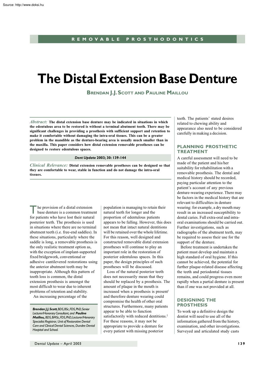
R E M O V A B L E R ES MT OHVO AB RO S TS H O D O N T I C S P R O DLOE NP T I C The Distal Extension Base Denture BRENDAN J.J SCOTT AND PAULINE MAILLOU Abstract: The distal extension base denture may be indicated in situations in which the edentulous area to be restored is without a terminal abutment tooth. There may be significant challenges in providing a prosthesis with sufficient support and retention to make it comfortable without damaging the intra-oral tissues. This can be a greater problem in the mandible as the denture-bearing area is usually much smaller than in the maxilla. This paper considers how distal extension removable prostheses can be designed to restore edentulous spaces. Dent Update 2003; 30: 139-144 Clinical Relevance: Distal extension removable prostheses can be designed so that they are comfortable to wear, stable in function and do not damage the intra-oral tissues. T he provision of a distal extension base denture is a common treatment for patients who
have lost their natural posterior teeth. The prosthesis is used in situations where there are no terminal abutment teeth (i.e free-end saddles) In these situations, particularly where the saddle is long, a removable prosthesis is the only realistic treatment option as, with the exception of implant-supported fixed bridgework, conventional or adhesive cantilevered restorations using the anterior abutment teeth may be inappropriate. Although this pattern of tooth loss is common, the distal extension prosthesis is amongst the most difficult to wear due to inherent problems of retention and stability. An increasing percentage of the Brendan J.J Scott, BDS, BSc, FDS, PhD, Senior Lecturer/Honorary Consultant, and Pauline Maillou, BDS, BMSc, FDS, PhD, Lecturer/Honorary Specialist Registrar, Unit of Restorative Dental Care and Clinical Dental Sciences, Dundee Dental Hospital and School. Dental Update – April 2003 population is managing to retain their natural teeth for longer and the
proportion of edentulous patients appears to be falling. However, this does not mean that intact natural dentitions will be retained over the whole lifetime. For this reason, well designed and constructed removable distal extension prostheses will continue to play an important role in the restoration of posterior edentulous spaces. In this paper, the design principles of such prostheses will be discussed. Loss of the natural posterior teeth does not necessarily mean that they should be replaced by a prosthesis. The amount of plaque in the mouth is increased when a prosthesis is present1 and therefore denture wearing could compromise the health of other oral structures. Furthermore, many patients appear to be able to function satisfactorily with reduced dentitions.2 For these reasons, it may not be appropriate to provide a denture for every patient with missing posterior teeth. The patients’ stated desires related to chewing ability and appearance also need to be considered carefully
in making a decision. PLANNING PROSTHETIC TREATMENT A careful assessment will need to be made of the patient and his/her suitability for rehabilitation with a removable prosthesis. The dental and medical history should be recorded, paying particular attention to the patient’s account of any previous denture-wearing experience. There may be factors in the medical history that are relevant to difficulties in denture wearing: for example, a dry mouth may result in an increased susceptibility to dental caries. Full extra-oral and intraoral examinations should be carried out Further investigations, such as radiographs of the abutment teeth, may be required to assess their use for the support of the denture. Before treatment is undertaken the patient must develop and maintain a high standard of oral hygiene. If this cannot be achieved, the potential for further plaque-related disease affecting the teeth and periodontal tissues remains, and could progress even more rapidly when a partial
denture is present than if one was not provided at all. DESIGNING THE PROSTHESIS To work up a definitive design the dentist will need to use all of the information gathered from the history, examination, and other investigations. Surveyed and articulated study casts 139 R E M OVA B L E P R O S T H O D O N T I C S a b Figure 1. Casts to show the support area available (shaded in green) on the tissues in the maxilla and mandible related specifically to the distal extension saddles. (a) The denture base can be extended over a wide area for a bilateral distal extension saddle in the maxilla. Further support may be gained by extending the prosthesis anterior to the shaded area. (b) The morphology of the mandible restricts the available base coverage for a lower bilateral distal extension saddle denture. will be useful to the dentist in the design process. Factors that need to be considered in the denture design include: l saddles to be replaced; l path of insertion of prosthesis; l
support – tooth/mucosa or mucosa only; l retention – direct and indirect; l bracing; l connectors; l overview for prosthesis stability and hygienic design. At this stage any sites at which tooth preparations are necessary should be identified before making the working impressions. Such preparations may be required to form appropriate contours to some tooth surfaces for siting particular components of a partial denture – e.g preparation of occlusal rest seats. It may also be necessary to carry out tooth preparation in the arch opposing the one for which a denture is planned, in order to create sufficient space to ensure that the components of a partial denture do not interfere with the occlusion. Examples of such modifications include modest reductions of overerupted teeth. Saddles The saddles are the parts of the denture that carry the artificial teeth. The base of the saddle will rest on the underlying tissues and will need adequate extension over the denture-bearing area 140
(see below). The artificial teeth should be sited to ensure that the denture will not tip under occlusal loading. The polished surfaces should be designed so that the denture is well retained and tolerated. The shape of the denture base becomes more critical when teeth have been missing for many years with associated resorption of bone. Furthermore, in some circumstances, the space available for a lower prosthesis may be limited because of changes in activity of the tongue musculature (tongue spread). It may therefore be necessary to narrow the denture teeth or even omit one or more (e.g second molars) to allow the patient to tolerate the prosthesis more easily. It is important to consider the status and prognosis of the teeth that will act as abutments to the saddles. Critical abutment teeth are those that are essential to the stability of the prosthesis: for example, the most posterior tooth in the arch that would offer support, retention and bracing against posterior movement of the
prosthesis. If a single distal abutment tooth remains on the side of the mouth opposite to a distal extension saddle, all efforts must be made to retain it. Loss of such a tooth would result in an arch that was originally a unilateral distal extension saddle becoming one with bilateral distal extension saddles. Almost certainly this would result in any prosthesis becoming significantly less stable as there would no longer be any posterior teeth present. The patient should be advised of the particular importance of cleaning abutment teeth at the posterior aspects of the arch – areas that can easily be neglected. Aids such as a finger bandage may be useful in cleaning single standing posterior abutment teeth. Support Unlike a bounded saddle prosthesis, in which it is possible to gain full support from the abutment teeth, occlusal loading of the saddle area of a distal extension denture will result in some force being directed towards the tissues of the edentulous ridge directly
underneath. The size and shape of the ridge, as well as the thickness and density of the overlying fibrous connective tissue and mucosa, will also influence the support offered. Generally these factors are more favourable in the maxilla, where the soft tissues may be thicker and less displaceable than those in the mandible. The amount and type of the underlying alveolar bone should also be considered. The opportunity in the maxilla to achieve greater coverage of the prosthesis on the alveolar ridges and across the hard palate allows more favourable loading than in the mandible, where the available support area is reduced (Figure 1). Therefore distal extension dentures are potentially less damaging in the maxilla. Support becomes a much greater problem if there is a knife-edge form to the ridge or if the pattern of alveolar resorption results in mobile fibrous tissue overlying the alveolar bone, particularly in the mandibular denture-bearing area. In a distal extension base denture,
optimum support is gained by using the natural teeth where possible. However, all distal extension base dentures will derive at least part of their support from the tissues underlying the saddle. When considering the component of mucosal support the objective is to minimize the load per unit area being transferred to the underlying ridge. This is usually done by extending the denture base maximally without interfering with structures that Dental Update – April 2003 R E M OVA B L E P R O S T H O D O N T I C S Figure 2. A design for a bilateral distal extension mandibular denture with the occlusal rests (shown by the arrows) sited on the mesial aspects of the mandibular teeth. influence movement of the border tissues. 3 l In the mandible the base should extend over the alveolar ridge, on to the buccal shelf and retromolar pad and fully into the functional sulci. l In the maxilla, base extension over a wide area of the alveolar ridge and palate will allow the most favourable load
distribution. The force to the ridge from occlusal loading can be minimized by reducing the size of the occlusal table by omitting denture teeth (e.g second molars), using smaller teeth or by narrowing their buccolingual width. It may be that the denture will be supported only on the soft tissues overlying the alveolar ridge (mucosal support). This is generally less damaging in the maxilla as it is usually possible to gain adequate support from the palatal mucosa. Great care should be taken when embarking on this course in the mandible as further alveolar ridge resorption could result in this becoming a ‘gum stripper’ denture. However, there are occasions in which this may be the only option – for example, for a transitional partial denture. Tooth support should be used where possible on the distal extension denture, especially in the mandible. Clearly this will depend on the periodontal support of the natural teeth being satisfactory: where possible, there should be support on
the abutment tooth adjacent to the saddle. There is less chance of distal tipping of the abutment tooth if the rest is Dental Update – April 2003 placed on the mesio-occlusal aspect (Figure 2), although this assumption has been challenged.4 In a unilateral saddle, cross-arch tooth support on the opposite side is desirable. Even if support is shared between the teeth and mucosa, occlusal loading may result in displacement of the distal extension saddle. As the underlying mucosa may be much more displaceable than a tooth supported by its periodontal ligament, the denture may rotate around the occlusal rest on the abutment tooth in function. In the mandible this can be a particular problem and may be exacerbated if there is further alveolar ridge resorption. The approaches used to address this range from specific impression techniques for the saddle (e.g altered cast technique) to allowing the saddle part of the denture to move independently by means of a flexible connector (stress
broken design). Stress broken designs are rarely used as it is very difficult to predict how the position of the hinge joint or flexible connector will influence the loading on the tissues of the edentulous area.5 The Altered Cast Technique The aim of the altered cast technique is to produce an impression of the mucosa of the saddle area such that movement of the denture base in function will be minimized.6 Detailed accounts of the clinical and laboratory stages are given elsewhere.6–8 The procedure is usually carried out when the casting is fitted. A close-fitting, non-spaced impression tray is attached to the cobalt chromium a framework. A mucodisplacement impression can be recorded with the close-fitting impression tray and an appropriate impression material (e.g zinc oxide/eugenol). Following the impression, in which care should be taken to avoid any direct pressure to the tray while ensuring the casting is seated properly around the teeth, the saddle is sectioned away from the
original master cast. The metal framework and associated impression are seated and a new area of the cast formed by casting stone into the impression (Figure 3). The use of this technique may help prevent the denture from tilting when the saddle is loaded occlusally. Retention and Stability Retentive forces are those that resist vertical movement of the denture away from the underlying ridge. When designing a partial denture the objective is to provide components that resist the displacing forces that may be applied to the denture during function. In the case of the distal extension prosthesis, this is usually achieved by siting retentive components close to (direct retention) and distant to (indirect retention) the saddle. Additional ways of achieving retention include physical forces from the denture base coverage of the mucosa, and the design of the polished surfaces to harmonize with a patient’s muscular control.5 In the maxilla the opportunity to spread the denture base widely
can help the retention of the prosthesis. However, retention problems may be b Figure 3. The laboratory stages of the altered cast technique An impression has been recorded in a close-fitting impression tray attached to the casting.The original distal extension saddle area of the master cast has been sectioned. (a) The casting secured to the original dentate portion of the master cast by means of yellow wax overlying the occlusal rests on teeth /5 and 5/. (b) The impression surface to which new stone will be poured. 141 R E M OVA B L E P R O S T H O D O N T I C S Figure 4. A design for a unilateral distal extension mandibular denture to show the principle of indirect retention.The tendency of the distal part of the saddle to lift away from the tissues rotating around the fulcrum axis (AA) is resisted by the premolar occlusal rest (B) and the lingual plate connector (C). encountered with a mandibular distal extension denture: adhesive and cohesive forces between the mucosa,
saliva and denture base may not make a major contribution to retention, especially if the available denture-bearing area is small. Furthermore, there is only one abutment tooth at the anterior end of the saddle that can offer direct retention – often either a premolar or a canine, which have small mesiodistal dimensions. If an occlusally approaching clasp is used it will need to be made from a material that is sufficiently flexible (e.g stainless steel) to allow the undercut to be engaged but this may distort compared with a cast material (e.g cobalt chromium) Where aesthetics are important, gold or toothcoloured acetyl resin may be the material of choice. The position of the survey line, together with the patient’s feedback on aesthetics, will determine whether the clasp will be occlusally approaching or gingivally approaching. A gingivally approaching clasp may have less potential for causing unwanted movement of the abutment tooth when the saddle moves under occlusal load as it
will disengage from the undercut. The RPI system is a form of component design with the objective of preventing or minimizing the potential for damage to the abutment tooth during loading of the distal extension saddle. The occlusal rest (R) is placed on the 142 mesial aspect of the abutment tooth. The plate (P) is in contact with a small guide plane on the distal aspect of the abutment tooth and is designed so that it is able to move towards the tissues, therefore disengaging from the tooth surface, when the saddle is loaded. Similarly the I-bar, which engages an undercut on the buccal surface of the tooth, will move towards the tissues and away from the tooth when the saddle is loaded. This system of design has the advantage of reducing the potentially damaging torquing forces to the abutment tooth due to occlusal loading of the saddle. More detailed descriptions of this concept are given in the literature.5,9 The absence of a distal abutment tooth may cause the saddle to lift away
from the tissues during function. For this reason components that offer indirect retention need to be included in the denture. An indirect retainer is a support component that rests on a firm structure on the side of the clasp axis opposite to the saddle (Figure 4). When the distal extension saddle attempts to lift during function, the supporting components become the fulcrum of movement instead of the clasp axis, increasing the force required to dislodge the denture. The support component should be positioned as far from the axis of rotation through the clasps as possible to optimize the indirect retention. In addition, the clasp axis should be as close to the saddle as possible. It may be necessary to change tooth contours to achieve more effective retention. The addition of composite a resin to the buccal aspect of a tooth provides a simple solution to the problem of an unsuitable abutment shape. In some circumstances it may be necessary to construct a crown for an abutment tooth,
in which case the crown can be designed to optimize the components on the prosthesis. Features include siting an appropriate rest seat, a well shaped bulbosity for clasping, and parallel surfaces to avoid undercuts between the prosthesis and abutment tooth (Figure 5). However, some consideration should be given to the possible torquing forces that might arise around the tooth from occlusal loading on a distal extension base denture. This could compromise the abutment, e.g if there is associated periodontal attachment loss. The occlusal relationships will need to be considered in relation to the stability of the prosthesis. The denture should be designed so that no interferences with the relationship of the natural teeth are created. Eccentric interferences during lateral or protrusive excursions of the mandible could destabilize the prosthesis. Overerupted, drifted and tilted teeth can contribute to this type of problem and therefore the occlusion should be carefully assessed before
commencing treatment. Additional Methods of Securing Retention Swinglock dentures (Figure 6) can be constructed if there are particular difficulties in retention.10 These consist of a hinged labial bar and locking device. Some patients may not tolerate the labial b Figure 5. Features that can be incorporated into a crown on an abutment tooth adjacent to a distal extension saddle. (a) The occlusal rest seat and a mesiobuccal undercut on the crown (b) A cobalt-chromium-based denture in place.The occlusal rest fits over the rest seat and the terminal part of the gold clasp is sited in the undercut area. Dental Update – April 2003 R E M OVA B L E P R O S T H O D O N T I C S a b Figure 6. A mandibular swinglock denture (a) The hinged labial flange and locking segment (b) The prosthesis in place to show the position of the hinge. a b to be carefully planned. As well as the features considered above, it is important that the patient is comfortable in function and is able to
clean the teeth and dentures well. Patients should be reviewed at regular intervals to identify any early signs of damage to intra-oral tissues. This allows the opportunity for appropriate modifications to be made before more serious problems arise. ACKNOWLEDGEMENTS We would like to thank Dr Kenneth Tyson for composing the line drawings for Figures 2 and 4. REFERENCES 1. Figure 7. A mandibular distal extension denture retained by extracoronal precision attachments on the abutment teeth.The prosthesis had been worn for over 10 years (a) The extracoronal attachment linked to a crown on the abutment tooth. (b) The locking pin in the denture that engages the attachment. bar and every attempt should be made to keep the bulk of the appliance to a minimum. It is also possible that large forces will be applied to the natural teeth during occlusal loading. This problem would be exacerbated if further alveolar resorption in the saddle areas takes place as there would be a greater tendency
for the appliance to rock. An alternative approach is to use precision attachments on the abutment teeth into which matching components in the prosthesis can engage. Again there may be the potential for damage to the abutment tooth, although these can function for many years (Figure 7). Very careful thought is necessary before embarking on these types of designs. They are contraindicated if the tooth structure or periodontal tissues are compromised and unless the patient has excellent plaque control. CONNECTING THE COMPONENTS OF THE DENTURE An appropriate major connector is required to link the distal extension saddle with other components of the 144 prosthesis. Factors that will determine a suitable connector design include: l the space available in the anterior part of the mouth; l the health of the teeth and periodontal tissues; and l the need to consider its use in indirect retention. Many types of connector are described (e.g palatal plate, anterior palatal bar, lingual plate,
lingual bar, dental bar).5,8 To maintain periodontal health it is preferable that the connector avoids coverage of the gingival tissues (Figure 8). Some connectors, such as lingual bars, are an ineffective source of indirect retention on their own, even though there are advantages in leaving the gingival margins of teeth remote from the saddle uncovered. In such cases, other sources of indirect retention need to be found, provided that there are already components on the prosthesis to provide effective direct retention. CONCLUSIONS A distal extension base denture needs Bates JF, Addy M. Partial dentures and plaque accumulation. J Dent 1978; 6: 285–293 2. Elias AC, Sheiham A The relationship between satisfaction with mouth and number and position of teeth. J Oral Rehabil 1998; 25: 649–661 3. McGivney GP, Carr AB Support for the distal extension base. In: McCracken’s Removable Partial Prosthodontics, 10th ed. St Louis: Mosby, 2000; pp.337–354 4. Feingold GM, Grant AA, Johnson W
The effect of partial denture design on abutment tooth and saddle movement. J Oral Rehabil 1986; 13: 549–557 5. Davenport JC, Basker RM, Heath JR, Ralph JP, Glantz PO. A Clinical Guide to Removable Partial Denture Design. London: British Dental Association, 2000. 6. Barsby MJ, Schwarz WD Partial dentures with free-end saddles: the altered cast technique. Dent Update 1987; 14: 101–110. 7. Feit DB The altered cast impression technique revisited. J Am Dent Assoc 1999; 130: 1476–1481 8. Davenport JC, Basker RM, Heath JR, Ralph JP, Glantz PO. A Clinical Guide to Removable Partial Dentures. London: British Dental Association, 2000 9. Krol AJ RPI (Rest, Proximal Plate, I Bar) clasp retainer and its modifications. Dent Clin North Am 1973; 17: 631–649. 10. Chan MFW-Y, Adams D, Brudvik JS The swinglock removable partial denture in clinical practice Dent Update 1998; 25: 80–84. Figure 8. A dental bar connector which avoids coverage of the gingival margins.These can be well tolerated by
the patient and may have a role in indirect retention. Dental Update – April 2003
 Just like you draw up a plan when you’re going to war, building a house, or even going on vacation, you need to draw up a plan for your business. This tutorial will help you to clearly see where you are and make it possible to understand where you’re going.