A doksi online olvasásához kérlek jelentkezz be!
A doksi online olvasásához kérlek jelentkezz be!
Nincs még értékelés. Legyél Te az első!
Legnépszerűbb doksik ebben a kategóriában


Tartalmi kivonat
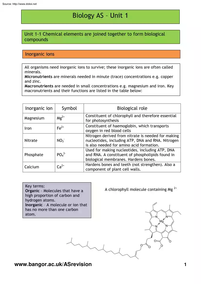
Source: http://www.doksinet Biology AS – Unit 1 Unit 1-1 Chemical elements are joined together to form biological compounds Inorganic ions All organisms need inorganic ions to survive; these inorganic ions are often called minerals. Micronutrients are minerals needed in minute (trace) concentrations e.g copper and zinc. Macronutrients are needed in small concentrations e.g magnesium and iron Key macronutrients and their functions are listed in the table below: Inorganic ion Symbol Magnesium Mg2+ Iron Fe2+ Nitrate NO3- Phosphate PO43- Calcium Ca2+ Biological role Constituent of chlorophyll and therefore essential for photosynthesis Constituent of haemoglobin, which transports oxygen in red blood cells Nitrogen derived from nitrate is needed for making nucleotides, including ATP, DNA and RNA. Nitrogen is also needed for amino acid formation. Used for making nucleotides, including ATP, DNA and RNA. A constituent of phospholipids found in biological membranes. Hardens bones
Hardens bones and teeth (not strengthen). Also a component of plant cell walls. Key terms: Organic – Molecules that have a high proportion of carbon and hydrogen atoms. Inorganic – A molecule or ion that has no more than one carbon atom. www.bangoracuk/ASrevision A chlorophyll molecule containing Mg 2+ 1 Source: http://www.doksinet Water Water is a polar molecule; the oxygen end of the molecule has a negative charge and the hydrogen atoms have a positive charge. This uneven distribution of charge is called a dipole. When two water molecules are in close contact the opposing charges attract each other forming a hydrogen bond. Individually hydrogen bonds are weak, but many hydrogen bonds (between many water molecules) form a lattice-like framework which is much stronger. This attraction between water molecules is called cohesion. Key terms: Dipole – A polar molecule which has a positive and negative charge, separated by a very small distance. Hydrogen bond – The weak
attractive force between a hydrogen atom (with a partial positive charge) and an atom with a partial negative charge, usually oxygen or nitrogen. www.bangoracuk/ASrevision 2 Source: http://www.doksinet Water Water’s properties make it essential for life, as we understand it. Property of water Function Water is a solvent The positive and negative parts of the water molecule attract other charged particles, such as ions and other polar molecules, such as glucose. Ions and polar molecules can dissolve in water. Non-polar molecules such as lipids do not dissolve in water. Water as a transport medium Chemical reactions take place in water Water has a high specific heat capacity Water has a high latent heat of vaporisation Cohesion Surface tension Density Blood is largely water and transports many dissolved substances around the body. Minerals dissolved in water are transported from the root to the leaves via the xylem in plants. Transport of ions and polar molecules allows
chemical reactions to take place when particles or molecules meet. A large amount of heat energy is needed to raise the temperature of water. This prevents large fluctuations in water temperature. This keeps the temperature of aquatic environments stable so that organisms do not have to endure extremes of temperature. This also allows enzymes within cells to work effectively. Due to cohesion between water molecules (caused by hydrogen bonding) a large amount of heat energy is needed to change water from a liquid to a vapour state (gas). This process of evaporation transfers heat energy and is a very effective way of cooling the body e.g sweating or panting. Evaporation of water from a surface causes cooling. The attraction between water molecules allows water to be transported, in long columns, up the xylem vessels of even the tallest trees. At ordinary temperatures water has the highest surface tension of any liquid except mercury. In a pond the cohesion between water molecules
supports organisms, such as pond skaters, allowing them to walk on water. Water has a maximum density at 4 oC; ice is less dense and therefore floats on the surface and insulates the water beneath it. This reduces the tendency for large bodies of water to freeze completely allowing organisms to survive. www.bangoracuk/ASrevision 3 Source: http://www.doksinet Carbohydrates - Monosaccharides Carbohydrates are organic compounds which contain the atoms carbon, hydrogen and oxygen. The basic unit of a carbohydrate is a monosaccharide Two monosaccharides form a disaccharide. Many monosaccharide molecules form a polysaccharide. A polysaccharide is a type of polymer Monosaccharides are sweet and soluble. They are the building blocks for the other larger carbohydrates. Monosaccharides have the general formula (CH2O)n and they can be grouped according to the number of carbon atoms they have. A triose sugar has three carbon atoms, a pentose sugar has five carbon atoms and a hexose sugar has
six carbon atoms. Triose (3C) Pentose (5C) Type of monosaccharide Hexose (6C) Function Triose Important in metabolism. Triose sugars are intermediates in the reactions of respiration and photosynthesis. Pentose Constituents of nucleotides e.g deoxyribose in DNA, ribose in RNA, ATP and ADP. Hexose Glucose is a hexose sugar. Glucose is a source of energy in respiration. Carbon-hydrogen and carbon-carbon bonds are broken to release energy, which is transferred to make adenosine triphosphate (ATP). www.bangoracuk/ASrevision 4 Source: http://www.doksinet Carbohydrates - Isomers Isomers have the same chemical formula and the same number of atoms; the atoms are simply arranged differently. The ring form of the monosaccharide glucose has two isomers α glucose and β glucose. They both have the same chemical formula C6H12O6, but the H and OH atoms are arranged differently at carbon 1. α glucose β glucose At carbon 1 α glucose has a hydrogen atom above and a hydroxyl
group (OH) below, but if you look at the β glucose molecule you will notice that carbon 1 has a hydroxyl group above and a hydrogen atom below. The H and OH atoms at carbon 1 have been flipped; this is the only difference between α glucose and β glucose. www.bangoracuk/ASrevision 5 Source: http://www.doksinet Carbohydrates - Disaccharides Disaccharides are composed of two monosaccharide sub-units bonded with the formation of a glycosidic bond and the elimination of water. This is an example of a condensation reaction. When two α glucose molecules are joined by condensation reaction the disaccharide maltose is formed. The diagram below shows water being removed between C1 of the first glucose molecule and C4 of the second (the atoms removed are shown in red). A 1-4 glycosidic bond is formed. Two molecules of α glucose Maltose and water The glycosidic bond can be broken by hydrolysis. During hydrolysis water is chemically added to break the glycosidic bond. Hydrolysis of
maltose is shown below The atoms added during hydrolysis are shown in red. Maltose and water www.bangoracuk/ASrevision Two α glucose molecules 6 Source: http://www.doksinet Carbohydrates - Disaccharides The table below summarises information about disaccharides. Disaccharide Component monosaccharide Biological role Maltose Glucose and Glucose In germinating seeds Sucrose Glucose and Fructose A product of photosynthesis which is transported in the phloem Lactose Glucose and Galactose Found in mammalian milk The reaction below shows the disaccharide lactose being hydrolysed. The glycosidic bond is broken and the monosaccharides glucose and galactose are formed. Benedict’s reagent is used to test for reducing sugars. Heat is needed for this reaction (80oC or above). Reducing sugars reduce blue copper ll sulphate forming copper l sulphate, which is a brick red precipitate. Examples include all the monosaccharides and the disaccharides lactose and maltose.
www.bangoracuk/ASrevision 7 Source: http://www.doksinet Carbohydrates – Testing for reducing sugars Sucrose is called a non-reducing sugar because it does not reduce copper ll sulphate. The Benedict’s test will not work; Benedict’s will remain blue. Sucrose must first be hydrolysed by boiling in dilute hydrochloric acid. Glucose and fructose are formed The acid must be neutralised with dilute sodium hydroxide before testing with Benedict’s reagent. This should now give a positive result; glucose and fructose are reducing sugars which readily donate an electron to reduce copper II sulphate to form the brick-red precipitate copper I sulphate. www.bangoracuk/ASrevision 8 Source: http://www.doksinet Carbohydrates - Polysaccharides Polysaccharides are large complex polymers. They are formed from very large numbers of identical monosaccharide units, which are their monomers, linked by glycosidic bonds formed by condensation reaction. Starch allows plants to store glucose.
Starch is made up of α glucose monomers, added one at a time by condensation reaction. Glucose can be easily added or removed Starch has two types of polysaccharide, amylose and amylopectin. Amylose is unbranched and coils; each α glucose monomer added forms a C1 – C4 glycosidic bond with the adjacent glucose molecule. Amylopectin is branched as it forms C1 - C4 gylcosidic bonds and C1 – C6 glycosidic bonds. Starch is compact and has no osmotic effect on the cell; it does not affect the water potential of the cell. Amylose is a polysaccharide component of starch. It is unbranched and coiled Amylopectin is a polysaccharide component of starch. Each branch point is formed by a C1 – C6 glycosidic bond. www.bangoracuk/ASrevision 9 Source: http://www.doksinet Carbohydrates - Polysaccharides Glycogen is the main storage product in animals. It is similar in structure to amylopectin. In glycogen the α glucose molecules are joined by C1 – C4 and C1 – C6 glycosidic bonds. The
main difference between amylopectin and glycogen is that glycogen has shorter C1 – C4 α glucose chains and there are more C1 – C6 branch points. Glycogen is more branched than amylopectin. Both starch and glycogen are easily hydrolysed to α glucose, which is soluble and can be transported to wherever energy is needed. www.bangoracuk/ASrevision 10 Source: http://www.doksinet Carbohydrates - Polysaccharides Cellulose is a structural polysaccharide found in plant cell walls. Cellulose consists of many long, parallel chains of β glucose units. The β glucose monomers are joined by C1 – C4 glycosidic bonds. The β bond rotates adjacent glucose molecules by 180o; this allows hydrogen bonds to form between OH groups of adjacent cellulose chains. Between 60 and 70 cellulose molecules become tightly cross-linked to form bundles called microfibrils. Microfibrils are bunched together in bundles to form fibres Cellulose is unreactive and stable (due to being unbranched) and has a
high tensile strength (due to the formation of microfibrils and fibres). Alternate β glucose molecules are rotated by 180o. This allows hydrogen bonds to form between adjacent cellulose molecules. www.bangoracuk/ASrevision 11 Source: http://www.doksinet Carbohydraye - Polysaccharides Chitin has a similar structure to cellulose. It is a structural polysaccharide found in the exoskeleton of arthropods, such as insects, and fungal cell walls. Chitin is composed of long chains of β glucose molecules linked by C1 – C4 glycosidic bonds. Chitin differs from cellulose in that each monomer has a group derived from amino acids added, called an acetylamine group. Like cellulose alternate glucose molecules are rotated by 180o; this allows hydrogen bonds to form between the OH groups of adjacent chitin chains. The cross-linked parallel chains form microfibrils. Chitin is strong, waterproof and lightweight. Key Terms: Polymer – A large molecule comprising of repeated, identical units
(monomers) bonded together. Condensation reaction – Water is chemically removed to form a bond between adjacent monomers. Hydrolysis – Water is chemically added to break a bond between monomers. www.bangoracuk/ASrevision 12 Source: http://www.doksinet Lipids - Triglycerides The most common types of lipid are triglycerides; these are the fats and oils. Like carbohydrates, lipids contain carbon, hydrogen and oxygen atoms (the oxygen content is very low). Triglycerides are insoluble in water as they are non-polar; they are soluble in other solvents such as ethanol, chloroform and ether. Triglycerides are formed by condensation reaction between glycerol and fatty acids. Glycerol is a type of alcohol. Fatty acids are organic molecules which have a –COOH group attached to a long hydro-carbon tail. Three molecules of water are released The bond formed is called an ester bond. The ester bond can be broken by hydrolysis A triglyceride has three ester bonds. You must be able to
circle the atoms which make up an ester bond. www.bangoracuk/ASrevision 13 Source: http://www.doksinet Lipids – Fatty acids Unsaturated fatty acids have double bonds between neighbouring carbon atoms e.g – C=C-C-C-. Unsaturated fatty acids do not contain the maximum possible number of hydrogen atoms. Double bonds make fatty acids and lipids melt more easily; most oils are unsaturated. If there is only one double bond between carbon atoms the fatty acid is monounsaturated. When there are two or more double bonds between carbon atoms the fatty acid is polyunsaturated. A monounsaturated fatty acid is shown below Top tip - It’s not enough to state that an unsaturated fatty acid has one or more double bonds; you must make it clear that the double bonds are between the carbon atoms in the hydrocarbon chain. Saturated fatty acids have no double bonds between neighbouring carbon atoms in the hydrocarbon tail. A saturated fatty acid carries the maximum possible number of hydrogen
atoms. Saturated fatty acids are solid Animal lipids tend to be saturated There is a possible link between the consumption of saturated fatty acids and heart disease. A saturated fatty acid is shown below www.bangoracuk/ASrevision 14 Source: http://www.doksinet Lipids – Saturated fatty acids and heart disease The main causes of heart disease are fatty deposits in the coronary arteries (atherosclerosis) and high blood pressure (hypertension). A diet that is high in saturated fatty acids, smoking, lack of exercise and ageing are all contributory factors. When food has been absorbed at the small intestine, lipids and proteins combine to make lipoproteins, which travel around the body in the blood stream. If the diet is high in saturated fats, low-density lipoproteins (LDL) build up. Fatty material called atheroma is deposited in the coronary arteries, restricting blood flow and, therefore, oxygen delivery to the heart tissue. This restricted blood flow can result in angina. If
the coronary arteries become completely blocked a myocardial infarction or heart attack occurs. If the diet has a high proportion of unsaturated fats, the body makes more highdensity lipoproteins (HDL), which carry harmful fats to the liver for disposal. The higher the ratio of HDL:LDL in a person’s blood, the lower the risk of cardiovascular and coronary heart disease. The inner wall of the artery has a smooth endothelial lining. Atheroma is deposited on the endothelium, reducing the available volume for blood flow. The atheroma may completely block the artery’s lumen. www.bangoracuk/ASrevision 15 Source: http://www.doksinet Lipids – Phospholipids Phospholipids are a special type of lipid. One of the three fatty acid tails is replaced by a phosphate group. The phosphate group is polar and therefore soluble in water Phospholipids have hydrophilic heads and two hydrophobic fatty acid tails. In water the hydrophobic fatty acid tails turn inwards (to avoid the water)
forming a micelle (a fatty droplet). If there are enough phospholipid molecules a bilayer (double layer) is formed – polar (hydrophilic) phosphate heads point outward (into water), hydrophobic fatty acid tails pointing inwards (away from the water). The phospholipid bilayer forms the basis of all cell membranes. www.bangoracuk/ASrevision 16 Source: http://www.doksinet Lipids – Comparing triglycerides and phospholipids Triglycerides Phospholipids 3 fatty acid tails 2 fatty acid tails No phosphate group Phosphate group Non polar (completely hydrophobic) Polar head is hydrophilic, fatty acid tails are hydrophobic Lipids – Test for fats and oils To determine whether a substance contains lipid it is mixed thoroughly with absolute ethanol; any lipid present in the sample will dissolve in the ethanol. An equal volume of water is added and the sample is shaken. Any dissolved lipids come out of solution, forming an emulsion; this turns the sample cloudy white. Top tip -
Lipids are insoluble in water and when mixed with water form tiny droplets called an emulsion. The emulsion is cloudy white and indicates that lipids are present. www.bangoracuk/ASrevision 17 Source: http://www.doksinet Lipids – Function summary Function Description Energy reserve (store) in plants and animals Triglycerides contain more carbon-hydrogen bonds than carbohydrate. One gram of fat, when oxidised, yields approximately twice as much energy as the same mass of carbohydrate. In animals fat is stored under the skin and around organs, in plants triglycerides are stored as oils in seeds. Thermal insulator When stored under the skin it acts as a thermal insulator which reduces heat loss. Protection Fat is often stored around delicate organs such as the kidneys. Metabolic water source Triglycerides produce a lot of metabolic water when oxidised. This is essential for desert animals such as the kangaroo rat which never drinks water and survives on metabolic water
from its fat intake. Waterproofing Fats (being non-polar) are insoluble in water and are important in land organisms such as insects where the waxy cuticle reduces water loss. Leaves also have a waxy cuticle to reduce water loss by evaporation from the leaf surface. Low density and buoyancy Fat has a fairly low density and helps animals such as polar bears float in water; it increases their buoyancy. Seeds which store oils can also be easily dispersed as they are light. Nerve transmission Triglycerides form the myelin sheath which surrounds the axon of nerve cells (neurones) in vertebrates; the myelin sheath seeps up nerve transmission. www.bangoracuk/ASrevision 18 Source: http://www.doksinet Lipids – Function summary (continued) Function Description Steroids and cholesterol Steroids, which include the sex hormones (testosterone and oestrogen), are lipids. They have a ring structure rather than a long chain structure. Cell membrane formation Phospholipids form a
bilayer which is the basis of all cell membranes. The phospholipid bilayer allows for the transport of non-polar molecules across cell membranes by simple diffusion. Top tip – A form of short hand is often used to denote the long hydrocarbon tail of a fatty acid; the capital letter R can be used or a zig-zag line. The monoglyceride (molecule A) and triglyceride (molecule B) below show the hydrocarbon tails of the fatty acids as zig-zag lines. X is the ester bond molecule A www.bangoracuk/ASrevision 19 Source: http://www.doksinet Proteins – Amino acids Proteins differ from carbohydrates and lipids in that, in addition to carbon, hydrogen and oxygen atoms, they also always contain nitrogen atoms. Many proteins also contain sulphur and phosphorus atoms too. Proteins are polymers made of monomers called amino acids. A chain of amino acids is called a polypeptide. There are 20 different amino acids There are thousands of different proteins and their shape is determined by the
specific sequence of amino acids in the chain. The shape of a protein determines its function All amino acids have the same basic structure. Attached to a central carbon atom are: An amino group (-NH2), which is basic or alkaline. A carboxyl group (-COOH), which is acidic. A hydrogen atom. The R-group, which is a variable group of atoms. There are 20 different R-groups. The simplest amino acid is glycine which has a hydrogen atom as its R-group. The basic structure of an amino acid is shown below (molecule A): Top tip – Remember the structure of each amino acid is basically the same. The R-group varies and is describes as a variable group of atoms. Don’t confuse the R-group of an amino acid with the R-group of a fatty acid, which is a long hydrocarbon chain. Amino acids can be essential or non-essential. Essential amino acids cannot be synthesised by our bodies, and must be provided by our diet. Non-essential amino acids can be synthesised by our bodies.
www.bangoracuk/ASrevision 20 Source: http://www.doksinet Proteins – Dipeptides and polypeptides Proteins are linear sequences of amino acids. The amino group of one amino acid reacts with the carboxyl group of another by condensation reaction; water is eliminated and a peptide bond is formed. The resulting compound is a dipeptide More amino acids can be added in this way to form a polypeptide molecule which is a type of polymer. The polypeptides can be further modified to form protein molecules each with specific structures and functions. www.bangoracuk/ASrevision 21 Source: http://www.doksinet Proteins – Four levels of protein structure Level of protein structure Primary Secondary Tertiary Quaternary Description The primary structure is the sequence of amino acids in a polypeptide chain. The sequence of amino acids is determined by DNA; one gene codes for one polypeptide. The bond between each amino acid is a peptide bond. The secondary structure is the shape
that the polypeptide chain forms due to hydrogen bonding. Hydrogen bonds twist and fold the polypeptide forming an alpha helix or a less common beta pleated sheet. The alpha helix of a secondary protein structure is further folded and twisted to give a more complex, compact 3D structure. The shape is maintained by disulphide, ionic, covalent hydrophobic and hydrogen bonds. Enzymes have a tertiary protein structure. The bonds maintain the shape of the enzyme’s active site. The quaternary structure arises from a combination of two or more polypeptide chains in tertiary form. These are associated with non-protein groups and form large complex molecules such as haemoglobin. Haemoglobin has four polypeptide chains. Four genes are needed to code for haemoglobin; one gene for each polypeptide. Top tip – You must be able to describe the four levels of protein structure, including the names of the chemical bonds. You must also be able to recognise an alpha helix (look right). X is a
hydrogen bond www.bangoracuk/ASrevision 22 Source: http://www.doksinet Proteins – A closer look at the tertiary structure This is the enzyme lysozyme. It has a tertiary structure The disulphide bonds connect different parts of the polypeptide molecule together. This maintains the shape of the active site, allowing enzyme-substrate complexes to form. www.bangoracuk/ASrevision 23 Source: http://www.doksinet Proteins - Classification Proteins can be classified into globular and fibrous proteins. Globular proteins have functions such as enzymes, antibodies and hormones. Globular proteins are compact and folded into 3D spherical molecules. They are soluble in water Haemoglobin is a globular protein (see the diagram below); it transports oxygen to the body tissues. Fibrous proteins perform structural functions. They consist of polypeptides in parallel chains or sheets with numerous cross linkages to form long fibres e.g keratin in hair Fibrous proteins are insoluble in water,
strong and tough. Collagen provides the properties needed in tendons (tendons attach muscle to bone); a single fibre consists of three identical polypeptide chains twisted together like a rope. These chains are linked by cross-bridges, making a very stable molecule. Collagen is shown below www.bangoracuk/ASrevision 24 Source: http://www.doksinet Proteins – Comparing globular and fibrous proteins Top tip – You must be able to compare globular and fibrous proteins (like for like). It is also good practice to give examples and describe their functions. Haemoglobin and collagen are compared in the table below. You could use this comparison to help you model other answers. Haemoglobin Collagen 4 polypeptide molecules 3 polypeptide molecules Each polypeptide molecule is different (4 genes are needed to code for haemoglobin) Each polypeptide is the same (only one gene is needed to code for collagen) Haemoglobin is associated with non-protein groups (haem groups) Collagen is
not associated with non-protein groups The highest level of protein structure is quaternary The highest level of protein structure is secondary Proteins – Biuret test The Biuret Test is a chemical test used for detecting the presence of peptide bonds (between amino acids). In the presence of peptides, a copper ll ion forms a violet coloured complex in an alkaline solution (Biuret reagent turns from blue to violet). The intensity of the colour is directly proportional to the protein concentration (or number of peptide bonds). At low protein concentrations the colour change may not be obvious (a colorimeter will be able to detect the colour change). Top tip – As with any biochemical test you must describe the colour change, not just the final colour. You must also be clear what a positive result means. In this case the colour change from blue to violet indicates the presence of proteins (peptide bonds). www.bangoracuk/ASrevision 25 Source: http://www.doksinet Unit 1-2 –
Cell structure and organisation Cell theory The cell theory states that all organisms are composed of cells; the cell is the basic unit of life. Organisms can be unicellular, such as amoeba and bacteria, or multicellular such as plants and animals. New cells arise from pre-existing cells; specialised cells arise from undifferentiated stem cells. Advances in microscopy have allowed us to understand the ultrastructure of cells. Eukaryotic cells – Animal cells Eukaryotic cells have a nucleus and membrane bound organelles. Eukaryotic cells include plant and animal cells. A diagram of an animal cell is shown below www.bangoracuk/ASrevision 26 Source: http://www.doksinet Eukaryotic cells – Plant cells Plant cells have additional organelles and structures e.g chloroplasts for photosynthesis and cellulose cell walls for support and to maintain turgor pressure. Top tip – you must be able to recognise and clearly describe the function of each organelle and
structure. www.bangoracuk/ASrevision 27 Source: http://www.doksinet Eukaryotic cells – Organelle function summary (part 1) Structure Function Nucleus Contains DNA which codes for or controls protein synthesis. DNA replication occurs here. Transcription produces mRNA templates. Nuclear pores Nuclear envelope or double membrane Allow the transport of mRNA and ribosomes out of the nucleus. Separates the contents of the nucleus form the cytoplasm. Nucleolus Produces rRNA, tRNA and ribosomes. Chromatin Condenses before cell division to form chromosomes. Rough endoplasmic reticulum Smooth endoplasmic reticulum Golgi body/apparatus Packaging and storing proteins. Producing transport vesicles which merge to form the Golgi body. Produce, package and transport steroids and lipids. Packaging proteins for secretion from the cell. Modification of proteins e.g by adding carbohydrate chains to form glycoproteins. Producing lysosomes and digestive enzymes (tertiary structure).
www.bangoracuk/ASrevision 28 Source: http://www.doksinet Eukaryotic cells – Organelle function summary (part 2) Structure Function Lysosomes Contain powerful digestive enzymes to break down worn out organelles or cells. Phagocytes use lysosomes to digest engulfed bacteria. Centrioles Form the spindle during cell division. They are not present in higher plant cells. Mitochondria ATP synthesis by aerobic respiration. Chloroplasts Contain photosynthetic pigments which trap light energy for photosynthesis. Vacuole Contains cell sap and stores solutes such as glucose. Swells due to osmosis for turgidity. Ribosomes Protein synthesis. Primary protein structure is formed at the ribosome. Plasmodesmata Connects cells via cytoplasm filled canals, which pass through cell walls. Allows transport via the symplastic pathway Cell wall Mechanical strength due to the high tensile strength of cellulose microfibrils. Transport of solutes via the apoplastic pathway. Cell to cell
communication via the plasmodesmata www.bangoracuk/ASrevision 29 Source: http://www.doksinet Eukaryotic cells – Recognising structures and organelles on an electron micrograph An electron micrograph is an image taken using an electron microscope. You must be able to recognise all the main structures and organelles on an electron micrograph. Here are some images from WJEC past papers. A Mitochondrion B Nucleus Double membrane C or nuclear envelope D Nucleolus www.bangoracuk/ASrevision A Mitochondrion B Golgi body/apparatus 30 Source: http://www.doksinet Eukaryotic cells – Organelles are interrelated (they work together) Although organelles are described separately, their functions are interrelated. Protein synthesis and secretion is a great example of how organelles work together. Ribosomes are produced in the nucleolus; they leave the nucleus via the nuclear pores and take up their positions on the rough endoplasmic reticulum (ER). The nuclear pores also
allow mRNA molecules (formed from DNA templates by transcription) to leave the nucleus. The mRNA molecules attach to the ribosomes on the rough ER. Protein synthesis takes place at the ribosome. The mRNA molecule contains the code for the primary structure of a protein; the order of amino acids in a polypeptide chain. The rough ER transports the polypeptides via transport vesicles, which merge with the Golgi body. The polypeptides are modified in the Golgi body and converted to their tertiary structure e.g enzymes The enzymes are packaged into secretory vesicles and transported to the cell membrane. The secretory vesicles merge with the cell membrane and release the enzymes by exocytosis. www.bangoracuk/ASrevision 31 Source: http://www.doksinet Eukaryotic cells – Comparing mitochondria and chloroplasts Mitochondria and chloroplasts have many similar features. You must be able to list similarities and differences. Similarities include:
Both have double membrane Both have highly folded inner membranes Both have a circle of DNA for self-replication Both have ribosomes Both produce ATP Differences include: Mitochondria have cristae, but chloroplasts have thylakoid membranes. Chloroplasts contain photosynthetic pigments to absorb light energy, mitochondria do not. Mitochondria have an inner matrix, but chloroplasts have a stroma. www.bangoracuk/ASrevision 32 Source: http://www.doksinet Prokaryotic cells Bacteria do not have membrane bound organelles in their cells – no nucleus, rough endoplasmic reticulum, Golgi apparatus, mitochondria or chloroplasts; these cells are prokaryotic cells. You must be able to draw a simple prokaryotic cell and label each structure correctly. An electron micrograph of a prokaryotic cell is show below. www.bangoracuk/ASrevision 33 Source: http://www.doksinet Prokaryotic cells – Comparing prokaryotic cells with eukaryotic cells Prokaryotic cells Eukaryotic cells
Small cells 1-10 µm Larger cells 10-100 µm Ribosomes smaller and free in cytoplasm Ribosomes larger and bound to the rough endoplasmic reticulum No membrane bound organelles Membrane bound organelles are present DNA free in cytoplasm DNA contained within the nucleus No nuclear envelope (double membrane) Nucleus has a double membrane Plasmids present No plasmids Cell wall is composed of peptidoglycan Cell wall (when present) is composed of cellulose No mitochondria, uses a mesosome (a folded region of the cell membrane) for aerobic respiration Mitochondria are used for aerobic respiration. There is no mesosome Top tip – You must remember to compare like for like (describe each row in the table above in turn). Make it clear which type of cell you’re describing, eg prokaryotic cells are smaller (1 - 10 μm), but eukaryotic cells are larger (10 - 100 μm). Always compare as fully as you can. www.bangoracuk/ASrevision 34 Source: http://www.doksinet Viruses
Viruses do not fit the cell theory; they have no cell membrane, no cytoplasm, no organelles and no chromosomes. Viruses can only reproduce with the help of a host cell Viruses are composed of a protein coat or capsid which surrounds DNA, RNA or simply a few genes; the HIV virus has only 9 genes. A bacteriophage and an Ebola virus are shown above. Notice the units are in nm Viruses are extremely small; they can only be viewed using an electron microscope. Outside a living cell a virus exists as an inert viron. When they invade the cell they are able to take over the cell’s metabolism and reproduce within the cell. Viruses cause a variety of infectious disease in humans, animals and plants. www.bangoracuk/ASrevision 35 Source: http://www.doksinet Levels of organisation – Types of mammalian tissue Atoms to systems: Atoms are arranged into molecules. Molecules form cells. Cells work together to form tissues. Tissues form organs and organs form systems. Cells
adjacent to each other in the embryo often differentiate in the same way to form a tissue. A tissue is defined as a group of similar cells working together to perform a particular function. Mammals have several different tissue types The ones you must learn are described in the table below. Type of tissue Epithelial tissue – This type of tissue forms a continuous layer, covering or lining the internal or external surfaces of the body. Epithelia have no blood vessels, but may have nerve endings. The cells sit on a basement membrane, made of collagen and protein and they vary in shape and complexity. The often have a protective or secretory function. Example Cuboidal epithelium lines the kidney tubules and the small intestine. Cuboidal epithelium cells are cube shaped Ciliated epithelium is composed of cells which transport substances like mucus in the bronchi and ova in the fallopian tubes/oviducts. The cilia move and sweep substances along These cells are columnar (they look like
columns). Squamous epithelium consists of flattened cells on a basement membrane. They form the walls of the alveoli and line Bowman’s capsule in the kidney nephron. Cuboidal epithelium found in the kidney tubules. Ciliated columnar epithelium found in the oviducts, trachea and bronchi. www.bangoracuk/ASrevision 36 Source: http://www.doksinet Levels of organisation – Types of mammalian tissue (continued) Type of tissue Example Muscle tissue – Muscle tissue comes in three main types, each with a different structure and function. Skeletal muscle is attached to bones and moves the skeleton. It has bands of long cells called fibres, which can contract powerfully, but tire easily. You can choose whether or not to contract these muscles, so they are called voluntary muscles. The fibres form a striped pattern which can be viewed under the microscope; this is why skeletal muscle is often referred to as striped or striated muscle. Smooth muscle has individual spindle shaped
cells that can contract rhythmically, but less powerfully than skeletal muscle. They occur in the skin, in the walls of the blood vessels and in the digestive and respiratory tracts. You cannot control these muscles, so they are called involuntary muscles. They do not have stripes and so are also called unstriped or unstriated muscle. Cardiac muscle is only found in the heart. Its structure and properties are somewhat in between skeletal and smooth muscle. The cells have stripes, but lack the long fibres of skeletal muscle. They contract rhythmically, without any stimulation from nerves or hormones, although these can modify their contraction. Cardiac muscle does not tire Top tip – Not all cells differentiate to become specialised tissue cells, some remain unspecialised and are called stem cells. Stem cells can divide by mitosis to become new stem cells or they differentiate into new specialised cells. For example, stem cells in the bone marrow can differentiate into any type of
blood cell. Specialised cells can no longer divide; when a specialised cell needs to be replaced a stem cell must differentiate into a new specialised cell. www.bangoracuk/ASrevision 37 Source: http://www.doksinet Levels of organisation – Types of mammalian tissue (continued) Type of tissue Example Areolar tissue is found under the skin and connects organs and tissues together. Connective tissue – Connective tissue connect, support or separate tissues and organs. It contains elastic and collagen fibres in an extracellular fluid or matrix. Between the fibres are fat storing cells (adipocytes) and cells of the immune system. Collagen forms tendons which connect muscles to bones. Ligaments which connect bones are elastic tissues. Adipose tissue is composed of fatty cells and is found just under the skin and around organs. It functions as an energy store, thermal insulator and protects delicate organs. Top tip – Did you know that blood, bone and cartilage are also
classified as connective tissues? www.bangoracuk/ASrevision 38 Source: http://www.doksinet Levels of organisation – Organs and systems Organs are comprised of several tissues working together, performing a specific function, in humans, for example, the eye contains nervous, connective, muscle and epithelial tissue. In plants, the leaf contains epidermal tissue, vascular tissue, mesophyll (photosynthetic) tissue and parenchyma (packing) tissue. Organ systems are groups of organs working together with a particular role. Some examples of mammalian organ systems are shown in the table. System Some organs Digestive Stomach, ileum Excretory Kidney, bladder Skeletal Cranium, femur Circulatory Heart, aorta Reproductive Ovary, testis Respiratory Trachea, lung Nervous Brain, spinal cord The diagram above shows some of the organs of the excretory system. www.bangoracuk/ASrevision 39 Source: http://www.doksinet Calculating the true size of a structure from a magnified
image The magnification of the image shown on the electron micrograph below is x 32500. Follow the step by step guide to calculate the actual width of the mitochondrion between points A and B. Step by step guide: Measure the A to B with a ruler in mm. Divide this by the magnification (this will be given in the question, but is x 32500 in this case). Then multiply by 1000 to convert mm to μm. Remember to give your answer to 3 significant figures. www.bangoracuk/ASrevision 40 Source: http://www.doksinet Unit summary SI stands for Système Internationale, the system which defines which units are used for scientific communication. These are the SI units for length: Measurement Symbol Number per metre Number of metres Objects measured Kilometre km 0.001 103 Ecosystems Metre m 1 1 Larger organisms Millimetre mm 1000 10-3 Tissues Micrometre (micron) µm 1 000 000 10-6 Cells and organelles Nanometre nm 1 000 000 000 10-9 Molecules Top tip -
Units are easy to remember: 1000 nm = 1 µm 1000 µm = 1 mm 1000 mm = 1 m 1000 m = 1 km You must be able to convert units confidently. www.bangoracuk/ASrevision 41 Source: http://www.doksinet Unit 1-3 – Cell membranes and transport The cell membrane (plasma membrane) The cell membrane appears under the electron microscope as a double line. The width of the cell membrane does not vary between organisms it is 7-8 nm (as measured with an electron microscope). Functions of the cell membrane: The cell surface membrane or plasma membrane is the boundary that separates the living cell from its non-living surroundings. The membrane also controls which substances pass into and out of the cell. The cell membrane controls the uptake of nutrients. It allows waste products to pass out of the cell. It’s responsible for secreting substances such as enzymes and glycoproteins. Cell recognition. Structure of the cell membrane - The cell membrane is
made up of almost entirely phospholipids and proteins. Phospholipids - Phospholipids can form bilayers, with one sheet of phospholipid forming over another. The phosphate head of the phospholipid is a polar molecule (hydrophilic) and is attracted to other polar molecules such as water. The 2 fatty acid tails of the phospholipid are non-polar (hydrophobic) and repel water. This phospholipid bilayer forms the basis of membrane structure. The phospholipid component allows lipid-soluble (non-polar) molecules to enter and leave the cell, but prevents water soluble (polar) molecules from doing so. www.bangoracuk/ASrevision 42 Source: http://www.doksinet The cell membrane (plasma membrane) Membrane proteins -In the membrane the proteins are arranged randomly in contrast to the more regular patterns of phospholipids. Extrinsic proteins occur on the surface of the bilayer, or are partly embedded in it. They provide structural support They also form recognition sites by identifying cells.
Intrinsic proteins span (go right through) the phospholipid bilayer; some act as channels or carriers to facilitate the diffusion of polar (water soluble) molecules, such as ions, across the cell membrane. Other intrinsic proteins form pumps and carry out active transport against a concentration gradient. The scientists Singer and Nicholson proposed a model to describe the arrangement of phospholipids and proteins in cell membranes in 1972. The model was called the fluid mosaic model. The phospholipids are fluid as each molecule can move in relation to the others within the membrane. The proteins form a mosaic pattern within the phospholipid bilayer. www.bangoracuk/ASrevision 43 Source: http://www.doksinet The cell membrane – Function of each part Part of model Description and function Phospholipid bilayer Forms the basis of the cell membrane and allows transport of small non-polar molecules into and out of the cell by simple diffusion e.g oxygen and carbon dioxide.
Extrinsic proteins These proteins do not span the membrane. They are charged (polar) and associate with the hydrophilic heads of the phospholipids. They are found above or below the membrane. Many are receptor sites and bind with proteins such as hormones or neurotransmitters. These proteins span the membrane. They have polar and non-polar regions; which correspond with hydrophilic and hydrophobic regions of Intrinsic proteins the bilayer. Their function is transport Channels and carriers take part in facilitated diffusion. Pumps take part in active transport Movement (fluidity) Mosaic pattern Cholesterol Glycolipids Glycoproteins The phospholipid layer is capable of movement. Components of the membrane are free to move with respect to each other. This is why Singer and Nicholson’s model is called the Fluid Mosaic model. The proteins are dotted throughout the phospholipid bilayer in a mosaic arrangement. Cholesterol is found in animal cells. It fits between the phospholipid
molecules, increasing the rigidity and stability of the membrane. Glycolipids are lipids which have combine with polysaccharide; they are found in the outer layer of the membrane and are involved in cell to cell recognition. Glycoproteins (proteins combined with polysaccharide) also stick out of some membranes. www.bangoracuk/ASrevision 44 Source: http://www.doksinet The cell membrane – Permeability The membrane as a barrier - The cell surface membrane is selectively permeable to water and some solutes. Lipid soluble (non-polar) substances can move through the membrane more easily than water-soluble (polar) substances Non-polar molecules - Small uncharged molecules, such as oxygen and carbon dioxide, freely pass through the membrane by simple diffusion. Lipid soluble molecules such as glycerol can also pass through the membrane, through the phospholipid bilayer. Polar molecules - The hydrophobic core of the membrane impedes the transport of ions and polar molecules. Charged
particles, such as ions, and relatively large charged molecules such as glucose cannot diffuse across the non-polar (hydrophobic) centre of the phospholipid bilayer as they are insoluble in lipid. Intrinsic proteins (proteins which extend across both phospholipid layers) allow these particles to cross the membrane. Channels and carriers allow facilitated diffusion (diffusion helped by an intrinsic protein). Pumps carry out active transport. Many factors can affect the permeability of the cell membrane and tonoplast (the membrane which surrounds the vacuole in plant cells). The permeability of cell membranes can be investigated using beetroot. Beetroot cells contain red pigments called betalains. The rate at which betalains diffuse out of the cells is determined by a number of factors including temperature and NaCl concentration. These cuvettes contain samples of external bathing medium surrounding beetroot discs which had been incubated at different temperatures. The absorbance of
each solution was determined using a colorimeter set at 550nm/AU. www.bangoracuk/ASrevision 45 Source: http://www.doksinet The cell membrane – Factors affecting cell membrane permeability Factor Increasing temperature Increasing ethanol concentration Increasing sodium chloride concentration Increasing detergent concentration Explanation The cell membrane and tonoplast are stable up to a temperature of 40 oC. At temperatures above 40 oC the cell membrane and tonoplast become increasingly unstable. Increased heat energy leads to increases kinetic energy. The phospholipids vibrate more and more and move further apart. This increases the permeability of the membrane The proteins within the membrane denature at high temperatures, this also allows betalains to diffuse out of the cells more readily. As the temperature increases cell membrane and tonoplast permeability increases due to increased disruption of the membranes. Organic solvents such as ethanol dissolve phospholipids.
The greater the concentration of ethanol the more permeable the membranes become. Sodium ions (Na+) attach to the oxygen atoms on the hydrophilic (phosphate) heads of the phospholipid bilayer. This reduces mobility of the phospholipid molecules so less betalain is released. As sodium chloride concentration increases the permeability will decrease. Detergents reduce surface tension of phospholipids and disperse the membrane. As the concentration of detergent increases the permeability of the membranes increase. www.bangoracuk/ASrevision 46 Source: http://www.doksinet Transport across cell membranes – Diffusion Molecules or particles in a liquid or gas move randomly, but if they are highly concentrated in one area there will be a net movement away from that area until equilibrium is reached (uniform distribution). This process is called diffusion; it is a passive process. Diffusion is the movement of molecules or ions from a region where they are in high concentration to a
region of lower concentration until they are equally distributed. Molecules move down a concentration gradient. This is a passive process, which needs no ATP from the cell. High concentration Top tip – A concentration gradient is the difference in concentration between two areas, e.g outside and inside a cell. Cells can maintain a concentration gradient as the substances transported into the cell by diffusion are either used up or transported somewhere else. A concentration gradient can be represented as a triangle. The arrow represents the direction of net movement. Lower concentration Unless the molecule is used up by the cell equilibrium will be reached, which means the concentration of molecules is equal either side of the membrane. At equilibrium molecules and particles continue to cross the membrane in both directions, but there is no net movement in a particular direction. www.bangoracuk/ASrevision 47 Source: http://www.doksinet Transport across cell membranes –
Factors affecting the rate of diffusion Factor Concentration gradient Effect on rate of diffusion The greater concentration gradient (the difference in concentration of ions of molecules in two areas) the greater the rate of diffusion. The steeper the triangle the faster diffusion will occur. Distance of travel The shorter the distance of travel the greater the rate of diffusion. The surface area of the membrane The larger the surface area the greater the rate of diffusion. The thickness of membrane The thinner the membrane the greater the rate of diffusion (the diffusion path is short). An increase in temperature An increase in temperature increases molecular kinetic energy and therefore increases the rate of diffusion. Particle size Small particles diffuse faster than larger molecules. Diffusion of non-polar molecules such as oxygen and carbon dioxide occurs across the phospholipid bilayer, this is called simple diffusion. Polar molecules cannot cross the phospholipid
bilayer and therefore must use an intrinsic (membrane spanning) protein to facilitate transport across the membrane, this is called facilitated diffusion. Facilitated diffusion can be limited by the number of available intrinsic proteins. www.bangoracuk/ASrevision 48 Source: http://www.doksinet Transport across cell membranes – Simple diffusion Remember simple diffusion occurs across the phospholipid bilayer. It involves the transport of non-polar molecules such as oxygen and carbon dioxide. Simple diffusion can be represented as a graph. You must be able to recognise and describe the graph fully When concentration gradient is plotted against rate of diffusion the resulting line graph will always be linear (a straight line). As the concentration gradient increases the rate of diffusion will also increase; the rate of uptake is directly proportional to the concentration difference across the membrane. Stopping respiration or killing the cell with a toxin, such as cyanide (a
respiratory inhibitor, which stops ATP production), will not stop diffusion as it needs no ATP from the cell. www.bangoracuk/ASrevision 49 Source: http://www.doksinet Transport across cell membranes – Facilitated diffusion Charged particles or ions and large molecules such as glucose do not readily pass through the cell membrane because they are insoluble in lipid. In the cell membrane intrinsic protein molecules span the membrane from one side to the other and help such particles to diffuse in or out of the cells. There are two types of proteins which facilitate (help) diffusion – channels and carriers. Channel proteins consist of pores lined with polar groups (hydrophilic). This allows charged ions to pass through (such as Na+). Each channel protein is specific for one type of ion. They can also open and close depending on the needs of the cell (these are called gated channels). Carrier proteins allow the facilitated diffusion across the membrane of larger polar molecules
such as sugars and amino acids. A particular molecule attaches to a carrier protein at its binding site and causes the carrier protein to change shape or rotate within the membrane; this action releases the molecule on the other side of the membrane. www.bangoracuk/ASrevision 50 Source: http://www.doksinet Transport across cell membranes – Facilitated diffusion Carrier proteins and channel proteins increase the rate of diffusion along the concentration gradient without the need for energy in the form of ATP from respiration. Look at the graph above. As you can see there is an initial increased rate of diffusion as the concentration gradient becomes steeper. This is due to the channel and carrier proteins facilitating (helping) the process. Rate of diffusion levels off at higher concentration differences. This is due to the channel or carrier proteins being occupied – this limits the rate of diffusion. Facilitated diffusion is not affected by respiratory inhibitors (which stop
ATP production) as ATP is not required. www.bangoracuk/ASrevision 51 Source: http://www.doksinet Transport across cell membranes – Facilitated diffusion Co-transport is a type of facilitated diffusion that brings molecules and ions into cells together on the same carrier protein. Sodium-glucose co-transport is significant in absorbing glucose and sodium ions across cell membranes and into the blood in the ileum (small intestine) and kidney nephron. Look at the diagram above. At X sodium ions and glucose attach to a carrier protein in the cell membrane. The carrier protein changes shape and deposits the sodium ion and glucose molecule into the cell. The sodium ion and glucose molecule diffuse separately across the cell to the opposite membrane. At Y sodium ions are pumped out of the epithelial cells by active transport. This lowers the sodium ion concentration inside the cell maintaining the concentration gradient needed for the diffusion of sodium ions from the gut lumen into
the cell. At Z glucose leaves the epithelial cells by facilitated diffusion and enters the blood in the capillaries. www.bangoracuk/ASrevision 52 Source: http://www.doksinet Transport across cell membranes – Active transport Active transport is an ATP requiring process in which ions and molecules are moved across membranes against a concentration gradient. Ions and molecules can move in the opposite direction to diffusion. Higher concentration Top tip – Active transport allows for the uptake of important solutes even when they are present in very low concentrations e.g the transport of nitrate from soil water into root hair cells. Transport is against the concentration gradient and requires ATP. Low concentration The molecule or ion, which needs to be transported, combines with a specific intrinsic protein called a pump. ATP transfers a phosphate group to the pump on the inside of the membrane. This causes the pump to change shape and transports the ion or molecule across
the membrane. The molecule or ion is released into the cell Processes involving active transport include – protein synthesis, muscle contraction, nerve impulse transmission and absorption of minerals, such as nitrates by plant root hair cells. Top tip – A phosphate group from the nucleotide ATP is transferred to the pump. This causes the pump to change shape or rotate within the membrane allowing the ion or molecule to be transported against its concentration gradient. Cyanide stops active transport as it stops ATP production. www.bangoracuk/ASrevision 53 Source: http://www.doksinet Transport across cell membranes – Active transport Look at the graph below. As you can see there is an initial increased rate of uptake as the concentration gradient increases. This is due to the pumps actively pumping ions and molecules across the cell membrane. Rate of active transport levels off at higher concentration differences. This is due to the pumps being full – this limits the rate
of active transport. Active transport is affected by respiratory inhibitors (which stop ATP production) as ATP is required for this process; rate of transport sharply drops after addition of a respiratory inhibitor such as cyanide. www.bangoracuk/ASrevision 54 Source: http://www.doksinet Transport across cell membranes – Osmosis Most cell membranes are permeable to water and certain solutes only. In biological systems osmosis is a special form of diffusion which involves the movement of water molecules only. Biologists use the term water potential (ψ) to describe the tendency of water molecules to move from a high to a low concentration (of water). Osmosis is the passage of water from a region of higher water potential to a region of lower water potential, through a partially permeable membrane. Pure water has the highest water potential of zero. At high concentrations (of water molecules) water has a greater potential energy i.e the water molecules are completely free
to move about When a solute, such as sugar, is dissolved in water there are proportionally fewer water molecules to move about and the water potential of the solution is lowered (becomes more negative). All water potentials (except that of pure water) have a negative water potential value. The more concentrated a solution (the more solutes dissolved in it) the more negative the water potential i.e the fewer free water molecules there are A higher water potential implies a greater tendency of water to leave a system by osmosis. Water will diffuse from a region of higher (less negative) to lower (more negative) water potential. Remember if water crosses a semi-permeable or selectively permeable membrane this type of diffusion is called osmosis. In plant cells the following equation is used to describe the relationship between the forces: ψcell = ψs + ψp ψcell = ψs + ψp ψcell ψs ψp Water potential of the cell Solute potential Pressure potential
www.bangoracuk/ASrevision 55 Source: http://www.doksinet Transport across cell membranes – Water potential (osmosis) The presence of solute molecules in the vacuole of a plant cell lowers the cells water potential. The concentration of dissolved substances inside the cell vacuole is called the solute potential (ψs); the solute potential (ψs) is always a negative value. When water enters a plant cell vacuole by osmosis a hydrostatic pressure is set up and pushes outwards on the cell wall. As the outward pressure builds up the cell wall develops an opposing force called the pressure potential (ψp); the pressure potential (ψp) is usually positive. Top tip – The solutes in the vacuole and cytoplasm contribute to the solute potential (ψs). The hydrostatic pressure generated when the cell contents push against the cell wall (due to water entering the cell by osmosis) is called the pressure potential (ψp). Water will move from a high water potential (less
negative) to a lower water potential (more negative) by osmosis. Look at the diagram below, water will move from cell C to cell B and then to cell A. Top tip - Cell C has the highest water potential and cell A has the lowest water potential. www.bangoracuk/ASrevision 56 Source: http://www.doksinet Calculating water potential The water potential of a cell is equal to the solute potential and pressure potential. Exam questions often ask you to calculate ψcell, ψs or ψp. This equation is usually given: ψcell = ψs + ψp Look at the diagram above. The pressure potential (ψ p) of cell G is 900 kPa and the solute potential (ψs) is -1600 kPa. Calculate the water potential (ψcell) of cell G and write it in the space. Then add arrows to show the direction of water movement by osmosis Remember water will move from a high water potential to a lower water potential, down a water potential gradient, across a partially permeable membrane. Apply what you’ve learnt to the table
above. In which direction will the water move? Add an arrow between the letters P and Q. www.bangoracuk/ASrevision 57 Source: http://www.doksinet Describing the water potential of the external medium (ψext) A hypotonic external medium - If the water potential of the external solution is hypotonic (higher than the solution inside the cell) water will move into the cell by osmosis. This will cause the cell to swell Animal cells may burst as they have no cell wall to prevent bursting; this is called lysis. Plant cells will become turgid (firm) as the cell contents pushes against the cell wall – turgidity of cells helps support plant tissues. Top tip – Plant cells are bathed in a hypotonic medium. The cytoplasm and vacuole swells and the cell contents pushes against the rigid cell wall. The hydrostatic pressure generated is called turgor pressure. Turgid cells support the plant tissues. If the water potential of the external medium is hypertonic (has a lower water potential
than the cell), water will move out of the cell by osmosis; this will cause the cell to shrink. In animal cells the cell will simply shrink In plant cells plamolysis occurs; the vacuole and cytoplasm shrink causing the cell membrane to pull away from the cell wall (this is usually fatal to plant cells). A plant cell in this condition is said to be plasmolysed and flaccid (floppy); the whole plant will wilt. Plasmolysed cells are shown below Key terms: Turgid – Fully turgid plant cells hold as much water as possible. Further entry of water is prevented as the cell wall cannot expand further. Plasmolysis – When water leaves a plant cell by osmosis the cytoplasm and vacuole shrink and the cell membrane pulls away from the cell wall. www.bangoracuk/ASrevision 58 Source: http://www.doksinet Describing the water potential of the external medium (ψext) An isotonic external medium –An isotonic external medium has the same water potential as the cell contents. There will be no net
movement of water by osmosis Animal cells are in an isotonic medium naturally; homeostasis maintains this situation to prevent water loss or gain. Plant cells in an isotonic medium have a pressure potential (ψ p) of 0 kPa; the water potential (ψcell) of the cell is equal to the solute potential (ψs) of the cell when this occurs. Plant cells in an isotonic medium are flaccid Type of external medium Animal cell Hypotonic Cells swell and may lyse (burst). Red blood cells in a hypotonic external medium burst; this is called haemolysis. Hypertonic The cell shrinks. Isotonic This is an animal cells optimal bathing medium. The water potential of the cell is equal to the water potential of the external medium at this point. www.bangoracuk/ASrevision Plant cell The cytoplasm and vacuole swell and push against the cell wall; the cells become turgid. Turgid plant cells support the plant tissues and structures (this is optimal for plant cells). The cytoplasm and vacuole shrink causing
the cell membrane to pull away from the cell wall. This process is called plasmolysis and cells in this condition are said to be plasmolysed. Cells become flaccid. This is the point of incipient plasmolysis. 59 Source: http://www.doksinet Osmosis in potato cylinders Plant cells need a hypotonic external medium to maintain turgor. The internal concentration of a cell can be calculated using potato cylinders. Potato cylinders bathed in a range of sucrose concentrations will undergo changes in mass due to osmosis. A full scientific explanation for three selected points is given below: The potato cells are fully turgid Ψcell = 0 Ψp = ψs Incipient plasmolysis Ψp = 0 Ψcell = ψs The potato cells are fully plasmolysed Ψp = 0 Ψcell is greater than ψs of the external medium Sucrose concentration (M) 0.2 0.3 1.0 Description and explanation An increase in mass is caused by water entering the potato cells by osmosis, from a high water potential in the external medium to a lower
water potential in the potato cells; down a water potential gradient, across a partially permeable membrane. There is no change in mass at this point (where the plotted line crosses the x-axis). The water potential either side of the cell membrane must be equal. There is no net movement of water The solute potential of the external medium is equivalent to the solute potential of the cell. This is the point of incipient plasmolysis. A decrease in mass is caused by water leaving the potato cells by osmosis, from a high water potential in the cells to a lower water potential in the external medium; down a water potential gradient, across a partially permeable membrane. www.bangoracuk/ASrevision 60 Source: http://www.doksinet Determining the water potential in beetroot cells The exam board often comes up with unusual and challenging water potential questions. You must learn your work thoroughly and be ready to apply it to an unfamiliar problem. The following method can be used to
determine the water potential in beetroot cells. In hypotonic or hypertonic external media the beetroot cells will change the water potential of the external medium as water enters or leaves the beet cells by osmosis. Adding or removing water will affect the density of the sucrose solutions. If the external sucrose solution is isotonic there will be no change in density as there will be no net movement of water. www.bangoracuk/ASrevision 61 Source: http://www.doksinet Determining the water potential in beetroot cells (continued) 1. Droplets that move downward have a greater density than the original bathing medium, this suggests that water has left the bathing medium and entered the beet cells by osmosis. 2. Droplets that move upwards must have a lower density than the original external medium, this suggests that water has left the beet cells and entered the bathing medium by osmosis. 3. Droplets that do not move upwards or downwards have not changed in density, this indicates
that the beet cells were in an isotonic medium and no net movement of water occurred. Let’s look at the droplet in a 0.6M sucrose solution as an example The droplet is less dense than the original sucrose solution and moves upwards. Water must have entered the external medium by osmosis, making it less dense. The 06M sucrose solution is hypertonic, this causes water to leave the beet cells by osmosis, from a high water potential (in the cells) to a lower water potential (in the external bathing medium), across a partially permeable membrane. Try the question yourselves, you’ll find it in the May 2012 BY1 paper (WJEC). Top tip – When the cells are in an isotonic medium the droplet will not change in density and will not move upwards or downwards. At this point the water potential of the cell is equal to the water potential of the external medium. You can use this to determine the water potential of the cell by using the conversion table on the left. www.bangoracuk/ASrevision 62
Source: http://www.doksinet Bulk transport by exocytosis and endocytosis Large particles enter cells by endocytosis. The cell membrane engulfs particles or liquid forming a vesicle which enters the cytoplasm. There are two types of endocytosis – phagocytosis (which means cell eating) and pinocytosis (which involves the entry of liquid into the cell). Certain types of white blood cell, called phagocytes, engulf microbes by phagocytosis. Phagocytosis is shown below as an amoeba engulfs a paramaecium. Exocytosis was described fully back on page 31. Substances leave the cell after being transported through the cytoplasm in transport vesicles (from the rough endoplasmic reticulum) to the Golgi body and then to the cell membrane via secretory vesicles. Secretory vesicles fuse with the cell membrane and the contents are secreted outside the cell. www.bangoracuk/ASrevision 63 Source: http://www.doksinet Unit 1-4 – Biological reactions are regulated by enzymes Enzymes – Lock and
key hypothesis Enzymes combine with substrate molecules at the active site to produce a product. All enzymes are tertiary proteins where the polypeptide chain is folded back on itself into a spherical globular shape. Enzymes are biological catalysts that speed up the rate of metabolic reactions. Each enzyme reacts with particular substrate molecules – enzymes are specific. Each enzyme has its own special 3D globular shape maintained by tertiary protein bonding. The substrate molecule fits into and binds to an active site within the enzyme to form an enzyme-substrate complex. The original lock and key hypothesis suggests that there is an exact fit between the substrate and the active site of the enzyme; X-ray diffraction studies of the enzyme lysozyme support this. Anabolic enzymes build larger products from smaller substrate molecules. Catabolic enzymes break large substrate molecules into smaller products. An anabolic reaction is illustrated below: Lysozyme is an
enzyme found in tears and other secretions. Its function is to destroy pathogenic bacteria by breaking down their cell walls. The bacterial cell wall is a polysaccharide consisting of chains of amino sugars. Lysozyme destroys the cell wall by breaking glycosidic bonds between the amino sugars. X-ray diffraction has shown that there is a groove on one side of the lysozyme molecule. A section of polysaccharide, six amino sugars long, fits into the groove. The substrate is held in place by hydrogen and ionic bonds. The polysaccharide is broken at a specific site each time This is a catabolic reaction. www.bangoracuk/ASrevision 64 Source: http://www.doksinet Enzymes – Induced fit hypothesis Recent research suggests that the active site may not be exactly the right shape to begin with. Scientists believe that the substrate molecule changes the shape of the active site; the active site changes to fit the substrate molecule perfectly. This is called the induced fit hypothesis. The fact
that a substrate can mould the enzyme to its own shape means that several different substrates can react with the same enzyme. This may explain the broad specificity of some enzymes e.g lipase The disulphide bonds in the diagram above link different parts of the polypeptide molecule and help maintain the 3D globular shape of the enzyme, in particular the active site. The active site is the groove in the molecule The substrate has a complimentary shape and fits into the active site. www.bangoracuk/ASrevision 65 Source: http://www.doksinet Enzyme properties and activation energy Enzymes are specific; each enzyme will catalyse only one particular reaction. Enzymes are very efficient and have a high turnover number; this means that they can convert many molecules of substrate into product per unit time. Chemical reactions need energy to start them off; this is called activation energy. The activation energy is the energy needed to break existing chemical bonds inside
molecules. In the body enzymes lower the activation energy of a reaction. This reduces the input of energy needed to allow reactions to take place; which means they can take place at lower temperatures. The graph below shows the energy changes that take place during a chemical reaction. The activation energy needed to begin the reaction is represented by the peaks. An enzyme lowers this activation energy (look at the blue curve below) www.bangoracuk/ASrevision 66 Source: http://www.doksinet Factors affecting enzyme activity - Temperature Changing the following factors can affect enzyme activity: Temperature pH Substrate concentration Enzyme concentration An increase in temperature gives molecules greater kinetic energy. Enzyme and substrate molecules move around more quickly, increasing the chance of molecules colliding; this leads to the formation of more successful enzyme-substrate complexes. Increasing the temperature of an enzyme controlled reaction results in
an increase in rate of reaction (product is formed at an increased rate). As a general rule, the rate of reaction doubles for each 10oC rise in temperature. This will continue until the optimum (best) temperature is reached. For most enzymes the optimum temperature is 40oC At 25 oC kinetic energy is low. The enzyme and the substrate molecules collide less often. Fewer successful enzymesubstrate complexes form. The product is produced slowly. Enzyme activity is low At 37 oC kinetic energy is higher. The enzyme and the substrate molecules collide more often. More successful enzyme-substrate complexes form. The product is produced more quickly (the curve is steeper between 0 and 20 minutes). Enzyme activity levels off between 20 and 60 minutes) as substrate concentration becomes a limiting factor (substrate molecules have been converted into product). www.bangoracuk/ASrevision At 60 oC product is initially formed very quickly due to very high kinetic energy levels. The enzymes quickly
become denatured as vibrations break hydrogen bonds within the active site of the enzyme, causing the shape of the active site of the enzyme to change. Less product is formed as successful enzymesubstrate complex cannot form. Unconverted substrate molecules remain. 67 Source: http://www.doksinet Factors affecting enzyme activity - pH and substrate concentration Enzymes have a narrow optimum pH range. Small changes in pH (within this range) can affect the rate of reaction without affecting enzyme structure. Small changes outside the optimum range can cause reversible changes in enzyme structure; this results in inactivation. Extremes of pH can denature an enzyme Top tip – Different enzymes have different pH optima; this is one of the reasons why our digestive system has different regions. To form an enzyme-substrate complex the charges on the amino acid side-chains of the active site must attract charges on the substrate molecule. The charges of the enzyme’s active site are
affected by free hydrogen (H+) and hydroxyl (OH-) ions. If, for example, there are too many H+ ions (too acidic) the active site and substrate may end up with the same charge. The enzyme active site and substrate would repel one another If the enzyme concentration remains constant, the rate of reaction will increase as the substrate concentration increases. The reaction will level off once all the active sites are occupied; the number of available active sites becomes a limiting factor at higher substrate concentrations. Top tip – Initially the substrate concentration is the limiting factor. As soon as the curve levels off the substrate is no longer limiting the rate of reaction; another factor becomes a limiting factor e.g enzyme concentration. When the enzyme concentration becomes a limiting factor all the active sites are occupied, having formed successful enzyme-substrate complexes. www.bangoracuk/ASrevision Enzyme concentration is limiting Substrate concentration is limiting
68 Source: http://www.doksinet Factors affecting enzyme activity – Enzyme concentration Once a product leaves the active site, the enzyme molecule can be re-used, so only a low enzyme concentration is needed to catalyse a large number of reactions. The number of substrate molecules that one enzyme molecule can turn into products in a given time is called the turn-over number. One of the fastest-acting enzymes is catalase, with a turnover number of 40 million molecules per second! Catalase breaks down the highly toxic waste, hydrogen peroxide. As the enzyme concentration increases, there are more active sites available and therefore the rate of reaction increases. Top tip – If temperature and pH are optimal and there is an excess of substrate, the rate of reaction is directly proportional to the enzyme concentration. Key term: Limiting factor – A factor is limiting when an increase in its value causes an increase in the rate of reaction. Top tip - Catalase is an enzyme found
in all living cells; catalase breaks down the toxic waste product hydrogen peroxide into harmless water and oxygen. You will investigate the factors which affect catalase activity in your lab-book. Remember to revise the contents of your lab-book too! www.bangoracuk/ASrevision 69 Source: http://www.doksinet Enzyme inhibitors An enzyme inhibitor is any substance which decreases the rate of an enzyme catalysed reaction or stops it. Enzyme inhibiters are either competitive inhibitors or noncompetitive inhibitors Competitive inhibitors are structurally similar to the substrate molecule; it can fit in the active site instead of the substrate molecule. A competitive inhibitor prevents enzymesubstrate complexes forming. Top tip – Never state that the competitive inhibitor is the same shape as the substrate molecule, it is not! It has a similar shape allowing it to fit into the enzyme’s active site, thus preventing successful enzyme – substrate complexes forming. Also the inhibitor
does not compete for the active site; it simply may collide with the enzyme due to random kinetic movement of the molecules. Increasing the substrate concentration will decrease the effect of the inhibitor as the enzyme is more likely to collide with a substrate molecule and form a successful enzymesubstrate complex. Non-competitive inhibitors do not bind to the active site; they bind to any other part of the enzyme. This alters the overall shape of the enzyme molecule, including the active site. The substrate molecule can no longer fit into the active site. Increasing the substrate concentration will not increase the rate of reaction in this case as the substrate can no longer fit into the enzyme’s active site. Successful enzyme-substrate complexes cannot form. www.bangoracuk/ASrevision 70 Source: http://www.doksinet Enzyme inhibitors (continued) You will need to be able to identify which type of inhibition is shown by curve A and curve B on the graph below. The x-axis shows
increasing substrate concentration Only competitive inhibition can be reduced by increasing the substrate concentration – so this must be curve A. Increasing the substrate concentration will not affect non-competitive inhibition – so this must be curve B. www.bangoracuk/ASrevision 71 Source: http://www.doksinet The industrial use of enzymes Enzymes are used on a wide commercial scale in the food, pharmaceutical and agrochemical industries. Immobilised enzymes are fixed, bound or trapped on an inert matrix. An example is alginate beads Enzymes can also be immobilised on a membrane. This is often preferable to using alginate beads as the enzyme can make direct contact with the substrate allowing the reaction to take place more quickly. Substrate molecules must diffuse into the jelly matrix of alginate beads (the product also needs time to diffuse out), so the reaction takes longer. You may have used alginate beads to immobilise lactase or pectinase in school. There are many
advantages to using immobilised enzymes. No Advantages of using immobilised enzymes 1 The enzyme does not contaminate the product. 2 The immobilised enzymes can be recovered and reused. 3 Only a small quantity of enzyme is needed. 4 The enzymes have greater stability and denature at higher temperatures. 5 Immobilised enzymes can catalyse reactions over a wider range of pH. 6 More than one enzyme can be used; enzymes can be added and removed. 7 Greater control over the process. 8 They can be used in a continuous process. www.bangoracuk/ASrevision 72 Source: http://www.doksinet The industrial use of enzymes – Immobilised enzymes About three-quarters of the world’s human population are intolerant to lactose (milk sugar) in adulthood. The lactose content of milk can be reduced by using the enzyme lactase. Lactase breaks the disaccharide lactose into glucose and galactose As the milk flows through the column the substrate (lactose) diffuses into the alginate
matrix and forms an enzyme-substrate complex with the lactase. The monosaccharides glucose and galactose diffuse out of the alginate beads and leave the column with the rest of the milk. Flow rate can be decreased to allow more contact time between enzyme and substrate, allowing more successful enzyme-substrate complexes to form. Smaller beads can be used to increase the surface area allowing diffusion to take place quicker. Top tip – Remember immobilised enzymes cannot move. This reduces the frequency of successful collision as the substrate is the only molecule moving. Free enzymes will therefore always have greater activity provided the temperature is not greater than the optimum. www.bangoracuk/ASrevision 73 Source: http://www.doksinet The industrial use of enzymes – Interpreting a graph Free enzyme - Between 20 – 40 oC the free enzyme has the greatest activity. Both enzyme and substrate are free to move and are therefore more likely to collide. As the temperature
increases the kinetic energy of the molecules increases, allowing more successful collisions between enzyme and substrate and the product is produced quickly. Between 40 – 60 oC the volume of fruit juice decreases sharply as increased vibrations break hydrogen bonds in the active site, this changes the shape of the active site and the enzymes become denatured. Enzyme immobilised in alginate beads – Enzyme activity continues to increase beyond the natural optimum (up to 60 oC). The alginate gel fills and supports the enzyme’s active site, maintaining the shape of the active site, allowing enzyme-substrate complexes to continue to form. Enzymes bound to a membrane – Membrane bound enzymes are in direct contact with the substrate and therefore the product is formed faster than with enzymes immobilised in alginate. A fruit juice manufacturer would select the membrane bound method at 60 oC to produce the greatest yield of fruit juice. To further increase yield the membrane could be
folded many times to increase the number of active sites available, the flow rate could also be reduced to allow longer contact time between enzyme and substrate. www.bangoracuk/ASrevision 74 Source: http://www.doksinet The industrial use of enzymes – Biosensors Biosensors can detect biologically important molecules very rapidly, even at low concentrations. Biosensors can be used to measure blood glucose concentration in individuals suffering from diabetes. Biosensors use immobilised enzymes on a gel membrane. The biosensor detects a chemical change, as substrate is converted to product, and a transducer converts this chemical change into an electrical signal which can be amplified and viewed on a display. The biosensor above detects urea molecules. Small urea molecules diffuse across the partially permeable membrane and form enzyme-substrate complexes with immobilised urease. The product formed is ammonium ions (this is the chemical change); the transducer converts this into
an electrical signal. The signal is amplified and reading is shown on the display. www.bangoracuk/ASrevision 75 Source: http://www.doksinet Unit 1-5 – Nucleic acids and their functions Nucleotide structure Nucleotides are made up of three components that combine by condensation reaction. These are: One or more phosphate groups (A). A pentose sugar (B). An organic base which contains nitrogen (C). Adenosine triphosphate (ATP) is an example of a nucleotide. ATP is the major energy currency of the cell – It provides energy for most reactions in most cells. A block diagram of ATP is shown below. The phosphate group is represented by W (there are three of them in ATP). The pentose sugar, called ribose is Y. Z is an organic base called adenine. The bond X is formed by condensation reaction. W www.bangoracuk/ASrevision Top tip – When bond X is broken, by the enzyme ATPase, energy is released which can be used by the cell; this bond is between the
middle and terminal phosphate group. The enzyme ATPase catalyses the hydrolysis of this bond. 76 Source: http://www.doksinet Adenosine triphosphate (ATP) To release energy from ATP the enzyme ATPase breaks the bond between the middle and terminal phosphate group; this releases energy. Adenosine diphosphate (ADP) and a phosphate group (Pi) are formed too. ATP + Water ADP + Pi + Energy This is a reversible reaction. ADP and Pi can re-form ATP molecules, but energy is needed The energy comes from the breakdown of glucose during respiration or from photons of light exciting electrons during photosynthesis. Adding a phosphate group to ADP is called phosphorylation. ATP is formed by phosphorylation Energy + ADP + Pi ATP + Water X represents an ATP molecule. When the bond between the middle and terminal phosphate group is broken, 30 kJ mol -1 of energy is released. This is and exergonic reaction (energy is released). Y is an ADP molecule. To re-form ATP a phosphate group (Pi) is
added to ADP (Pi is not shown in this diagram). To build a new high energy bond between ADP and Pi 30 kJ mol -1 of energy is needed. This is an endergonic reaction (energy is needed). Top tip - ATP is produced in the cytoplasm, the mitochondria (matrix and inner membranes) and in chloroplasts (thylakoid membranes). www.bangoracuk/ASrevision 77 Source: http://www.doksinet ATP – Uses and advantages ATP provides energy for Description Metabolic processes To build large, complex molecules from smaller, simpler molecules e.g the synthesis of DNA from nucleotides, and polypeptides from amino acids. Active transport To change the shape of carrier proteins in cell membranes to allow molecules and ions to be transported against a concentration gradient. Movement For muscle contraction. Nerve transmission Sodium-potassium pumps actively transport sodium and potassium ions across the axon cell membrane. Secretion The packaging and transport of secretory products into vesicles in
cells. Advantages include: The hydrolysis of ATP to ADP involves a single reaction that releases immediate energy. The breakdown of glucose involves a number of intermediates and it takes much longer for the energy to be released. Only one enzyme (ATPase) is needed to release energy from ATP, while many are needed in the case of glucose. ATP releases energy in small amounts when and where needed, whereas glucose contains large amounts of energy that may not be needed immediately. ATP is soluble and easily transported e.g from companion cell to sieve element in phloem. ATP provides a common source of energy for many different chemical reactions, increasing efficiency and control by the cell. ATP is the universal intermediary molecule between energy-yielding and energy-requiring reactions in the cell. www.bangoracuk/ASrevision 78 Source: http://www.doksinet Nucleic acids, DNA and RNA There are two types of nucleic acid; both are built up of nucleotides.
Deoxyribonucleic acid (DNA). Ribonucleic acid (RNA). As you know individual nucleotides are made up of three parts that combine by condensation reaction. A – Phosphate group B – Pentose sugar C – Organic base containing nitrogen DNA nucleotides have the pentose sugar deoxyribose and the bases adenine, thymine, cytosine or guanine. RNA nucleotides have the pentose sugar ribose and the bases adenine, uracil, cytosine and guanine. Adenine and guanine are purine bases with a double ring structure. Thymine, uracil and cytosine are pyrimidine bases with a single ring structure. A pyrimidine base must bond with a purine base. Bases are complimentary to each other, adenine bonds with thymine or uracil (2 hydrogen bonds) and cytosine bonds with guanine (three hydrogen bonds). Deoxyribonucleic acid (DNA) DNA is a double stranded polymer of nucleotides or polynucleotide. Each polynucleotide may contain many million nucleotide units. The alternating phosphate groups and
pentose sugars form the backbone of the polynucleotide. The pentose sugar in DNA is always deoxyribose. There are 4 different bases, each contain nitrogen – adenine, guanine, cytosine and thymine. Purine bases bond with pyrimidine bases by hydrogen bonding. Adenine bonds with thymine and guanine bonds with cytosine (A-T and G-C); this is called complimentary base pairing. Base pairing links two polynucleotide chains. The polynucleotide chains are antiparallel to each other. The molecule is twisted to from a double helix. The shape of the twisted double helix is maintained by hydrogen bonding. www.bangoracuk/ASrevision 79 Source: http://www.doksinet DNA A short section of a DNA molecule is shown below. Notice the polynucleotide strands are antiparallel to each other. Complete the diagram by writing in the correct letter to represent the organic base. The bonds between each base are hydrogen bonds The sequence of bases forms the genetic code. A G DNA
is found in the nucleus of eukaryotic cells and has two functions – replication and protein synthesis. If a sample of DNA has 10% adenine it must also have 10% thymine, as the bases are complimentary and must pair up. The remaining 80% of bases belong to cytosine and guanine (40% each). www.bangoracuk/ASrevision 80 Source: http://www.doksinet Ribonucleic acid (RNA) Ribonucleic acid (RNA) • • • • • RNA is a single stranded polynucleotide. RNA contains the pentose sugar ribose. RNA contains the organic bases adenine, guanine, cytosine and uracil (uracil replaces thymine). RNA does not contain the base thymine. RNA is much shorter than DNA. Type of RNA Description and function mRNA is a long single-stranded molecule. It is synthesised in the nucleus and carries the genetic code from the DNA to the Messenger RNA (mRNA) ribosomes in the cytoplasm. Each strand of mRNA contains the genetic code for one gene. Each gene codes for a particular polypeptide. Ribosomal RNA
(rRNA) rRNA is found in the cytoplasm and is a component part of ribosomes. Ribosomes are made of rRNA and protein and are synthesised in the nucleolus of the nucleus (they leave the nucleus via the nuclear pores). Ribosomes are the site of protein synthesis by a process called translation. Transfer RNA (tRNA) tRNA is a small single stranded molecule folded into the shape of a clover leaf. Each tRNA molecule has an amino acid attachment site CCA. At the opposite end of the tRNA molecule there is a triplet of bases called an anticodon. tRNA molecules transport amino acids to the ribosomes. The anticodon bases form a complex with complimentary bases on the mRNA molecule (codon). This allows translation to take place. www.bangoracuk/ASrevision 81 Source: http://www.doksinet DNA replication DNA is copied during replication. Replication takes place during interphase Replication occurs as follows: Hydrogen bonds holding the base pairs together break and two halves of the DNA
molecule separate. DNA unwinds. As the DNA strands separate the enzyme DNA polymerase catalyses the addition of free nucleotides to the exposed bases; each chain acts as a template so that free nucleotides can be joined to their complimentary bases. This process results in the formation of two identical DNA molecules; each made up of one newly synthesised chain and one chain from the original molecule. Meselson and Stahl proposed the semi-conservative hypothesis of DNA replication. This hypothesis suggests that each DNA strand act as a template for new DNA. Each new strand of DNA formed is composed of an original strand and a newly synthesised strand. Experiments using DNA isolated from bacteria support this hypothesis You should be able to describe this experiment fully. Meselson and Stahl cultured the bacterium Escherichia coli, for several generations on a medium containing amino acids made with the heavy isotope 15N. The bacteria incorporated the 15N into their
nucleotides; nucleotides contain an organic base which contains nitrogen. After several generations all the DNA contained 15N www.bangoracuk/ASrevision 82 Source: http://www.doksinet The Meselson and Stahl experiment Meselson and Stahl’s experiment: The scientists extracted the bacterial DNA and centrifuged it. The DNA settled at a low point in the tube because it contained the heavy 15N isotope (a). The bacteria were washed, then transferred to a medium containing the normal lighter isotope 14N and were allowed to replicate once. When extracts of DNA from the first generation culture were centrifuged it was shown to have a mid-point density (positioned in the middle of the tube); half the strand was made up of 15N DNA and the other half was made up of new 14N DNA (b). When extracts of DNA were taken from the second generation grown in a 14N medium the DNA settled at mid-points and high-points in the tube after centrifugation (c). This provided evidence
which supported the semi-conservative hypothesis. www.bangoracuk/ASrevision 83 Source: http://www.doksinet DNA and the genetic code The sequence of bases which make up a gene carry the genetic information to build the primary structure of a single polypeptide. Three bases code for a single amino acid; this is called the triplet code or a codon. Protein synthesis requires the transcription of a gene into a mRNA molecule, from the original DNA template. The code within the mRNA molecule is then translated into a polypeptide by a ribosome. Transcription - DNA does not leave the nucleus; it acts as a template for the production of mRNA (messenger RNA). The mRNA is copied from a specific region of DNA called the cistron. The cistron is equivalent to a gene and codes for a specific polypeptide The DNA unwinds and unzips at a particular region to be copied; this is catalysed by an enzyme called helicase (helicase breaks hydrogen bonds between complimentary bases). The enzyme RNA
polymerase attaches to the DNA at the beginning of the sequence to be copied. Only one of the DNA strands acts as the template to be copied. Transcription occurs when free RNA nucleotides align themselves opposite complimentary nucleotides on the DNA strand. RNA polymerase moves along the DNA forming bonds that add nucleotides one at a time to the RNA. This results in the synthesis of a molecule of mRNA alongside the unzipped portion of DNA. Behind the RNA polymerase the DNA strands re-join to reform the double helix. The mRNA carries the DNA code out of the nucleus through a nuclear pore to the cytoplasm and attaches itself to a ribosome. Top tip – The NTPs in the diagram are free nucleotides. Remember the RNA bases are adenine, guanine, cytosine and uracil. There is no thymine base in an RNA molecule. www.bangoracuk/ASrevision 84 Source: http://www.doksinet Protein synthesis at the ribosome Translation begins when an mRNA molecule attaches itself to a
ribosome. The ribosome acts as a framework moving along the mRNA, reading the code. mRNA contains triplet codes or codons. Each codon codes for a different amino acid. tRNA (transfer RNA) molecules attach to specific amino acid molecules and carry them to the mRNA molecule. Complimentary anticodon - codon bases align and are held by together by the ribosome at an attachment site; a codon-anticodon complex is formed. Peptide bonds are formed between adjacent amino acids by condensation reaction. Look at the diagram above. X is the ribosome Molecule B is mRNA and molecule A is a tRNA molecule. Amino acid number 5 is attached to a tRNA molecule The tRNA molecule carrying amino acid 5 forms an anticodon-codon complex with complimentary bases on the mRNA molecule. A peptide bond forms between amino acid 4 and 5 by condensation reaction; amino acid number 5 is added to the polypeptide chain. The ribosome shifts three bases to the right and the free tRNA molecule is
released. The process is repeated over and over until the ribosome reaches a stop codon. The polypeptide is released and can be modified by the cell into a protein. www.bangoracuk/ASrevision 85 Source: http://www.doksinet tRNA and amino acid activation The sequence of bases on the anticodon of a tRNA molecule determines which amino acid it carries. If the anticodon sequence is CCC then the amino acid glycine will attach to the other end of the tRNA molecule. A CCC anticodon will combine with a GGG codon on the mRNA molecule. The mRNA codon GGG translates into the amino acid glycine Once tRNA is released from the ribosome it is free to collect another amino acid from the amino acid pool in the cytoplasm. Energy in the form of ATP is needed to attach the amino acid to the tRNA molecule; this is amino acid activation. Key terms: Initiation – A ribosome attaches to a start codon at one end of the mRNA molecule. Elongation – Two amino acids are close enough together for a peptide
bond to form between them; a new amino acid is added to the polypeptide chain. Termination - Amino acids are added until the ribosome reaches a stop codon. The ribosome detaches from the mRNA molecule and the polypeptide is released. Start and stop codons on the mRNA molecule tell the ribosome where to start and stop reading the genetic code. A group of ribosomes moving along the same mRNA molecule, one after the other, is called a polysome system. Each time a ribosome moves along a mRNA molecule a polypeptide molecule is produced. The polypeptide (primary protein structure) can be modified, folded and combined with other polypeptides to form secondary, tertiary and quaternary protein structures. Remember proteins are modified in the Golgi body. The one gene – one polypeptide hypothesis states that one gene codes for a single polypeptide. The quaternary protein haemoglobin has four different polypeptide chains, therefore four genes are needed to code for haemoglobin. Collagen has
three alpha helices (secondary protein structure), but they are identical, therefore one gene is sufficient. www.bangoracuk/ASrevision 86 Source: http://www.doksinet Unit 1-6 – Genetic information is copied and passed on to daughter cells Chromosomes and genes Chromosomes are long sections of DNA, proteins and a small amount of RNA. Genes run along the length of the chromosomes. It is only at the onset of cell division that chromosomes become visible. Shortly before cell division begins each DNA molecule makes a copy of itself. The single thread of DNA becomes two identical threads; these are called chromatids (A) and they lie parallel along most of their length. The chromatids are joined only in a specialised region called the centromere (B). The number of chromosomes in the cells of different species varies. Humans always have 46 chromosomes (a fruit fly has 8 chromosomes). Chromosomes are found in matching pairs, called homologous pairs. Humans have 23 pairs of homologous
chromosomes The total number of chromosomes is called the diploid number. Gametes (sex cells) have half the diploid number, this is called haploid; human gametes have 23 chromosomes. www.bangoracuk/ASrevision 87 Source: http://www.doksinet Mitosis Mitosis produces two daughter cells that are genetically identical to the parent cell. Dividing cells undergo a regular pattern of events known as the cell cycle; this includes interphase and the 4 stages of mitosis. The 4 stages of mitosis are PMAT: Prophase Metaphase Anaphase Telophase Interphase is not a part of mitosis, but is a very important part of the cell cycle. Top tip - The image above shows a nucleus during interphase. The chromosomes have not condensed yet and the chromatids are not visible. At any given time most cells will be in interphase as it is the longest part of the cell cycle. In plants mitosis only takes place in the meristems – tip of the root, tip of the shoot, buds and tree rings. Interphase
is not a part of mitosis, but plays an essential role in the cell cycle. During interphase the following occurs: Replication of DNA. Replication of organelles which have their own DNA – mitochondria and chloroplasts. Making new organelles (replication is not acceptable here, only organelles with DNA can be replicated). Synthesis of ribosomal material. Synthesis of ATP. Synthesis of proteins. Increase in cell size (not growth). www.bangoracuk/ASrevision 88 Source: http://www.doksinet The stages of mitosis Prophase Prophase is the first stage of mitosis. During prophase the DNA condenses (becomes shorter and thicker) forming chromosomes. Chromatids become visible In animal cells the centrioles move to opposite poles of the cell. Protein microtubules form from each centriole and the spindle develops, extending from pole to pole. Towards the end of prophase the nuclear membrane disintegrates and the nucleolus disappears. Pairs of chromatids can clearly
be seen lying free in the cytoplasm. Metaphase During metaphase the chromosomes arrange themselves at the centre or equator of the spindle. The chromosomes become attached to the spindle fibres at the centromere. Contraction of the spindle fibres draws the individual chromatids apart. Anaphase Anaphase is a very rapid stage. The centromere splits. The spindle fibres contract. The chromatids separate and are pulled to opposite poles of the cell; the centromeres lead the way. Telophase Telophase is the final stage of mitosis. The chromatids have now reached the poles of the cells and are referred to as chromosomes again. The chromosomes uncoil and lengthen. The spindle breaks down. The nucleolus reappears and the nuclear membrane reforms. www.bangoracuk/ASrevision 89 Source: http://www.doksinet Cytokinesis and DNA content In animal cells cytokinesis occurs by constriction of the centre of the parent cell from the outside inwards (cytokinesis means cytoplasm splitting). In plant
cells, a cell plate forms across the equator of the parent cell from the centre outwards and a new cell wall is laid down. The number of chromosomes remains constant throughout the cell cycle, but the amount of DNA present in the cell changes. Look at the cell cycle and the table below: www.bangoracuk/ASrevision 90 Source: http://www.doksinet Changes in DNA content and the importance of mitosis Look back at the cell cycle and the table on the previous page. G1, S and G2 are stages during interphase. The DNA is doubled during stage S to ensure that the DNA content is maintained after cytokinesis (stage C). Two genetically identical daughter cells are produced; genetically identical or clones of the parent cell. The graph below shows two complete cell cycles: Look at the graph above. The time periods A and B represent interphase Time period C is mitosis and D is cytokinesis. During time period B on the graph, the DNA content of the cell has doubled due to replication. During time
period D the DNA content is halved after cytokinesis. You must be able to interpret a graph like this in the exam Mitosis is essential for growth, the repair of tissues and the replacement of dead or worn out cells. Asexual reproduction takes place by mitosis Offspring produced asexually are genetically identical to the parent. An advantage of asexual reproduction is the ability to increase in numbers quickly to take advantage of an ideal environment. The disadvantage is the lack of genetic variation, leading to an inability to adapt if the environment changes. Top tip – Remember mitosis maintains the diploid chromosome number. The daughter cells are genetically identical to the parent cell. Each parent cell produces two new daughter cells. www.bangoracuk/ASrevision 91 Source: http://www.doksinet Calculating the mitotic index A – Anaphase B – Prophase C – Telophase D – Metaphase www.bangoracuk/ASrevision 92 Source: http://www.doksinet Calculating the mitotic index
and proportions Use the diagram on the previous page to calculate the mitotic index. The mitotic index is the percentage of cells in mitosis. For any sample it can be calculated as: Mitotic index = No of cells in prophase + metaphase + anaphase + telophase x 100 Total number of cells You will also be expected to calculate the proportion of cells at a particular stage. You should have prepared onion or garlic root tip squashes in order to calculate these for yourself. An example is given below: If a preparation of a root tip meristem has 40 cells with 36 in interphase the calculation is as follows Proportion of cell cycle spent in interphase = 36 ÷ 40 x 100 = 90% Therefore the proportion of the cell cycle spent in mitosis and cytokinesis = (100 – 90) = 10% You may be asked to estimate the time taken by a stage of the cell cycle. If 90% of the cell cycle is spent in interphase, and the cell cycle takes 24 hours: Time spent in interphase = 90 ÷ 100 x 24 = 21.6 hours Try this for
yourselves using the root meristem diagram on the previous page. www.bangoracuk/ASrevision 93 Source: http://www.doksinet Mitosis and disease Cancers are the result of uncontrolled mitosis. Cancerous cells divide repeatedly, out of control, with the formation of a tumour. A tumour is an irregular mass of cells; tumours prevent the normal function of body organs. Cancers are thought to be initiated when mutations (changes) occur in the genes that control cell division. www.bangoracuk/ASrevision 94 Source: http://www.doksinet Meiosis Meiosis takes place in the reproductive organs of both plants and animals. It results in the formation of gametes with half the normal chromosome number; this is the haploid number. Meiosis produces cells which have genetic variation and plays an important role in bringing about genetic variation in living organisms. Interphase is not a part of meiosis, but plays an essential role in the cell cycle. During interphase the following occurs:
Replication of DNA. Replication of organelles which have their own DNA – mitochondria and chloroplasts. Making new organelles (replication is not acceptable here, only organelles with DNA can be replicated). Synthesis of ribosomal material. Top tip – Interphase in meiosis is Synthesis of ATP. exactly the same as interphase Synthesis of proteins. mitosis! Increase in cell size (not growth). The cell to the left is in late interphase. The nucleus is large and the amount of DNA has doubled due to replication. The chromosomes and chromatids are not visible yet. This cell is about to begin meiosis I. The first stage of meiosis I is prophase I. www.bangoracuk/ASrevision 95 Source: http://www.doksinet Meiosis - Prophase I Prophase I - Prophase I is the first stage of meiosis, and is similar to prophase in mitosis. During prophase the DNA condenses (becomes shorter and thicker) forming chromosomes. Chromatids become visible In animal cells the centrioles move
to opposite poles of the cell. Protein microtubules form from each centriole and the spindle develops, extending from pole to pole. Paternal and maternal chromosomes associate as homologous pairs (this process is called synapsis); each pair is called a bivalent. Each bivalent has 4 strands, consisting of 2 chromosomes, each with 2 chromatids. These chromatids wrap around each other and then partially repel each other, but remain joined at certain parts. The points which are joined are called chiasmata. At the chiasmata the chromatids may break and recombine with a different but equivalent chromatid. This swapping of pieces of chromosomes is called crossing over and is a source of genetic variation. Towards the end of prophase the nuclear membrane disintegrates and the nucleolus disappears. www.bangoracuk/ASrevision 96 Source: http://www.doksinet Meiosis – Metaphase I and Anaphase I Metaphase I - During metaphase I the homologous chromosomes arrange themselves randomly on the
equator of the spindle. Chance determines how the homologous chromosomes are arranged on the equator. This random distribution and consequent independent assortment of chromosomes produces new genetic combinations. The homologs remain in their bivalent pairs at this stage. Top tip - Maternal and paternal chromosomes are arranged randomly to the left or right of the equator; this is a source of genetic variation. Anaphase I - Anaphase I is a very rapid stage. The spindle fibres contract. The chromosomes in each bivalent separate and are pulled to opposite poles of the cell. The random arrangement of homologous pairs at metaphase I means that each pole has a random mixture of paternal and maternal chromosomes. Top tip – Homologous pairs are separated. The centromere does not split and chromatids are not pulled apart. The chromosomes remain intact at this stage. www.bangoracuk/ASrevision 97 Source: http://www.doksinet Meiosis – Telophase I and cytokinesis Telophase I - Telophase
I marks the end of the first meiotic division. The chromosomes have reached opposite poles. The nuclear envelope reforms around each group of haploid chromosomes. The chromosomes remain in their condensed form In animal cells cytokinesis occurs after telophase I. Meiosis II follows on immediately Top tip – Remember A is the centromere and B is a chromatid. The chromosome number is halved after cytokinesis at the end of the first meiotic division. Cells are now haploid. Prophase II In animal cells a new spindle develops at right angles to the old spindle. Many plant cells do not need to form a new spindle as the old one remains. Metaphase II The chromosomes line up separately on the spindle fibres at the equator. Each chromosome is attached to the spindle by its centromere. Anaphase II Spindle fibres contract. The centromeres split Chromatids are pulled to opposite poles. Telophase II On reaching the poles the chromatids lengthen and are indistinct. The spindle disappears.
The nuclear membrane reforms At the end of telophase II cytokinesis takes place. www.bangoracuk/ASrevision 98 Source: http://www.doksinet At the end of meiosis II The result of these two meiotic divisions is that there are 4 haploid daughter cells. Each daughter cell has genetic variation; they are genetically different. Top tip – Each daughter cell is genetically different. Genetic variation is introduced due to homologous pairs carrying different genetic material, crossing over at chiasmata during prophase I and due to random assortment of maternal and paternal chromosomes during metaphase I. Top tip – You must be able to compare mitosis and meiosis like for like. Mitosis Meiosis One division resulting in 2 daughter cells Two divisions resulting in 4 daughter cells Number of chromosomes is unchanged Number of chromosomes is halved Homologous chromosomes do not associate in pairs Homologous chromosomes pair up to form bivalents Crossing over does not occur
Crossing over occurs and chiasmata form Daughter cells are genetically identical (no genetic variation) Daughter cells are genetically different ( genetic variation) www.bangoracuk/ASrevision 99 Source: http://www.doksinet Biology AS – Unit 2 Unit 2-1 All organisms are related through their evolutionary history Phylogenetic Phylogen classification The term phylogenetic means evolutionary relatedness. A phylogenetic method of classification (grouping organisms) reflects an organism’s evolutionary history; closely related organisms are grouped together. Organisms in the same group have a more recent common ancestor. If they are closely related they may show physical similarities The diagram below is a phylogenetic tree. Branch points represent common ancestors of the organisms in the branches above. Living organisms are shown at the tips of branches. Ancestral species (now extinct) would be shown in the trunk Top tip – The human and chimpanzee have the most recent common
ancestor and are therefore more closely related. All life can be represented in a phylogenetic tree. The common ancestor of all life is called LUCA (the last universal common ancestor). www.bangoracuk/ASrevision 100 Source: http://www.doksinet Classification is hierarchical Taxa are levels of classification. Large taxa contain smaller taxonomic groups Organisms become more closely related as you move down the taxonomic groups. The largest taxonomic groups are called Domains. The smallest taxonomic group is called Species Domain Kingdom Phylum Class Order Family Genus Species Taxa are discrete. An organism cannot belong to more than one taxon at any level For example humans belong to the phylum Chordata (vertebrates) we cannot belong to any other phylum. The praying mantis and orang-utan above belong to the Animal Kingdom (Animalia), but they belong to different phyla; the mantis belongs to the phylum Arthropoda, the orang-utan to Chordata. This is
why we need a phylogenetic classification system: A phylogenetic classification system allows us to infer evolutionary relationships. If two organisms are so similar that we put them in the same taxon, we infer that they are closely related. If a new animal is discovered with a beak and feathers, we predict some of its other characteristics, based on our general understanding of birds. When we communicate, it is quicker to say ‘bird’ than to say the ‘vertebrate, egg laying biped with a beak and feathers’. When describing the health of an ecosystem or the rate of extinction in the geological record, conservationists often find it more useful to count families than species. www.bangoracuk/ASrevision 101 Source: http://www.doksinet Domains and kingdoms A Domain is the largest taxon and all living thing belong to one of three Domains. Domains were originally defined on the basis of rRNA base sequences. More modern methods of analysis consider similarities in
the DNA base sequence. Domain Description Eubacteria These are familiar bacteria such as E. coli and Salmonella They are prokaryotes. Archaea These are bacteria, and often have unusual metabolism; for example some generate methane. They live in marginal habitats and are also prokaryotes. Eukaryota This domain includes Plantae, Animalia, Fungi and Protoctista. They are all eukaryotic organisms Domains contain Kingdoms. There are 5 main kingdoms largely based on morphological similarities between organisms not on DNA analysis. Kingdom Main features Prokaryota Includes all bacteria and cyanobacteria. Microscopic, single celled, organisms with no membrane bound organelles The cell wall is made out of peptidoglycan or murein (not cellulose). Protoctista Eukaryotic organisms. Single celled No tissue differentiation Fungi Heterotrophic eukaryotes with cell wall made up of chitin. They reproduce by spores. Planta Multicellular eukaryotes. Photosynthetic Cellulose cell walls
Animalia Multi cellular eukaryotes. Heterotrophic No cell wall Nervous coordination. www.bangoracuk/ASrevision 102 Source: http://www.doksinet Relatedness of organisms The diagrams below show examples of pentadactyl limbs. They show homologous features. Their similarities suggest a common ancestor The limbs below are an example of divergent evolution, where a common ancestral structure has evolved to perform different functions. A is a whale’s flipper for swimming. B is the wing of a bat for flying C is a seal’s flipper for swimming. They all have 5 digits and a similar bone arrangement They are homologous due to their structural similarities. Key terms Homologous structures – Have a similar arrangement of component parts and a similar developmental origin but different functions. Analogous structures – Have a corresponding function, but different developmental origins. Top tip – The flipper of a whale and the fin of a shark may have a similar function, but the
arrangement of bones is different. A shark does not have a pentadactyl limb. The flipper and fin are analogous structures. www.bangoracuk/ASrevision 103 Source: http://www.doksinet Genetic evidence for relatedness Method Description DNA base sequence During the course of evolution, species undergo changes in their DNA base sequences, which accumulate until the organisms are so different that they are considered to be a different species. More closely related species show more similarity in their DNA base sequence than those more distantly related. DNA analysis has confirmed evolutionary relationships, and corrected mistakes made in classification based on physical characteristics. DNA hybridisation This involves comparing the DNA base sequence of two species. To work out how closely related two species of primates are, e.g humans and the chimpanzee Pan troglodytes, DNA from both is extracted, separated and cut into fragments. The fragments from the two species are mixed and,
where they have complimentary base sequences, they hybridise together. This has shown that chimpanzees and humans have at least 95% of their DNA in common. Recent studies have also shown that the hippopotamus and whale are closely related. Amino acid sequence The sequence of amino acids in protein is determined by the DNA base sequence. The degree of similarity in the amino acid sequence of the same protein in two species will reflect how closely related they are. Part of the fibrinogen molecule of various mammal species has been compared and differences in the amino acid sequences have allowed scientists to propose an evolutionary tree for mammals. Immunology The proteins of different species can be compared using immunological techniques. If you mix the antigens of one species, such as the blood protein albumin, with specific antibodies of another, the antigens and antibodies form a precipitate. The closer the evolutionary relationship, the more the antigen and antibody react and
make more precipitate. www.bangoracuk/ASrevision 104 Source: http://www.doksinet Determining relatedness by comparing amino acid sequences The sequence of amino acids in the haemoglobin molecules of three species has been used to determine their evolutionary relationships. The results show the same sections of the haemoglobin molecules of three mammals. Each letter represents a different amino acid. B.acutorostrata P.vitulina M.gigas To tackle this you must compare the amino acid sequences in turn. Count how many amino acids each pair has in common. Mgigas and Pvitulina have the most amino acids in common (17); they must be the most closely related and have the most recent common ancestor. They belong at the end of the bottom two branches (as these branches stem from the most recent branch point or common ancestor). This tells us that Bacutorostrata must go at the end of the top branch. Now we need to determine whether Mgigas should be placed at the end of the middle or bottom
branch – well M.gigas has fewer amino acids in common (13) with B.acutorostrata than Pvitulina does (14), it must therefore be placed further away on the phylogenetic tree. www.bangoracuk/ASrevision 105 Source: http://www.doksinet The concept of species There are two definitions of the term species used by scientists: The morphological definition – if two organisms look very similar they are likely to be the same species. There may be differences, such as the presence of a mane on male lions but not females. This sexual dimorphism must be taken into account when deciding if two organisms are the same species. The reproductive definition – another way of defining species states that two organisms are in the same species if they can interbreed to produce fertile offspring. Dissimilar organisms may have a different number of chromosomes or incompatible physiology or biochemistry, so a hybrid would not be viable. Sterile hybrids - Closely related organisms may be able
to interbreed, but will not produce fertile offspring. A common example is the mule; the mule is a sterile hybrid of a male horse and a female donkey. www.bangoracuk/ASrevision 106 Source: http://www.doksinet The binomial system Taxonomy is the identification and naming of organisms. This area of study allows us to: Discover and describe biological diversity. Investigate evolutionary relationships between organisms. Classify organisms to reflect their evolutionary relationships. Many living organisms have common names, which differ from country to country; this can cause confusion. To overcome this problem a binomial system is used The binomial system was introduced by Carl Linnaeus in 1753. It is based on using Latin as an international language. Each organism is given two names, the name of its genus and the name of its species. The scientific name given to an organism is unique and specific Binomial names are accepted and understood worldwide; this would not be the
case if common names were used. The binomial system also allows biologists to recognise that two species are closely related e.g Panthera leo (lion) and Panthera tigris (tiger) These rules must be obeyed: The genus name is the first word and always has a capital letter. The species name always comes second and starts with a lower case letter (small). The first time a scientific name is used in a text it should be written out in full e.g Panthera tigris; the genus name can then be abbreviated e.g Ptigris Both names should be written in italics or underlined when hand written. www.bangoracuk/ASrevision 107 Source: http://www.doksinet Biodiversity The term biodiversity refers to two aspects of organisms in a given environment: The number of species; sometimes called species richness. The number of organisms within each species. The number of species and the number of organisms depend, in part, on the environment. Tropical rain forests and coral reefs are the
most bio-diverse habitats on the planet. The number of species, per square kilometre, decreases as you move from the equator towards the poles. Habitats with a high biodiversity are usually stable and productive Biodiversity can vary over time: Process Description Succession Over time a community of organisms changes its habitat, making it more suitable for other species. The change in the composition of a community over time is called succession. It increases animal biodiversity, but ultimately decreases plant biodiversity. Natural selection Natural selection can generate and change biodiversity. This will be discussed in detail later. Human influence Human activity has made the environment less hospitable to living organisms. It has decreased biodiversity and in many cases led to extinction. Extinction is the loss of species; the fossil record shows that most species are now extinct. Extinction can be caused by a change in climate or habitat, increased competition, new
predators and new diseases. Extinction is a natural process; the normal rate of extinction is one extinction per 1 million species per year. Human activity is accelerating this, increasing extinction rates between 1000 and 100 000 times. Human destruction of habitat is the single greatest threat to biodiversity on the planet. Top tip - According to the WWFN the black rhino is critically endangered. In general any dramatic decline in numbers is due to loss of habitat, poaching (illegal hunting by humans) and competition from introduced species. www.bangoracuk/ASrevision 108 Source: http://www.doksinet Conservation Conservation is defined as actively planning to protect a species or habitat (obviously the plan must also be carried out).There are a number of methods which have been used successfully: Conservation method Description Trade restrictions on endangered species (CITES) Ban the sale of endangered species and their parts or products. National parks and sites of special
scientific interest (SSSI) Protect habitats from over-development. Government agencies and other organisations e.g WWFN Educate, lobby governments, raise awareness and fund conservation projects. They also monitor changes in biodiversity and alert us to changes in an organism’s risk status. Captive breeding programmes (Zoos and Safari Parks) Breed endangered species in captivity, ensuring limited human contact. Then reintroduce the organisms into the wild and monitor their numbers. Seed banks (Kew Gardens) Carry out research into plant species and their genetic diversity. Collect and preserve seeds of all species Government legislation Pass legislation to protect habitats and species at risk. www.bangoracuk/ASrevision 109 Source: http://www.doksinet Species as important human assets and assessing biodiversity It is now recognised that each species may represent an important human asset. Many plants and animals are used to support human civilisation. A potential
source of food; a small number of plant species provide staple foods for humans worldwide e.g wheat and rice Essential raw materials such as cotton, rubber and wood. Useful chemicals and pharmaceuticals, e.g antibiotics, aspirin and many drugs used to treat heart disease. Disease resistant genes which can be spliced into new genomes to produce useful GM crops. The extinction of any plant species before their chemical properties have been investigated could amount to an incalculable loss; conservation is therefore essential. Assessing biodiversity at the population level produces a biodiversity index, which can be used to monitor the biodiversity of a habitat over time and to compare biodiversity between different habitats. Simpson’s index describes the biodiversity of motile organisms, such as the invertebrates in a stream. The commonest way of calculating it gives a numerical value and the higher the value, the higher the biodiversity. If you collect water samples from
a stream and identify and count all the organisms you can see. Simpson’s index can be calculated using the following formula: S = 1 - ∑n(n-1) N(N-1) N = the total number of organisms present and n = the number of each species To calculate S (Simpson’s index), the total number of organisms (N) is counted and N(N-1) can be calculated. For each species, n(n-1) is calculated and the values added to give ∑n(n-1). www.bangoracuk/ASrevision 110 Source: http://www.doksinet Simpson’s biodiversity index (a worked example) The table below shows counts made in the open water at Shirburn, a stream in Suffolk: Species Number of individuals (n) n(n-1) Flatworm 11 11(11-1) = 11x10 = 110 Fresh water shrimp 55 55(55-1) = 55x54 = 2970 Blackfly larva 1 1(1-1) = 1x0 = 0 Caddis fly larva 1 1(1-1) = 1x0 = 0 Mayfly nymph 7 7(7-1) = 7x6 = 42 Midge pupa 1 1(1-1) = 1x0 = 0 Stonefly nymph 4 4(4-1) = 4x3 = 12 Total N = 80 ∑n(n-1) = 3134 Simpson’s index = S = 1-
∑n(n-1) = 1 – 3134 = 1- 3134 = 1-3134 N(N-1) 80(80-1) 80x79 6320 Top tip – Remember the higher the numerical value the higher the biodiversity. = 1 – 0.4959 = 050 (2dp) www.bangoracuk/ASrevision 111 Source: http://www.doksinet Assessment of biodiversity using polymorphic loci An examination of genes and alleles gives an assessment of biodiversity at the genetic level. This approach focuses on all the alleles present in the gene pool of a population, not on individuals. Number of alleles - A gene’s position on a chromosome is its locus. A locus shows polymorphism if it has two or more alleles at frequencies greater than would occur by mutation alone. If a gene has more alleles, its locus is more polymorphic than if there were fewer. In some plants: Gene T controls height; there are two different alleles. Gene S controls whether or not pollen grains can germinate on the ripe stigma of a flower of the same species. In one species of poppy, gene S has 31 different
alleles. Gene S has a greater biodiversity than gene T as more phenotypes are possible for gene S than for gene T. Proportion of alleles - If we consider the whole gene pool, and 98% of all the alleles of a particular gene are the same recessive allele, there is low biodiversity for that gene. If only 50% of the alleles in the gene pool were recessive, 50% would be other alleles, so the biodiversity of that gene would be higher. An example of polymorphism in humans is the ABO blood grouping system, in which the I gene has three alleles IA, IB and IO. Among the indigenous populations of Central America, the frequency of IO is almost 100%, a low biodiversity. The indigenous population of New Guinea has more IA and IB alleles than the population of Central America and, for this gene, has a greater biodiversity. Indigenous population of Approx. % of allele in the gene pool Relative biodiversity IA IB IO Central America 0.1 0.1 99.8 Lower New Guinea 29 10 61 Higher
www.bangoracuk/ASrevision 112 Source: http://www.doksinet Molecular assessment of biodiversity by DNA profiling Genetic profile or fingerprint is the term for a pattern unique to each individual, related to the base sequences of their DNA. Organisms that are more closely related to each other have DNA base sequences that are more similar. The DNA of organisms does not all code for proteins; there are sections of non-coding DNA in-between coding sections. Like all DNA, non-coding sequences undergo mutation so individuals acquire different base sequences. Sometimes it is only one base that differs. These single base differences are called SNPs, pronounces ‘snips’, which stands for single nucleotide polymorphism. There are also regions of DNA that vary, generally about 20-40 base sequences long, often repeated many times. These unique lengths of non-coding DNA are called hyper variable regions (HVR) or short tandem repeats (STRs). These differences can be seen in DNA
profiles or fingerprints, including the number of times that the lengths of non-coding DNA are repeated. Comparing the number and position of the bands in the DNA profiles of a population indicates how similar or different their DNA sequences are. The more SNPs and HVRs a population has, the more differences there are in its DNA profiles. More differences indicate a greater biodiversity In a biodiverse population, DNA profiles show a lot of variation. The image to the left shows a typical genetic profile. The bands represent fragments of DNA. The DNA is cut using restriction endonucleases; enzymes which cut the DNA molecule at specific base sequences. The DNA is separated using a process called electrophoresis. The banding pattern formed can be compared and similarities and differences analysed. Key term Genetic or DNA profile or fingerprint – A pattern unique for each individual, related to the base sequences of their DNA. www.bangoracuk/ASrevision 113 Source:
http://www.doksinet Evolution Evolutionary history shows that biodiversity has gone through several bottlenecks called mass extinctions; a bottleneck is a sudden decrease in biodiversity. Mass extinctions are followed by radiations of new species. Evolution is the process by which new species are formed from pre-existing ones over very long periods of time. The theory of evolution was first put forward by Charles Darwin. During his visit to the Galapagos Islands Darwin accumulated geological and fossil evidence that supported the idea that life changes with time. In 1859 he proposed natural selection as the force that causes changes in populations. Key terms: Natural selection – This is the gradual process in which inherited characteristics become more or less common in a population, in response to a change in the environment and new selection pressures. Adaptive radiation – This is the formation of new species from a single common ancestor. A classic example of this is Darwin’s
Galapagos finches. When the finches first arrived on the islands there was no competition for food and nesting sites from other birds (as there were no other birds) and there were plenty of vacant niches for them to occupy. Darwin noticed how individual finches differed from one island to the next. The main differences were in the size and shape of their beaks and these were related to the different types of food eaten e.g insects, seeds and fruit. Darwin suggested that the finches had developed from a common ancestor and that the type of beak had developed over time and become specialised to feed on a particular food source. This is an example of adaptive radiation – several new species evolve from a common ancestor due to natural selection. www.bangoracuk/ASrevision 114 Source: http://www.doksinet Natural selection Darwin’s observations led him to propose the theory of natural selection: Stage Event Explanation 1 Mutation Changes in DNA form a new gene 2 Variation
Different physical appearance or behaviour 3 Competitive advantage Some organisms are more suited to the environment than others and out-compete them for resources 4 Survival of the fittest Those more suited to the environment are more likely to survive 5 Reproduction Those more suited to the environment have more offspring 6 Pass advantageous genes to offspring Offspring inherit the advantageous alleles, so they are also more suited to the environment As habitats change, for example increasing temperature, over many generations, individuals with alleles more suited to higher temperature will be selected for. These individuals will reproduce more successfully as they have an advantage and are better suited to the environment. Over time many individuals will inherit these features Should the environment change again, perhaps getting wetter, different features will confer an advantage and they will be selected for, while others are selected against. Once again, over many
generations the makeup of a population will change. Natural selection generates biodiversity and is the driving force behind evolution. Palaeontology is the study of plants and animals of the geological past, as represented by their fossil remains. By arranging extinct animals and plants into geological sequence, it is possible to suggest how one group may have evolved into another. The fact that fossils are formed in sedimentary rocks helps palaeontologists to do this. In the formation of sedimentary rocks, layers of silt harden, and form layers on top of each other. The resulting rock consists of a series of horizontal layers called strata. Each layer contains fossils typical of the time when it was laid down. The oldest rocks, and therefore the earliest fossils are contained in the lowest layers. Rocks can often be dated precisely by radiometric dating techniques. Fossils are dated by the radiocarbon method Knowing the age of the rocks and the study of the fossil record tells
scientists about the sequence and timing of the appearance of the major groups of living organisms. www.bangoracuk/ASrevision 115 Source: http://www.doksinet Missing links and creation Missing links - Darwin’s theory is based on the idea that species gradually change over long periods of time from one form to another; most biologists believe this is true. If this idea is correct, it would be expected that intermediate forms (missing links) would be found in successive rock layers between one fossil species and the next. Intermediate forms in the fossil record are surprisingly rare. Creation - Creationists believe that this rarity of intermediate forms is evidence of special creation of intelligent design rather than the evolution of species. Eldridge and Gould (American scientists) have put forward a different interpretation. They suggest that new species may arise rapidly, perhaps within a few thousand years, and then remain unchanged for millions of years before changing
again. This sudden evolution of a new species may take place at the edges of the area that the population inhabits and that only a few individuals are involved. Under these circumstances, it would be unusual to find a graduation between successive species in the fossil record. Adaptation The change in a species, as a useful characteristic becomes more common, is called adaptation. The useful characteristic is referred to as an adaptive trait Every aspect of an organism is subject to adaptation and adaptive traits may be seen in many features. Anatomical traits Sharks, dolphins and penguins have streamlined bodies. Without this body shape they would be less efficient at catching food or escaping predators. Some plants have flowers with honey or nectar guides, sometimes called beelines. They indicate the centre of the flower, the source of nectar and pollen for visiting insects. A flower without these lines would attract fewer pollinators Physiological traits Mammals and
birds are endothermic and must avoid wasting energy trying to maintain body temperature in the cold. During hibernation a polar bear resets its body thermostat to use less energy, and the body temperature drops to 2 oC, rather than staying at 37 oC. The leaves fall off deciduous plants when the temperature and light intensity decrease in autumn. This way they do not lose water by transpiration and risk dehydration throughout the winter when water may be frozen, and so they survive in cold weather. www.bangoracuk/ASrevision 116 Source: http://www.doksinet Adaptation (continued) Behavioural traits Like many plants the hawthorn, flowers in the spring, when its pollinating insects have emerged. If it flowered earlier, it would not be pollinated Mating rituals in animals include the displaying of a peacock’s tail feathers or the elaborate dances performed by birds such as flamingos. These behaviours increase an animal’s chance of reproducing successfully. The
penguin’s streamlined body is an anatomical adaptive trait. This trait may not be obvious when the penguin is waddling on land. Hedgehogs hibernate during the winter when food is scarce. The recent warm winter may have disrupted their hibernation pattern. Hedgehogs are in decline. Hibernation is a physiological adaptive trait. Peacocks display their magnificent tails in order to attract a mate. This is an example of a behavioural adaptive trait. Top tip – The process of ‘adaptation’ produces’ adaptive traits’. It is not considered correct to say that the characteristic itself is the adaptation. www.bangoracuk/ASrevision 117 Source: http://www.doksinet Unit 2-2 Adaptation for gas exchange Gas exchange All living organisms exchange gases with their environment. Gases are echanged across respiratory surfaces. A respiratory surface must be: Thin (short diffusion pathway) Permeable to gases Moist Have a large surface area Gases are always exchanged by
diffusion. Single celled organisms such as Amoeba have a large surface area to volume ratio and therefore gas exchange across the cell surface membrane is sufficient. Huge organisms such as an elephant cannot rely on diffusion alone, as they have a smaller surface area to volume ratio. They must have a ventilation mechanism and sometimes a circulatory system with specialised blood pigments to ensure that respiratory gases are exchanged with the body tissue rapidly. Top tip – The Amoeba has a large surface area and a short diffusion pathway which allows oxygen to diffuse throughout the organism quickly enough to accommodate its respiratory needs. Large multicellular organisms cannot rely on diffusion alone; they are adapted with specialised respiratory surfaces, circulatory systems and blood pigments to facilitate the transport of gases. www.bangoracuk/ASrevision 118 Source: http://www.doksinet Amoeba, flatworm and earthworm Small unicellular organisms exchange gases across the
cell surface. The surface area to volume ratio is large enough to supply their needs. Distances within the cell are small, so diffusion is rapid enough. Larger, multicellular organisms have a smaller surface area to volume ratio. They may still be small enough not to need a specialised gas exchange surface. Organism Amoeba Flatworm Earthworm Gas exchange Single cells have a large surface area to volume ratio. The cell membrane is thin so diffusion into the cell is rapid. A single cell is thin so diffusion distances into the cell are short. Gaseous exchange by diffusion across the cell surface is rapid enough to supply oxygen for respiration and to remove carbon dioxide. Flatworms are aquatic, and being flat, have a much larger surface area than spherical organisms. Their large surface area to volume ratio has overcome the problem of size increase as no part of the body is far from the surface and so diffusion paths are short. The earthworm is a terrestrial organism. It is
cylindrical and so its surface area to volume ratio is smaller than a flatworm’s. Its skin is the respiratory surface, which it keeps moist by secreting mucus. It has a low oxygen requirement because it is slow moving and has a low metabolic rate. Enough oxygen diffuses across the skin surface to reach the blood capillaries beneath. Haemoglobin present in the blood carries oxygen around the body in blood vessels. This maintains a steep concentration gradient at the respiratory surface. Carbon dioxide diffuses out across the skin, down a concentration gradient. Key terms: Metabolic rate – The rate of energy expenditure by the body. Terrestrial organism – An organism that lives on land. Top tip – Oxygen dissolves in the moisture on the earthworm’s surface before diffusing into capillaries. www.bangoracuk/ASrevision 119 Source: http://www.doksinet Gas exchange in fish Bony fish are larger and more active that the organisms considered so far. Their oxygen needs are far
greater. Bony fish have a specialised gas exchange surface – the gills Gills have a large surface area due to gill filaments. Gill filaments are a specialised respiratory area. Water is a dense medium with relatively low oxygen content. This means that water must be forced over the gill filaments. The density of the water prevents the gills from collapsing (maintaining the large surface area). Water is forced over the gills by a ventilating mechanism. Flow of water is one way – unidirectional. Ventilation in a bony fish allows water to be passed continuously across the gills even when the fish is resting. Ventilation is achieved by pressure changes in the buccal (mouth) and opercular (gill) cavities. There are three stages to the ventilation mechanism. Stage 1 The mouth opens and the floor of the buccal cavity is lowered. Volume of the buccal cavity increases and pressure decreases. The operculum remains closed. Water is pulled into the buccal cavity from the outside due to the
change in pressure. Stage 2 The mouth closes and the buccal cavity contracts, raising the floor of the buccal cavity. Water is forced across the gills Stage 3 Pressure in the gill cavity increases and forces the operculum (gill slit) open. Water leaves via the operculum www.bangoracuk/ASrevision 120 Source: http://www.doksinet The ventilation mechanism in bony fish The gills have an extensive network of capillaries to allow efficient diffusion of oxygen. The blood pigment haemoglobin and a circulatory system carry oxygen throughout the fish. www.bangoracuk/ASrevision 121 Source: http://www.doksinet Diffusion across the gills Gill filaments have gill plates or lamellae. Water flows between the gill plates (lamellae) in the opposite direction to the blood flow in the gill capillaries. The electron micrograph to the left shows gill filaments with gill plates or lamellae (A). Counter current flow increases the efficiency of diffusion by maintaining a steep concentration
gradient across the whole gill filament. Blood always meets water with relatively high oxygen content. Look at the diagram above. Water and blood flow in opposite directions across the gill plate. Water always has a higher oxygen concentration than the blood, so oxygen diffuses into the blood, down a concentration gradient, across the entire gill plate. Key term: Counter current flow – Blood and water flow in opposite directions across the gill plate. This maintains a concentration gradient for the efficient diffusion of oxygen into the blood. www.bangoracuk/ASrevision 122 Source: http://www.doksinet Recognising counter current and parallel flow on a graph The graphs below shows oxygen concentration in water and blood passing across gill plates. The solid lines represent the oxygen content of the water The dashed lines represent the oxygen concentration in the blood. The graph on the left shows counter current flow; water and blood flow in opposite directions across the gill
plate. Notice as distance along the gill plate increases the oxygen content of the blood also increases. The entire gill plate is used for gaseous exchange. Equilibrium is not reached. Cartilaginous fish such as sharks have a more inefficient system. Water and blood flow in the same direction across the gill plate. This is called parallel flow. Parallel flow is shown on the graph on the right. The concentration gradient is not maintained. Diffusion is not optimal and does not continue across the entire gill plate, as equilibrium is reached. www.bangoracuk/ASrevision 123 Source: http://www.doksinet Comparing counter current and parallel flow Counter-current flow Parallel flow Water flows across the filament (through Water flows across the filament (through the gill plates) in the opposite direction the gill plates) in the same direction as to blood flow in the gill capillaries. blood flow in the gill capillaries. A steep oxygen concentration gradient is maintained allowing
diffusion of oxygen across entire gill plate. The oxygen concentration gradient is not maintained. Equilibrium is reached between the water and the blood. Diffusion of oxygen from the water to the blood is occurs across the entire gill plate. Diffusion of oxygen from the water to the blood does not occur across the entire gill plate Rate of diffusion is high. Rate of diffusion is lower and decreases as equilibrium is reached. A greater amount of oxygen is absorbed into the blood. The percentage oxygen saturation will be higher. Less oxygen is absorbed into the blood. There will be lower percentage oxygen saturation of the blood. www.bangoracuk/ASrevision 124 Source: http://www.doksinet Amphibians, reptiles and birds The respiratory surface in amphibians, reptiles and birds share the following characteristics: Large surface area for the rapid diffusion of respiratory gases. Moist surface to facilitate rapid diffusion of gases. Short diffusion pathway (thin walls).
Circulatory system with blood pigments to carry oxygen e.g haemoglobin Internal lungs to minimise the loss of water and heat (this does not apply to amphibians as they are aquatic). Ventilation mechanism which forces the respiratory medium (air) to and from the respiratory surface; this ensures oxygen is brought to and carbon dioxide is removed from the gas exchange surface. Amphibians Reptiles Birds An inactive amphibian uses its moist skin for gas exchange. Active amphibians use simple lungs. Frog lungs are simply a pair of hollow sacs Their surface is highly folded, which increases the surface area. Compared with human lungs the surface area of frog lungs is relatively small. The tadpole stage uses gills. Reptilian skin is impermeable to gases and cannot be used as a respiratory surface. Reptiles have more efficient lungs than amphibians Gaseous exchange occurs exclusively in the lungs. Reptilian lungs are sac-like and have more complex folding than
amphibian lungs. Reptiles have ribs, but no diaphragm. Ventilation is aided by the movement of the ribs by the intercostal muscles. Birds are warm-blooded and have a high respiration rate; efficient gas exchange is essential. Bird lungs are small and compact, composed of numerous branching air tubes called bronchi. The smallest air tubes, the parabronchi, have an extensive blood capillary network – it is here that gaseous exchange takes place. The parabronchi end in large, thin walled air sacs which help in ventilation (they act like bellows). Ventilation of the lung is brought about by movement of the ribs. During flight the action of the wing muscles ventilates the lungs. www.bangoracuk/ASrevision 125 Source: http://www.doksinet Insects Most insects are terrestrial. In common with all terrestrial organisms water evaporates from the body surface; this may cause dehydration. Efficient gas exchange requires a thin, permeable surface with a large surface area – this conflicts
with the need to reduce water loss. To reduce water loss insects have evolved a rigid waterproof exoskeleton which is covered by a cuticle. Insects have a relatively small surface area to volume ratio and cannot use their body surface to exchange gases by diffusion. Gas exchange in insects occurs through paired holes, called spiracles, running along the side of the body. The spiracles lead into a system of branched, chitin lined airtubes called tracheae. The spiracles can open and close like valves; this allows gaseous exchange to take place and reduces water loss. Resting insects rely on diffusion to take in oxygen and remove carbon dioxide. During periods of activity, such as during flight, movements of the abdomen ventilate the tracheae. The ends of the tracheae are called tracheoles. Gas exchange takes place at the end of the tracheoles. Oxygen passes directly to the cells; this is very rapid. www.bangoracuk/ASrevision 126 Source: http://www.doksinet Insects at rest and
during flight The muscle fibres connected to the tracheoles never exceed 20μm in diameter. This provides a short diffusion path for gaseous exchange. As a result diffusion is rapid enough to supply sufficient oxygen to the cells and tissues. Fluid levels in the tracheoles decrease during flight; this provides more surface area for gas exchange. It also further shortens the diffusion pathway. www.bangoracuk/ASrevision 127 Source: http://www.doksinet Ventilation of the tracheal system Compression and expansion of the abdomen ventilates the tracheal system. Ventilation carries the respiratory medium (air) to the respiratory surface at the end of the tracheoles. Spiracles open and close to allow air in and out of the tracheal system Look carefully at the graphs above. When the abdomen is expanded the thorax spiracles are open and the abdominal spiracles are closed; air enters the tracheal system through the thorax spiracles. As the abdomen is compressed the thorax spiracles close
and the abdominal ones open; air leaves the tracheal system via the abdominal spiracles. The expansion and compression of the abdomen ventilates the tracheal system – air is drawn in via the spiracles in the thorax and expelled via the spiracles in the abdomen. www.bangoracuk/ASrevision 128 Source: http://www.doksinet The human respiratory system The lungs are enclosed within an airtight compartment called the thorax. A diagram of the lungs within the thorax is shown below: A B C D E F G H Intercostal muscles Pleural membranes Bronchioles Larynx (with trachea below) Alveoli Rib Chest cavity or thorax Diaphragm The trachea transports air to the bronchi. The bronchi transport air to the bronchioles and the bronchioles transport air to the alveoli. The alveoli are the respiratory surface and the site of gaseous exchange by diffusion. The lungs are not muscular and need a ventilation mechanism. Ventilation is a mechanism which moves the respiratory medium (air) to and from the
respiratory surface; the respiratory surface is the alveoli. Mammals ventilate their lungs by negative pressure breathing, forcing air down into the lungs. During inspiration the intercostal muscles contract, raising the ribcage upwards and outwards. The diaphragm also contracts and flattens The volume of the thorax increases and the pressure decreases. Air enters the lungs and the lungs expand During expiration the intercostal muscles relax, which moves the ribcage inward and downward. The diaphragm also relaxes and curves upwards The volume of the thorax decreases and the pressure increases. The air is forced out of the lungs www.bangoracuk/ASrevision 129 Source: http://www.doksinet Gaseous exchange across the walls of the alveoli The gas exchange surface in mammals is the alveoli. They provide a large surface area relative to the volume of the body. They have a moist surface for gases to dissolve. Thin walls, which provide a short diffusion path for diffusion.
Each alveolus is covered by an extensive capillary network. Oxygenated blood is carried away form the alveolus and blood rich in carbon dioxide returns – this maintains a steep concentration gradient for diffusion. A Alveoli B Blood capillaries C Pulmonary artery D Pulmonary vein A surfactant (anti-sticking chemical) covers the surface of each alveolus. This prevents the alveoli collapsing when breathing out (by reducing surface tension). Surfactants are often given to premature babies to prevent the alveoli in their immature lungs sticking together. www.bangoracuk/ASrevision Top tip – Look at the diagram below, you may be asked to identify these cell types. A is a red blood cell. B is a squamous epithelial cell of the alveoli wall. 130 Source: http://www.doksinet Composition of the air in the lungs Gas Inspired air Alveolar air Expired air Oxygen 20.95 13.80 16.40 Carbon dioxide 0.04 5.50 4.00 Nitrogen 79.01 80.70 79.60 Water vapour Variable
Saturated Saturated Top tip - The percentage oxygen in the alveolus is lower than in inspired air. This is because it mixes with air already in the lungs, which has lower percentage oxygen content. Gas Approximate percentage of gas (%) Inspired air Reason Expired air Oxygen 21 16 Carbon dioxide 0.04 4 Nitrogen 79 79 Water vapour Variable Saturated www.bangoracuk/ASrevision Oxygen is absorbed into the red blood cells at the alveoli and used in aerobic respiration. Carbon dioxide produced by respiration diffuses from the blood plasma into the alveoli. Nitrogen is neither absorbed nor used so all that is inhaled gets exhaled. The water content of the atmosphere varies. Alveoli are permanently lined with moisture; water evaporates from them and is exhaled. 131 Source: http://www.doksinet Calculating oxygen absorption Humans inhale air that has approximately 20% oxygen and the air they exhale contains approximately 16% oxygen. Therefore they absorb (20-16) = 4% of
inhaled oxygen % oxygen extracted = % oxygen absorbed x 100 % of air that is oxygen = 4 ÷ 20 x 100 = 20% Bony fish remove about 80% of the oxygen passing over their gills and humans absorb 20% of the oxygen in their alveoli. Therefore the gills are 80 ÷ 20 = 4 times more efficient than humans at extracting oxygen from the respiratory medium. Gas exchange in plants Plants need to exchange gases for respiration and photosynthesis. The main gas exchange surface is the leaf. The structure of a leaf is related to its function The leaf blade (lamina) is thin and flat, with a large surface area. Diffusion pathways for gases are short. You need to be able to label each part of a flowering plants leaf (angiosperm) and describe its function. Waxy cuticle Upper epidermis Palisade mesophyll Spongy mesophyll Vascular bundle Guard cell Stomata www.bangoracuk/ASrevision 132 Source: http://www.doksinet Function and adaptations (of the leaf) Structure Waxy cuticle Upper epidermis Palisade
mesophyll Spongy mesophyll and air spaces Vascular bundles (xylem and phloem) Guard cells Stomata Function Reduces water loss from the leaf surface by evaporation. Transparent cells which allow light to pass to the mesophyll tissue. The epidermal cells also synthesise and secrete the waxy cuticle. Contain many chloroplasts for photosynthesis. The palisade layer is the main photosynthetic tissue. Spongy palisade cells also carry out photosynthesis as they contain chloroplasts. The air spaces between the cells allow for the circulation of gases. Contain xylem for water and mineral transport and phloem for the transport of the products of photosynthesis (sucrose and amino acids). Guard cells become turgid and flaccid due to changes in water potential; this opens and closes the stomatal pore. Stomata allow gaseous exchange. Adaptations for gaseous exchange: The spongy mesophyll tissue allows for the circulation of gases. The plant tissues are permeated by air
spaces. Stomatal pores allow gases to enter and leave the leaf. Gases diffuse through the stomata down a concentration gradient. Gases then diffuse through the intercellular spaces between mesophyll cells. Gases dissolve in the moist layer which covers each cell and diffuse inside. Adaptations for photosynthesis: Leaves have a large surface area to capture as much light as possible. Leaves can orientate themselves so that they are held at an angle perpendicular to the sun to expose the surface to as much light as possible. Leaves are thin to allow light to penetrate to lower cell layers. The cuticle and epidermis are transparent to allow light to penetrate to the mesophyll. Palisade mesophyll cells are elongated and densely arranged in layers. Palisade cells are packed with chloroplasts and arranged with their long axes perpendicular to the surface. www.bangoracuk/ASrevision 133 Source: http://www.doksinet Chloroplasts Chloroplasts can rotate and move
within the mesophyll cells; this allows them to arrange themselves in the best possible position for light absorption. Stomata and guard cells Pores called stomata (one is a stoma) allow the exchange of gases. Water is also lost through the stomata. Each pore is bounded by two guard cells. The guard cells have chloroplasts (the other epidermal cells do not).Guard cells have a thick inner wall (and a thin outer wall). The thick inner wall causes the cell to become a curved sausage shape when it swells – this opens the stomatal pore. The guard cells can change shape to open and close the stomata; this helps control gas exchange and water loss. Top tip –Plants wilt (cells become flaccid) if they lose too much water. Light strikes the upper leaf surface. Most stomata are found on the lower leaf surface. This reduces water loss (stomata are in the shade which reduces evaporation).In most plants the stomata close at night. This prevents the plant needlessly losing water when the light
intensity is too low for photosynthesis. www.bangoracuk/ASrevision 134 Source: http://www.doksinet Opening and closing stomata Guard cells change shape due to a change in turgor. When water enters the guard cells by osmosis the guard cells swell (become more turgid).This opens the stomatal pore; the thicker inner wall causes the guard cell to curve. When water leaves the guard cells (by osmosis) they become flaccid. This closes the stomatal pore. Stomatal opening During the day (if light intensity is sufficient) potassium ions (K+) are pumped, by active transport, into the guard cells. As a result stored starch is converted to malate. This lowers the water potential (ψcell becomes more negative). Water enters by osmosis. The guard cells become turgid and curve apart because their outer walls are much thinner than their inner walls. This opens the stomatal pore, allowing gas exchange. www.bangoracuk/ASrevision Stomatal closing When light intensity
is too low for photosynthesis, potassium ions diffuse, down a concentration gradient, out of the guard cells. Malate is converted back into starch by condensation reaction. The water potential of the guard cells increases (ψcell becomes less negative). Water leaves the guard cells by osmosis. The guard cells become flaccid; this closes the stomatal pore. This prevents gas exchange, but also reduces water loss. 135 Source: http://www.doksinet Unit 2-3 Adaptations for transport (animals) Transport systems Transport systems are needed to carry oxygen, nutrients, carbon dioxide, and waste products to and from the exchange surfaces. Transport systems generally have the following features in common: A suitable medium (blood) in which to carry materials. A system of vessels, forming a branching network to distribute the blood to all parts of the body. A pump, such as the heart, to move the blood within the vessels. Valves to maintain flow in one direction.
A respiratory pigment (this is absent in insects) which increases the volume of oxygen that can be transported. Circulatory systems Circulatory systems can be open or closed. You must be able to compare an open and a closed circulatory system. Open circulatory system e.g insect Open blood system. Blood is pumped at low pressure by a long, dorsal (top) tube shaped heart running the length of the body. Blood is pumped out of the heart into spaces collectively called haemocoel, within the body cavity Blood bathes the tissues directly where exchange of materials takes place. Closed circulatory system e.g earthworm Closed blood system. Blood is pumped at high pressure by a series of five muscular pseudohearts. Blood circulates in a continuous system of blood vessels – dorsal (top) and ventral (bottom) – which run the length of the body. Organs and tissues are not bathed directly by the blood, but are bathed by tissue fluid which seeps out of thin-walled capillaries Little
control over direction of circulation. Direction of flow is controlled. Blood slowly returns to the heart. Blood flow is fairly rapid. Valves and waves of muscle contraction move the blood forward to the head region where the open circulation is started again. No respiratory pigment – the blood does not transport oxygen. Oxygen is transported directly to the tissues by the tracheae. www.bangoracuk/ASrevision Blood moves by the pumping action of the pseudohearts. The blood contains a respiratory pigment (haemoglobin) which carries oxygen. 136 Source: http://www.doksinet Circulation in insects and earthworms In insects the heart pumps blood through the aorta towards the head. Blood empties into the body cavity, called the haemocoel and makes direct contact with the organs. The ostia are pores which allow blood back into the heart. Insect blood contains no respiratory pigments; oxygen is not transported by the blood, it is transported directly to the tissues through the
tracheal system. Earthworms have a closed circulatory system. The blood does not make direct contact with the organs. The blood contains the respiratory pigment haemoglobin to transport oxygen. www.bangoracuk/ASrevision 137 Source: http://www.doksinet Closed circulatory systems Closed circulation systems are of two types – single or double. Single circulations – blood passes through the heart once during one circuit of the body. Double circulations – blood passes through the heart twice during one circuit. Fish have a single circulation system. The heart pumps deoxygenated blood to the gills. Oxygenated blood is then carried to the tissues before returning to the heart. Blood goes once through the heart per circulation of the body. Mammals have a double circulation – pulmonary (to the lungs) and systemic (to the body). The right side of the heart pumps deoxygenated blood to the lungs. Oxygenated blood then returns to the heart. The left side of the heart pumps
oxygenated blood to the body tissues. Deoxygenated blood then returns to the heart. In each circuit the blood passes through the heart twice; once through the right side and once through the left. Double circulation advantages: Separate circulation to the body and the lungs. Oxygenated and deoxygenated blood is separated. High blood pressure is maintained to the body tissues (systemic circulation), which leads to greater oxygenation of tissues. Lower blood pressure to the lungs (pulmonary circulation), which prevents hydrostatic pressure forcing tissue fluid (plasma) into the alveoli; accumulation of tissue fluid in the alveoli would reducing gas exchange efficiency. www.bangoracuk/ASrevision 138 Source: http://www.doksinet The human circulatory system The human circulatory system is a closed double system. The system consists of: A pump (the heart) to sustain high pressure. Valves to control the direction of flow. Vessels to distribute the blood.
There are three types of blood vessels: Blood vessel Function Artery Transport blood from the heart to the body tissues Transport blood from the body tissues back to the heart Facilitates the exchange of substances between the blood and body tissues Vein Capillary Arteries and veins have the same basic structure. The endothelium is a one cell thick inner layer, which provides a smooth lining to reduce friction, and ensures minimum resistance to blood flow. The middle layer or tunica media is made up of elastic fibres and smooth muscle; this layer is thicker in arteries to accommodate changes in blood flow and pressure. The outer layer, tunica externa, is made up of collagen fibres which are resistant to overstretching. www.bangoracuk/ASrevision 139 Source: http://www.doksinet Blood vessels Veins have larger diameters and thinner walls than arteries as the pressure and flow is reduced. Veins also have semi-lunar valves along their length to prevent backflow of blood; this
ensures blood flows in one direction. Top tip - The massaging effect of skeletal muscles pushes blood upward towards the heart. When these muscles relax blood will fall downward due to gravity. The valve pockets fill with blood forcing the valve to close. This prevents the backflow of blood. Capillaries take blood as close as possible to the cells. This allows rapid exchange of substances between blood and cells. Capillaries form a network throughout every tissue except the cornea and cartilage. This network is called the capillary bed Capillaries are thin walled – only a layer of endothelium. Tiny gaps (fenestrations) between individual cells allow some components of blood to leak out into the surrounding tissue (this is tissue fluid). Capillaries are permeable to water and dissolved substance like glucose www.bangoracuk/ASrevision 140 Source: http://www.doksinet Blood pressure changes in the blood vessels Aorta and arteries - The highest pressure is in the aorta and
arteries. A rhythmical rise and fall in pressure is found here which corresponds to the contraction and relaxation of the ventricles in the heart. Arterioles - Friction with vessel walls causes a pressure drop. Arterioles have a large surface area and are narrow leading to a substantial drop in pressure. Arterioles can adjust their diameter to control blood flow. Pressure here depends on whether the arterioles are dilated (wide) or contracted (narrow). Arterioles are also further away from the heart. Capillaries - Capillaries have a huge cross-sectional surface area. This further reduces pressure and therefore slows blood flow. This allows time for the exchange of substances. Pressure also drops in the capillaries due to the leakage of substances into the tissues. Veins - The return flow to the heart is non-rhythmical. Pressure in the veins is low Pressure can be increased by the massaging effect of muscles. www.bangoracuk/ASrevision 141 Source: http://www.doksinet The heart The
heart is situated in the thorax between the two lungs. It’s a highly specialised organ, composed of cardiac muscle. The coronary arteries transport oxygen, glucose and other metabolites to the cardiac muscle tissue. Waste products are also passed from the cardiac tissue to the blood in the coronary arteries. If the heart is cut open vertically it can be seen that it contains four chambers. The chambers on the right are completely separated from those on the left by a wall of muscle called the septum. Deoxygenated blood returns to the heart through the Vena Cava and enters the right atrium. Once the right atrium has filled with blood the wall of the atrium contracts; this increase in blood pressure forces the tricuspid valve open and blood enters the right ventricle. Once the right ventricle is full of blood, the wall of the ventricle contracts from the apex (bottom) upwards; this forces blood upwards. The tricuspid valve shuts and the semi-lunar valve at the opening to the pulmonary
artery is forced open. Blood is transported to the lungs. Oxygenated blood returns to the heart via the pulmonary vein. The left atrium fills with blood and then contracts. The bicuspid valve is forced open allowing blood to fill the left ventricle. Once the left ventricle is full the ventricle contracts and forces blood upwards This increase in blood pressure closes the bicuspid valve and forces the semi-lunar valve, at the opening to the aorta, open. Blood is forced into the aorta and onwards to the body at high pressure. www.bangoracuk/ASrevision 142 Source: http://www.doksinet Valves Valves are forced open due to an increase in blood pressure as the atria or ventricles contract. Tendons are attached to the valves to prevent the valve turning inside out A Tricuspid valve B Bicuspid valve C Semi-lunar valve D Coronary blood vessels www.bangoracuk/ASrevision 143 Source: http://www.doksinet The cardiac cycle The heart beats around 70 times per minute. The cardiac
cycle is the sequence of events which makes up one heartbeat. Atrial systole Atria contract. Blood flows through the atrio-ventricular valves into the ventricles. The pressure developed during this contraction is not very great due to the thin atrial walls. Backflow is prevented by the valves closing. Ventricular systole Ventricles contract (about 0.1 seconds after atrial systole) Atrio-ventricular valves close (due to greater pressure in the ventricles). Semi-lunar valves in the aorta and pulmonary artery open. Blood flows into the arteries. Ventricular systole lasts about 0.3 seconds Thick muscular walls generate greater pressures in the ventricles. The left ventricular wall is particularly thick and strong as it has to pump blood around the entire body. Ventricular diastole The heart muscle relaxes and pressure in the ventricles drops. Semi-lunar valves snap shut to prevent backflow of blood from the arteries (into the
ventricles). Top tip – Draw in the valves on the Diastole diagrams below. You may be asked The whole of the heart muscle relaxes during diastole. to do this in the Blood from the veins flows into the atria. exam. The cardiac cycle begins again. Atrial systole Y = Open X = Closed Ventricular systole Y = Closed X = Open www.bangoracuk/ASrevision Ventricular diastole Y = Open X = Closed 144 Source: http://www.doksinet Changes in pressure within the heart 4 Atrial systole. The walls of the atria contract; this increases the pressure in the atria. Blood is forced through the atrio-ventricular valves into the ventricles Atrio-ventricular valves close as pressure in the ventricles begins to increase. Ventricular systole. The wall of the ventricle contracts and pressure rises sharply. The semi-lunar valves open. High blood pressure forces the valves open 5 Pressure increases in the aorta as blood is forced in. 6 Semi-lunar valves close due to a drop in
ventricular pressure. 7 Elastic recoil of the aorta wall causes pressure to increase. 8 Pressure continues to drop in the ventricles. 9 Atrio-ventricular valve opens as pressure in the ventricle drops. 10 Passive filling of the atria as blood returns from the veins. 11 During this period the ventricle wall relaxes; this is called ventricular diastole. 12 During this period ventricles contract during ventricular systole. 1 2 3 www.bangoracuk/ASrevision 145 Source: http://www.doksinet Control of heartbeat Cardiac muscle is myogenic (beats on its own). Contraction is stimulated from within the cardiac muscle itself. It does not need impulses from nerves to make it contract Cardiac cells contract and relax rhythmically all by themselves. Individual heart cells cannot be allowed to beat at random; the heart simply wouldn’t function as a pump. The heart has its own built in controlling and coordinating system. The cardiac cycle is initiated by a specialised patch of
muscle in the wall of the right atrium called the sino-atrial node (SAN). The sino-atrial node (SAN) acts like a pacemaker. The muscle cells of the SAN set the rhythm for all other cardiac muscle cells. The SAN is shown on the diagram to the right. The SAN contracts slightly faster than the rest of the heart muscle. It sets up a wave of electrical activity which spreads out rapidly over the whole of the atrial walls. This causes contraction of the atrial wall at the same rhythm as the SAN. Muscle in both atria contract simultaneously. Top tip – Hormones such as adrenaline and exercise can affect the rate of the heartbeat. The ventricles contract after the atria. This essential delay is caused by a node between the atria and ventricles; this is the atrioventricular node (AVN). The AVN picks up the excitation wave as it spreads through the atria. After a delay of 01 second, the AVN passes the impulse down the bundle of His; the bundle of His branches, forming Purkinje tissue. The
electrical impulse is transmitted to the base of the septum between the ventricles; this is called the apex. The electrical impulse spreads outwards and upwards from the apex, through the ventricle walls – causing the ventricles to contract; this happens very quickly. Contraction from the base upwards allows all the blood to be pumped out of the ventricles and into the arteries. The AVN is X on the diagram to the left. www.bangoracuk/ASrevision 146 Source: http://www.doksinet Electrocardiograms The electrical activity taking place in the heart as it beats can be measured using an electrocardiogram (ECG). The ECG is a quick and important means of collecting information to diagnose problems affecting the heart. Electrodes are attached to the patient’s chest and the electrical activity is displayed as an electrocardiograph by means of a chart recorder. The ECG shows the electrical activity that takes place in the heart muscle as the heart beats. The activity is related to the
electrical impulses that pass through the heart tissue. The ECG of a healthy person is shown below: The Q, R and S part of the ECG trace show the electrical impulse passing to the base of the ventricles, through the ventricle wall. The ventricles contract immediately after the QRS spike. P shows atrial systole (contraction) T shows ventricular diastole (relaxation). You may be asked to calculate the average resting heart rate per minute. This is how you should go about it using the diagram below: Count the number of spikes; this is the number of heart beats in 4 seconds. Multiply the number of spikes by 15 (60/4 = 15). This will give you the average number of heart beats per minute. Top tip - During and shortly after exercise the ECG spike would be exactly the same shape, however the spikes would be more frequent. www.bangoracuk/ASrevision 147 Source: http://www.doksinet Comparing and ECG trace to a heart pressure graph P corresponds to number 1 on the blood
pressure graph – atrial systole. QRS occurs just before ventricular contraction – just before 2. T corresponds to ventricular diastole (relaxation) – after 5. www.bangoracuk/ASrevision 148 Source: http://www.doksinet Blood Red blood cells are very small; having a diameter of 7 μm (average liver cell is 40 μm). They therefore have a large surface area to volume ratio and a short diffusion path. Red blood cells are shaped like a biconcave disc. This further increases the surface area to volume ratio. Red blood cells have no nucleus, no mitochondria and no endoplasmic reticulum. This means there is more room for haemoglobin molecules, which maximises the amount of oxygen which can be carried by each cell. Top tip - Red blood cells are also called erythrocytes (meaning red cells). Their colour is caused by the respiratory pigment haemoglobin. Haemoglobin is a globular protein with a quaternary structure. Its main function is to transport oxygen from the lungs to the
body tissues. Oxygen is transported around the body inside red blood cells. Oxygen combines with a molecule called haemoglobin to form oxyhaemoglobin. Each haemoglobin molecule can combine with 4 oxygen molecules (8 oxygen atoms). Haemoglobin can readily pick up oxygen in the lungs and then release it to respiring tissues. 4O2 + Hb Hb.4O2 An oxygen dissociation curve illustrates how haemoglobin behaves at different partial pressures of oxygen. www.bangoracuk/ASrevision 149 Source: http://www.doksinet Oxygen dissociation curves The maximum amount of oxygen which a sample of haemoglobin can carry is 100%. Haemoglobin which has combined with the maximum possible amount of oxygen is saturated. A dissociation curve has a characteristic s-shape The curve shows that at lower partial pressures of oxygen, the percentage saturation of haemoglobin is very low; haemoglobin has combined with very little oxygen. At high partial pressures of oxygen, the percentage saturation is very high;
haemoglobin has combined with a large amount of oxygen. In the lungs the partial pressure of oxygen is high. Haemoglobin will be 95-98% saturated with oxygen. Almost every haemoglobin molecule will be combined with 8 atoms of oxygen. In actively respiring tissues the partial pressure of oxygen will be low. Haemoglobin will be about 20-25% saturated; carrying only a quarter of the possible oxygen. www.bangoracuk/ASrevision 150 Source: http://www.doksinet Interpreting the dissociation curve Region of graph Description and advantage to the organism A Flat region of the curve. At high partial pressures of oxygen a drop in partial pressure does not lead to a corresponding drop in haemoglobin saturation. In other words at high oxygen partial pressures (in the lungs) haemoglobin will not release oxygen readily. B Linear region of the curve. At lower partial pressures of oxygen e.g in the body tissues A small drop in oxygen partial pressure leads to a large decrease in haemoglobin
saturation. This means the oxygen is readily released to the tissues. A B Key terms: Saturation – When haemoglobin is combined with the maximum possible number of oxygen atoms, it is said to be fully saturated. Partial pressure of oxygen (KPa) – The concentration of oxygen in the lungs or body tissues. The partial pressure of carbon dioxide can also affect haemoglobin saturation. A high partial pressure of carbon dioxide causes haemoglobin to release more oxygen; this is the Bohr Effect. The Bohr Effect allows haemoglobin to release oxygen even more readily when it reaches respiring tissues. The Bohr Effect shifts the dissociation curve to the right (B). A curve shifted to the right is less ready to pick oxygen up, but more ready to release it. www.bangoracuk/ASrevision 151 Source: http://www.doksinet The Bohr Effect explained Look at the diagram below. Carbon dioxide produced by respiring tissues diffuses into the blood plasma. Some of the carbon dioxide diffuses into the
red blood cells (1) Carbonic anhydrase catalyses the reaction between carbon dioxide and water to form carbonic acid (2). Carbonic acid dissociates to form H+ and HCO3- ions (3) The HCO3- ions diffuse out of the red blood cell into the plasma, here they combine with Na + ions to form sodium hydrogen carbonate (4). To balance the outward movement of negatively charged ions chloride ions Cl- diffuse into the red blood cell; this is called the chloride shift; this maintains electrochemical neutrality (5). Back in the red blood cell oxyhaemoglobin releases oxygen and combines with H+ ions to form haemoglobinic acid; haemoglobin has a higher affinity for hydrogen ions (7). Oxygen diffuses out of the red blood cell into the plasma and is passed to the respiring tissues by diffusion (8). The Bohr Effect allows haemoglobin to release oxygen more readily. Haemoglobin combining with H+ ions also has a buffering effect which maintains the pH of the red blood cells cytoplasm. Plasma transports
carbon dioxide, digested food products, hormones, plasma proteins, fibrinogen and antibodies. It also helps distribute heat Carbon dioxide is transported in 3 ways: In solution in plasma (5%) As sodium hydrogen carbonate (85%) In combination with haemoglobin as carbamino-haemoglobin (10%) www.bangoracuk/ASrevision 152 Source: http://www.doksinet Myoglobin and foetal haemoglobin Myoglobin is far more stable than haemoglobin. The dissociation curve is far to the left of haemoglobin. At each partial pressure of oxygen, myoglobin has a higher percentage oxygen saturation than haemoglobin. In other word myoglobin picks up oxygen more readily and holds onto it. Myoglobin does not release oxygen as readily as haemoglobin However, at very low oxygen partial pressures (during exercise) oxymyoglobin releases oxygen to the muscle tissues. Myoglobin acts as an oxygen reserve in respiring muscle tissue. Top tip - Even when the partial pressure of oxygen drops as low as 3 KPa the
saturation of myoglobin remains very high. Below 3 KPa myoglobin dumps its oxygen, releasing it very rapidly. At partial pressures below 3 KPa a small decrease in the partial pressure of oxygen leads to a dramatic decrease in haemoglobin saturation – look at the steepness of the curve! The blood of a foetus and the mother flow closely together in the placenta, but rarely mix. Foetal haemoglobin is slightly different compared to an adults curve. Two of the polypeptide chains making up the haemoglobin molecule are altered. This structural difference makes the dissociation curve shift to the left. The foetal haemoglobin has a greater affinity for oxygen (combines more readily). This allows oxygen to pass from the mother’s blood to the foetus. www.bangoracuk/ASrevision 153 Source: http://www.doksinet Organisms living in low oxygen partial pressure environments The chemical composition of haemoglobin is not the same in all animals. Some animals have become adapted to living in
habitats with low oxygen levels. The lama lives at high altitudes and the lugworm lives under the sand – these are both low oxygen environments. The oxygen dissociation curves of these organisms will be shifted to the left. Curves shifted to the left tell us that haemoglobin in organisms living in low oxygen environment has a higher affinity for oxygen; the haemoglobin becomes fully saturated at lower partial pressures of oxygen. The oxygen is only released at very low partial pressures of oxygen. Top tip – You may be asked to draw in typical dissociation curves. www.bangoracuk/ASrevision 154 Source: http://www.doksinet Intercellular fluid The capillaries are the site of exchange between the blood and the cells; they have thin, permeable walls. Capillaries have a large surface area for exchange of materials Blood flows slowly to allow time for exchange. Blood consists of the fluid plasma; it carries blood cells, dissolved materials and large molecules called plasma proteins.
Blood is contained in a closed system, but fluid from the plasma can escape through the walls of the capillaries - this is called tissue fluid. Tissue fluid bathes the cells, supplying them with glucose, amino acids, fatty acids, salts and oxygen. Tissue fluid also removes waste materials such as carbon dioxide and urea from the cells. At the arterial end (A) the hydrostatic pressure (push out) is greater than the osmotic forces (pull in). So the net movement of fluid is out of the capillary to the tissues. A diffusion gradient allows movement of glucose, oxygen and ions out of the capillary into the tissue fluid. The gradient is maintained due to the use of these substances during metabolism. A Top tip – There are two types of capillary shown in the diagram above. X is a blood capillary. Y is a lymph capillary. Some excess tissue fluid passes into the lymphatic system. Always make it clear to the examiner which type of capillary you’re describing. www.bangoracuk/ASrevision V
At the venous end (V) blood is at lower hydrostatic pressure (less fluid is pushed out). Water passes back into the capillaries by osmosis (negative water potential is maintained by the plasma proteins); there is a net inflow of water (into the capillaries). Carbon dioxide and urea diffuse into the capillary network down a concentration gradient. 155 Source: http://www.doksinet Unit 2-3 Adaptations for transport (plants) Water and mineral uptake by roots Large quantities of water are lost through the stomata via the transpiration stream; this water must be replaced from the soil. A specialised region of root, called the root hair zone absorbs most water. Root hair cells are adapted to their function: Large surface area for the absorption of water by osmosis Thin cell walls (short diffusion pathway) Soil water is very dilute; containing a low concentration of mineral salts; the water potential is high. The vacuole of root hair cells has a high concentration of solutes (cell
sap) and therefore a low water potential. Water passes from a high to low water potential, down a water potential gradient, into the root hair cell by osmosis. Xylem tissue transports water and minerals throughout the plant. The xylem tissue is found at the centre of the root (C). It is surrounded by a single layer of cells called the endodermis (B). A is the cortex Water can be transported via three pathways: C www.bangoracuk/ASrevision Apoplast – through the cell wall. Symplast – through the cytoplasm and plasmodesmata. Vacuolar – from vacuole to vacuole. 156 Source: http://www.doksinet The endodermis, Casparian strip and root pressure Tissue type Function Xylem (A) Transport water and minerals Phloem (B) Endoder mis (C) Transport the products of photosynthesis e.g sucrose and amino acids Contains a waterproof Casparian strip, which prevents further transport via the apoplast Top tip – You must be able to recognise these structures and describe
their functions. Look at the diagram below. The endodermis (A) is impregnated with a waxy material called suberin. This forms a distinctive band called the Casparian strip (C); suberin is waterproof. On the diagram below B is the cortex, A is the endodermis and D is the epidermis. Water is transported along the apoplast pathway to the endodermis The Casparian strip prevents further transport through the apoplast; water must enter the cell by osmosis and enter the symplast pathway at this point. Root pressure - There is some evidence that salts are actively pumped into the vascular tissue from the endodermal cells. This makes the water potential of the xylem more negative, causing water to enter the xylem by osmosis from the root cortex. The water potential gradient produced creates a force known as root pressure. www.bangoracuk/ASrevision 157 Source: http://www.doksinet Mineral uptake Minerals are taken up by the root by active transport from the soil solution. Once absorbed the
minerals move along the apoplast pathway (carried in solution by the water) in the transpiration stream. When minerals reach the endodermis the Casparian strip prevents further movement via the apoplast. Mineral ions must enter the cytoplasm and are transported from cell to cell via diffusion or active transport. Nitrogen enters the plant as nitrate or ammonium ions. These ions diffuse along the concentration gradient into the apoplastic pathway. They enter the symplastic pathway by active transport against the concentration gradient. At the endodermis ions must enter the symplastic pathway by active transport to by-pass the Casparian strip (from non-living apoplast to living symplast). This allows the plant to selectively take up ions at this point Xylem structure Xylem transports water and mineral salts from the root to the leaves. Phloem transports soluble products of photosynthesis (sucrose and amino acids) from the leaves to other parts of the plant. www.bangoracuk/ASrevision
158 Source: http://www.doksinet Xylem structure Xylem is made up of four different types of cells: Vessels Tracheids Fibres Xylem parenchyma Vessels and tracheids (above) are dead cells. Lignin is deposited on the cellulose cell walls rendering them impermeable to water and solutes (this kills the cell). Vessels and tracheids form a system of tubes through which water can travel. They also provide mechanical strength and support to the plant. A xylem vessel is labelled A above and B to the right. Its main function is to transport water and mineral salts. Xylem also provides mechanical support. www.bangoracuk/ASrevision 159 Source: http://www.doksinet Vascular tissue in stems, leaves and roots In stems xylem occurs as peripheral vascular bundles. This organisation gives flexible support and resistance to bending strain. In leaves the arrangement of vascular tissues in the midrib and network of veins gives flexible strength and resistance to tearing strains.
In roots the central arrangement of vascular tissue is ideal for resistance to vertical stresses (pull); this helps anchor the plant. The vascular tissue, surrounded by the endodermis is called the stele. A stele is shown on the right. www.bangoracuk/ASrevision 160 Source: http://www.doksinet Transpiration Water travels in the xylem through the stem to the leaves. Water travels via the apoplast, symplast and vacuolar pathways through the leaf. Most of the water is lost as it evaporates from the internal leaf surface and passes out as water vapour, into the atmosphere. This loss of water from the surface of the leaves, by evaporation through the stomata, is called transpiration. As water molecules leave xylem cells in the leaf, they pull up other water molecules. This pulling effect is known as the transpiration pull Transpiration pull is possible because of large cohesive forces between water molecules. Also adhesive forces exist between the water molecules and the hydrophilic
lining of the xylem vessels. These two factors combine to maintain the column of water in the xylem even in the tallest trees. This theory is called the Cohesion-Tension theory Capillary is another force that may contribute to the rise of water in the xylem. Water rises up narrow tubes by capillary action, but is probably only relevant in small plants. The rate of transpiration is affected by 4 factors: Factor Temperature Humidity Air movement Light intensity Effect on transpiration rate A rise in temperature provides additional kinetic energy for the movement of water molecules. This increases the rate of evaporation from the walls of the mesophyll cells and, if the stomata are open, speeds up the rate of diffusion of water vapour into the surrounding air. The water potential of the air becomes lower as its temperature is raised and it can hold more moisture. The air inside the leaf is saturated with water vapour, but the humidity of the air surrounding a leaf varies, with
values rarely exceeding 70% in Britain. A water potential gradient between the leaf and air is always present and when the stomata are open water vapour rapidly diffuses out of the leaf. The greater the humidity the lower the rate of transpiration. Transpiration in still air allows water vapour to accumulate around the leaf surface. This decreases the water potential gradient between the leaf and the air and therefore decreases the rate of transpiration. Movement of the surrounding air (wind) removes the layer of saturated air, increasing the water potential gradient and increasing the rate of transpiration. Light intensity controls the degree of stomatal opening. The higher the light intensity the greater number of stomata will be open; this increases the rate of transpiration. www.bangoracuk/ASrevision 161 Source: http://www.doksinet Potometer The rate of transpiration can be measured using a potometer. A potometer actually measures rate of water absorption, but if the cells are
fully turgid water absorption and rate of transpiration should be the same. Air bubble – movement of the bubble indicates the volume of water taken up by shoot. Reservoir (sometimes a syringe) – returns the bubble to zero/starting point. To set up a photometer correctly: Cut the shoot under water (to prevent air bubbles forming in the xylem). Keep the leaves dry. Set up apparatus under water. Ensure all joints are airtight. www.bangoracuk/ASrevision 162 Source: http://www.doksinet Translocation The products of photosynthesis are transported in the phloem, away from the site of synthesis in the leaves (the source) to all other parts of the plant. The products of photosynthesis are used for growth or storage (the sink). In plants the transport of soluble organic materials, sucrose and amino acids, is called translocation. There are four types of cells in phloem tissue: Sieve tubes Companion cells Phloem fibres Phloem parenchyma B Sieve plates
containing pores allows bidirectional flow from element to element throughout the plant. The cytoplasm is thin, with no large organelles, which allows the products of photosynthesis to flow without obstruction. Plasmodesmata are present which allows the transport of ATP and other molecules from the companion cell into the sieve tube element. A sieve tube is made of sieve element or sieve cells. Its function is the transport of organic materials such as sucrose and amino acids (the products of photosynthesis). Sieve tubes are formed from cells called sieve elements (see A to the left) placed end to end. The ends of the walls do not break down, but are perforated by pores. These areas are called sieve plates. B is a companion cell. Cytoplasmic filaments containing phloem protein extend from one sieve cell to the next through the pores in the sieve plate. Sieve tubes do not contain a nucleus and most other cell organelles disintegrate during sieve tube development. Companion cells have
dense cytoplasm, large centrally placed nuclei, many mitochondria, rough endoplasmic reticulum and Golgi body. They are connected to the sieve tube elements by plasmodesmata. Companion cells make proteins and ATP for the sieve tube cells/elements. www.bangoracuk/ASrevision 163 Source: http://www.doksinet Evidence to support translocation in the phloem Early evidence to support translocation in the phloem was obtained by ringing experiments where cylinders of outer bark tissue were removed from all the way around a woody stem, in a ring. This removed the phloem Analysis revealed that sucrose, which is a product of photosynthesis, accumulated above the cut ring. The tissue would swell due to osmosis as sucrose decreases the water potential of the tissues allowing water to enter the cells. This provided evidence that sucrose was transported to this region of the stem by translocation. No sucrose accumulated in the tissues below the ringed area. Sucrose could not be transported here
as the phloem tissue had been removed. Top tip – The leaves are described as the source. The products of photosynthesis are produced at the source by photosynthesis. A sink is any region of the plant which stores or uses the products of photosynthesis; it could be a storage root such as a carrot or a meristem such as a bud. www.bangoracuk/ASrevision 164 Source: http://www.doksinet Evidence to support translocation in the phloem (continued) These experiments provide more convincing evidence that the phloem is responsible for translocation. Phloem sampling - Aphids have hollow, needle-like mouthparts called a stylet. The stylet is inserted directly into the sieve tube allowing the aphid to feed on the sugary sap. The stylet can be cut off (using a laser) leaving it attached to the plant (it forms a useful micropipette). Sap exuding from the stylet is collected and analysed. Analysis shows that the sap contains the products of photosynthesis – sucrose and amino acids.
Radioactive labelling - Carbon dioxide labelled with radioactive carbon is supplied to an illuminated plant leaf. The radioactive carbon is fixed in the sucrose produced by photosynthesis and is translocated to other parts of the plant. This radioactive carbon in the sucrose can be traced using autoradiography. The source leaf and sink tissues are placed firmly on photographic film in the dark for 24 hours; when the film is developed the presence of radioactivity in parts of the plant tissue show up as fogging of the negatives. This technique has shown that sucrose is transported upward and downwards. Top tip – Transport in the phloem is bidirectional. Radioactivity was detected at points A, B and C (see the diagram to the left). This proves that transport of the products of photosynthesis by translocation happens in all directions. Point A is above the source leaf, which proves that translocation moves upwards, points B and C are below the source leaf, which proves that
translocation moves downwards too. www.bangoracuk/ASrevision 165 Source: http://www.doksinet Mass flow theory The main theory put forward to explain translocation is called the mass flow hypothesis (1937). The theory suggests that there is a passive mass flow of sugars from the phloem of the source leaf, which has the highest concentration of sugar to other areas of the plant, such as growing tissues, which have a lower sugar concentration. The theory states that translocation occurs from source to sink. When sugar is made at the source the water potential becomes more negative and water passes into the source cells by osmosis. As water enters the source cells, hydrostatic pressure increases forcing sugars and other products of photosynthesis into the sieve tubes – phloem sieve tubes are loaded. Mass flow occurs along the sieve tubes to the sink, the products of photosynthesis are forced along by the flow of water from a high to a low hydrostatic pressure.
Hydrostatic pressure will be lower at the sink because sugars are stored as starch or are used for respiration; this reduces the water potential. Water passes from the sink cells to the xylem to be returned to the source. Arguments against the mass flow theory 1 The rate of translocation is 10,000 times faster than it would be if the substances were moving by diffusion. 2 Sieve plates with tiny pores act as a barrier impeding flow. 3 Sucrose and amino acids move at different rates and in different directions in the same phloem tissue. 4 Phloem tissue has a high rate of oxygen consumption, and translocation is stopped when a respiratory poison such as potassium cyanide enters the phloem. 5 Companion cells contain numerous mitochondria and produce ATP, but the mass flow hypothesis fails to suggest a role for the companion cells. www.bangoracuk/ASrevision 166 Source: http://www.doksinet Hydrophytes, xerophytes and hydrophytes It is possible to classify plants according
to their adaptations to water availability. Hydrophytes Xerophytes Mesophytes Hydrophytes Xerophytes Mesophytes Hydrophytes are water plants. They grow submerged or partially submerged in water. An example is the waterlily. Xerophytes are plants which live in conditions where water is scarce. They are highly specialised. Examples include Ammophila (marram grass), cacti and pine trees. Mesophytes flourish in habitats with adequate water supply. Most plants of temperate regions are mesophytes. Most crops are mesophytes. Close stomata during the night to decrease water loss. Shed leaves in the winter to survive unfavourable times e.g frost. Underground organs survive winter e.g bulbs Annual mesophytes (plants which flower, produce seed and die in the same year) survive the winter as dormant seeds. www.bangoracuk/ASrevision 167 Source: http://www.doksinet Hydrophytes – Nymphea (waterlily) The waterlily has little
or no waxy cuticle; the plants are submerged or partially submerged in water, water loss is not a problem. The plant is surrounded by water and therefore there is little need for highly developed transport tissues, so xylem is poorly developed. Stomata are on the upper surface; the underside of the leaf is submerged. Stomata must be on the upper leaf surface to allow gas exchange. Stems and leaves have large air spaces; these structures form reservoirs of oxygen and carbon dioxide. The gases also provide buoyancy to the plant tissues when submerged. Water is a support medium; therefore little or no lignified support tissues are needed. www.bangoracuk/ASrevision 168 Source: http://www.doksinet Xerophytes - Ammophila arenaria (marram grass) Rolled leaves - Large, thin walled epidermal cells at the bases of the grooves shrink when they lose water from excessive transpiration, causing the leaf to roll inwards. This reduces the leaf area exposed to air, and so reduces
transpiration. Hairs - Stiff, interlocking hairs trap water vapour and reduce the water potential gradient. A thick waxy cuticle - A waxy covering which reduces water loss by evaporation from the epidermal tissue. Sunken stomata - Stomata are found in grooves on the inner side of the leaf. Sunken stomata allow water vapour to accumulate above the stomatal pore. This increases the humidity in the air chamber. The humid air is not blown away by the wind as it’s sheltered by the cuticle and leaf rolling. This reduces the water potential gradient between the inside of the leaf and the air chamber. This reduces the rate of transpiration. www.bangoracuk/ASrevision 169 Source: http://www.doksinet Unit 2-4 Adaptations for nutrition Nutrition Nutrition is the process by which organisms obtain energy to maintain life functions and matter to create and maintain structure. Energy and matter are obtained from nutrients There are two main types of nutrition: Autotrophic
Heterotrophic Autotrophic nutrition - Organisms which are able to manufacture complex organic compounds from simple inorganic molecules, such as carbon dioxide and water, are called autotrophs. Autotrophs are referred to as producers, as they synthesize their own complex organic compounds (they don’t need to eat or consume). Plants are autotrophic. Photosynthesis is a process which takes place in the chloroplasts of green plants and algae. Simple inorganic compounds (water and carbon dioxide) form complex organic compounds such as sugars and starches. Autotrophic bacteria are either photosynthetic or chemosynthetic. Photosynthetic bacteria use a pigment called bacteriochlorophyll, which is simpler than chlorophyll and comes in two forms – green and purple. The source of energy which drives photosynthesis in autotrophic bacteria is light. These bacteria differ from plants in that the hydrogen needed to reduce carbon dioxide does not come from water, but from hydrogen sulphide.
Chemosynthetic bacteria can synthesise organic compounds from inorganic materials in the absence of light. They use energy derived from special methods of respiration to synthesis organic food. Iron bacteria oxidise divalent iron salts The colourless sulphur bacteria live in decaying organic matter and oxidise hydrogen sulphide to sulphur. Hydrogen bacteria can oxidise hydrogen to form water. Nitrifying bacteria are chemosynthetic and are essential in the nitrogen cycle. www.bangoracuk/ASrevision 170 Source: http://www.doksinet Heterotrophic nutrition Heterotrophs cannot synthesize their own organic food. They have to consume complex organic food material produced by autotrophs. Since they eat or consume ready-made organic compounds they are known as consumers. Heterotrophs include animals, fungi, some types of protoctists and bacteria. Types of heterotrophic nutrition Description Holozoic feeders Includes almost all animals. They take food into their bodies and break it down
by the process of digestion. They have a specialised digestive system. Digested material is absorbed into the body tissues and used by the body cells. Animals which feed solely on plant material are called herbivores. Carnivores feed on other animals Omnivores feed on both plant and animal material. Detritivores are animals which feed on dead and decaying material. Saprophytes or saprobionts Include all fungi and some bacteria. They feed on dead and decaying matter and do not have a specialised digestive system. They feed by secreting enzymes such as proteases, amylases, lipases and cellulases onto the food material outside the body and then absorb the soluble products across the cell membrane by diffusion. This is known as extracellular digestion. Microscopic saprophytes are called decomposers and their activities are essential in the decomposition of dead plant and animal material and the recycling of nutrients, such as nitrogen. Parasites A parasite lives in or on another living
organism and causes harm to the host. The parasite feeds on the host Some parasites live inside the host, while others live on the surface. They are considered highly specialised organisms and show considerable adaptation. Examples include the tapeworm, potato blight (caused by a fungus) and Plasmodium (the malarial parasite). Mutualism or symbiosis This involves a close association between members of two different species, but in this case both derive benefit from the relationship. Cows and sheep feed mainly on grass, a high proportion of which is made up of cellulose cell walls. Herbivores do not secrete cellulase and cannot digest cellulose. Instead they have mutualistic bacteria which live in a specialised region of the gut, called the rumen. These bacteria produce cellulase which benefits the herbivore and the bacteria absorb digested products such as amino acids which allow them to grow and thrive. www.bangoracuk/ASrevision 171 Source: http://www.doksinet The gut Large
insoluble, organic molecules must be broken down by digestion and absorbed into the body tissues from the digestive system before utilisation in the body cells. Digestion and absorption take place in the gut, which is a long, hollow, muscular tube. The gut is organised to allow the movement of its contents in one direction only. In simple organisms, which feed on only one type of food, the gut is undifferentiated. In more advanced organisms with a varied diet, the gut is divided into various parts along its length; each part is specialised to carry out particular steps in the processes of mechanical and chemical digestion as well as absorption. Nutrition in unicellular organisms Unicellular organisms, such as amoeba, have no gut. They engulf food particles or other unicellular organisms using pseudopodia; a food vacuole forms as the pseudopodia fuse together. Lysosomes fuse with the food vacuole, releasing their digestive enzymes. Digestion is carried out intracellularly (inside the
cell). Nutrition in multicellular organisms - Hydra Hydra has a simple, undifferentiated, sac-like gut. The mouth (in the middle of the tentacles) is the only opening. The inner layer of cells is called the gastrodermis; the gastrodermis secretes digestive enzymes into the lumen of the gut. Digested food is absorbed by the gut wall. Undigested food is egested via the mouth www.bangoracuk/ASrevision 172 Source: http://www.doksinet A simple tube–like gut – The earthworm Earthworms have a tube-like gut with an opening at both ends; a mouth for ingestion and an anus for egestion. The gut has different regions eg an oesophagus, crop, gizzard and intestine, each with a specific function. The human gut The human gut is highly specialised with distinct regions, each with a specific function. Different regions allow for the digestion of different food substances. In the human the main regions of the gut are the mouth, oesophagus (gullet), stomach, small intestine (duodenum and
ileum), large intestine and anus. Top tip – You should be able to recognise and label the liver and pancreas too. They are not shown on this diagram. Mouth Oesophagus Stomach Small intestine Large intestine Rectum Anus www.bangoracuk/ASrevision 173 Source: http://www.doksinet Key digestion terms and definitions Food is processed as it passes along various regions of the gut. It is propelled along the gut by the process of peristalsis. You must learn the definitions of the following terms: Ingestion Taking food into the body through the mouth. Mechanical digestion Cutting or crushing action of the teeth, followed by rhythmical contractions of the gut. The gut wall, particularly the stomach has layers of muscle which contract and relax; these muscles are responsible for mixing the food with enzymes and pushing it along the gut (peristalsis). Chemical digestion The breakdown of large insoluble molecules into small soluble molecules using enzymes (chemical bonds are
broken). Absorption The passage of digested food through the gut wall into the blood. Egestion The elimination of undigested food from the body e.g cellulose cell walls of plants (fibre). www.bangoracuk/ASrevision 174 Source: http://www.doksinet The structure of the mammalian gut Throughout its length, from the mouth to the anus, the gut wall consists of five tissue layers surrounding the gut cavity or lumen: Serosa Longitudinal muscle Circular muscle Sub-mucosa Mucosa Longitudinal muscle Circular muscle Sub-mucosa Mucosa The outer serosa consists of a layer of tough connective tissue that protects the wall of the gut and reduces friction from other organs in the abdomen as the gut moves during the digestive process. The muscle layer consists of two layers of muscle running in different directions – longitudinal muscle (just below the serosa) and circular muscle (below the longitudinal muscle). Collectively these muscles cause waves of
muscular contractions, peristalsis, which propels food along the gut. Behind the ball of food the circular muscles contract and the longitudinal muscles relax, thus helping move the food along. The sub-mucosa (labelled S on the diagram) consists of connective tissue containing blood and lymph vessels to take away absorbed food as well as nerves that coordinate the muscular contractions involved in the process of peristalsis. The mucosa is the innermost layer and lines the wall of the gut. It secretes mucus which lubricates and protects the mucosa. In some regions of the gut the mucosa secretes digestive juices; in others it absorbs digested food. The epithelium is the outermost layer of cells of the mucosa, it is in direct contact with the food in the lumen (gut cavity); it secretes substances into the lumen. www.bangoracuk/ASrevision 175 Source: http://www.doksinet Glands Glands produce a large amount of secretions, some of which contain digestive enzymes. The glands
of the gut are of three types: Type of gland Examples Large glands found outside the gut with secretions passing through ducts (tubes) into the gut cavity. Salivary glands which secrete saliva into the mouth. Liver which secretes bile into the duodenum (bile is stored in the gall bladder, but produced in the liver). Pancreas which secretes pancreatic juice into the duodenum. Glands in the form of cells in the submucosa. Glands which secrete mucus into the duodenum. Glands in the form of cells in the mucosa. Gastric glands in the stomach wall which secrete gastric juice into the stomach (containing enzymes and hydrochloric acid). Glands found at the base of the villi in the small intestine, which secrete enzymes into the small intestine. Liver Gall bladder Stomach Pancreas Small intestine www.bangoracuk/ASrevision Top tip – The bile and pancreatic ducts are shown in the diagram on the left. They transport secretions to the duodenum, which is the
upper part of the small intestine. The lower part of the small intestine is called the ileum. 176 Source: http://www.doksinet Digestion The absorption of nutrients by the gut epithelial cells is only possible if the large molecules, carbohydrate, fats and proteins are first broken down or digested into smaller products by means of enzymes. Different enzymes are required to carry out the digestion of different food substrates and usually more than one type of enzyme is needed for the compete digestion of a particular food. Carbohydrates (polysaccharides such as starch) are first broken down into disaccharides and then into monosaccharides. The enzyme amylase hydrolyses starch into the disaccharide maltose. Another enzyme maltase breaks down maltose into the monosaccharide glucose. Polysaccharide Disaccharide Starch is hydrolysed by Maltose is hydrolysed by amylase forming maltose maltase to form glucose Monosaccharide Glucose can now be absorbed by the gut; digestion of
carbohydrate is completed. Proteins are broken down into polypeptides, then dipeptides, and finally amino acids. The general name given to the protein-digesting enzymes is peptidase. Proteins are extremely large molecules. Endopeptidases hydrolyse peptide bonds within the protein molecule to form shorter polypeptides. Then exopeptidases hydrolyse peptide bonds at the ends of the shorter polypeptides; releasing amino acids. Protein Polypeptides Amino acids Proteins are hydrolysed by endopeptidases (which act on the inner peptide bonds), forming shorter polypeptides Polypeptides are hydrolysed by exopeptidases (which act on the ends of the molecules) to release amino acids Amino acids can now be absorbed by the gut; digestion of protein is completed. www.bangoracuk/ASrevision 177 Source: http://www.doksinet Digestion (fat) Fats (triglycerides) are broken down by a single enzyme, lipase, which hydrolyses fats to form glycerol and fatty acids. Regional specialisation of the
mammalian gut The gut has highly specialised regions which perform different functions. Each region has different glands and secretions which maintain optimal conditions for the action of digestive enzymes. Many secretions also protect the lining of the gut The mouth - Mechanical digestion begins in the mouth when food is chewed using the teeth; food is broken into small pieces by chewing and moistened by saliva from the salivary glands. There are three pairs of major salivary glands and numerous minor ones The minor salivary glands secrete saliva continuously. The major salivary glands are stimulated by the sight, smell, taste or even the thought of food. Saliva is a watery mixture of mucus, the enzyme salivary amylase and minerals ions (mineral ions maintain a slightly alkaline pH in the mouth). Amylase breaks down starch into maltose Saliva moistens food as it is being chewed. The mucus binds the food together and lubricates it The ball of food which is formed is called a bolus.
Swallowing forces the bolus into the oesophagus (gullet). The epiglottis is a valve which prevents food entering the trachea The bolus is pushed down the oesophagus by localised contraction of circular muscles; this process is called peristalsis. Top tip – The wall of the oesophagus is muscular. The wall of the oesophagus above the bolus contracts and the wall below the bolus must remain relaxed; this pushes the bolus of food downward towards the stomach. The bolus is pushed downwards by a wave of muscular contraction. www.bangoracuk/ASrevision 178 Source: http://www.doksinet The stomach The stomach - The stomach is a wide sack-like structure. A ring of muscle or sphincter controls the entry of food into the stomach. The stomach is the widest part of the gut The mucosa and epithelium secrete gastric juices into the lumen of the gut; these secretions are produced in pits called gastric glands. Gastric juice contains hydrochloric acid that gives the stomach contents a pH of 2.0
This provides the optimum pH of the stomach enzymes and kills most bacteria in the food. Gastric juice also contains peptidase enzymes which hydrolyse protein to polypeptides. Food remains in the stomach for up to four hours, during this time the muscles of the stomach wall contract rhythmically and mix up the food with the gastric juice. Mucus lines the stomach wall, forming a protective barrier against the acid and enzymes. The mucus also lubricates and assists in the movement of food within the stomach. Specialised cell in the gastric gland Function Goblet (mucous) cells Produce mucus Oxyntic cells Produce hydrochloric acid Chief or peptic cells Produce pepsinogen which is an inactive precursor of the peptidase enzyme pepsin (pepsinogen is activated by HCl in the stomach lumen) www.bangoracuk/ASrevision 179 Source: http://www.doksinet The small intestine The small intestine is divided into two regions, the duodenum and the ileum. Relaxation of the muscle (sphincter) at
the base of the stomach allows small amounts of partially digested food into the duodenum a little at a time. The duodenum makes up the first 20 cm of the small intestine and receives secretions from both the liver and the pancreas. Secretion Function Bile Bile is produced in the liver and stored in the gall bladder. Bile passes into the duodenum via the bile duct. Bile contains no enzymes, but the bile salts are important in emulsifying lipids present in food. Emulsification is achieved by lowering the surface tension of the lipids, causing large globules to break up into tiny droplets. This enables the action of the enzyme lipase to be more efficient as the lipid droplets now have a larger surface area. Bile also helps to neutralise the acidity of the food as it comes from the stomach. Pancreatic juice The pancreatic juice is secreted from the exocrine glands in the pancreas and enters the duodenum through the pancreatic duct. It contains a number of different enzymes
Endopeptidase, which hydrolyse proteins to peptides. Amylase which breaks down remaining starch to maltose. Lipase, which hydrolyses lipids into fatty acids and glycerol. Bile duct Pancreatic duct www.bangoracuk/ASrevision 180 Source: http://www.doksinet The duodenum The duodenum - The walls of the duodenum contain glands that secrete alkaline juice and mucus (Brunner’s glands). The alkaline juice helps to keep the contents of the small intestine at the correct pH for enzyme action, and the mucus is for lubrication and protection. Enzymes secreted by cells at the tips of the villi (finger-like projections on the inner surface of the duodenum) complete digestion: Maltase hydrolyses maltose into two glucose molecules. Endopeptidases and exopeptidases complete the digestion of polypeptides to amino acids. The end products of carbohydrate digestion are all monosaccharides. The final stage of carbohydrate digestion is intracellular, as disaccharides are absorbed by
the plasma membrane of the epithelial cells before being broken down into monosaccharides. The ileum The ileum is adapted for absorption: In humans the ileum is very long and the lining is folded to give a large surface area compared to a smooth tube. On the folds are numerous finger-like projections called villi. On the surface of the villi are epithelial cells with microscopic projections called microvilli (forming a brush border). The microvilli increase the surface area of the cell membrane of the epithelial cells for absorption. At the base of the villi are glands called crypts of Lieberkuhn; the epithelial cells of the crypts produce digestive enzymes which complete digestion, often intracellularly. Absorption follows digestion and takes place mainly in the small intestine. Epithelial cells contain large numbers of mitochondria as ATP is needed for the active absorption of some of products of digestion (by active transport). Top tip – The villi are lined with
epithelial cells; the epithelial cells are in direct contact with the contents of the ileum. The epithelial cells are columnar and sit on a basement membrane. Each epithelial cell has a brush border of microvilli which greatly increases the surface area available for absorption. www.bangoracuk/ASrevision 181 Source: http://www.doksinet The structure of a villus Glucose and amino acids are absorbed across the epithelium of the villi by a combination of diffusion and active transport. They pass into the capillary network that supplies each villus. The blood from the venules (tiny veins – X on the diagram), which contains the dissolved food, eventually reaches the hepatic portal vein and is carried to the liver. Top tip – Y on the diagram to the right is a lymph capillary. It branches up into the centre of the villus to form a lacteal. Always make it clear to the examiner which type of capillary you’re talking about! Fatty acids and glycerol are passed into the lacteal. This
is a blind ending lymph capillary found in the centre of each villus. Fatty acids and glycerol are transported in the lymphatic system, which ultimately opens into the blood stream at the thoracic duct. The fluid transported in the lymphatic system is called lymph, it has a creamy colour. www.bangoracuk/ASrevision 182 Source: http://www.doksinet The ileum This is a section through the wall of the ileum. You must be able to identify the following structures and state their function: Mucus secreting goblet cell Villus Blood capillaries and lacteal Krypt of Lieberkuhn Circular muscle layer Absorption summary Products of digestion Fatty acids and glycerol Vitamins Glucose and amino acids Dipeptides Method of absorption Diffusion Top tip – Look back at the cotransport of glucose on page 52. Diffusion Requires energy in the form of ATP for absorption by active uptake into the epithelial cells. Diffusion out of the epithelial cells into the blood capillaries.
Requires energy in the form of ATP for absorption by active uptake. Dipeptides are then digested intracellularly into simple amino acids. Amino acids diffuse from the epithelial cells into the blood in the capillaries. www.bangoracuk/ASrevision 183 Source: http://www.doksinet The large intestine The large intestine is about 1.5 metres long and is divided into the caecum, the appendix, the colon and the rectum. Water and mineral salts are absorbed from the colon along with vitamins secreted by micro-organisms living in the colon. These bacteria are responsible for making vitamin K and folic acid. By the time it reaches the rectum, indigestible food is in a semi-solid condition. It consists of residues of indigested cellulose, bacteria and sloughed cells. The contents of the colon passes along the colon and is egested as faeces; this process is called defecation. Uses of the products of digestion Product of digestion Assimilation (use in the body) Glucose Glucose is
absorbed from blood by cells, for energy release in respiration. Excess glucose is stored as fat Amino acids Amino acids are absorbed for protein synthesis (new cells, tissues and enzymes). Excess cannot be stored so are deaminated in the liver (the amino group is removed and converted into urea and the remainder to carbohydrate and stored). Lipids Lipids are used for cell membranes and hormones. Excess are stored as fat. Fat is an energy store and an insulator. www.bangoracuk/ASrevision 184 Source: http://www.doksinet Adaptions to different diets Reptiles and amphibians swallow food whole as soon as it is caught, but, in mammals food is retained in the mouth whilst it is cut up and chewed. Mammals have a palate that separates the air path (nasal cavity) from the mouth. This allows food to be retained in the mouth rather than swallowed whole between breaths. The gut of a carnivore is short reflecting the ease at which protein is digested. A herbivore’s gut is long because
digestion of plant material is difficult. Since food is retained for cutting, crushing, grinding or shearing according to diet, mammals have evolved different types of teeth with each being specialised for a different function. Herbivores and carnivores have teeth specialised to suit their diets. Teeth are important in the mechanical digestion of food. Chewing food is important as it makes it easier to swallow and also increases the surface area for enzyme action. Humans are omnivore; they eat both plant and animal material. The teeth in humans are not particularly specialised, but there are four different types, each with a different function (adults have 32 teeth in total). Type of teeth Function Incisors (8) Chisel shaped for biting and cutting Canines (4) Premolars and molars (20) Pointed for tearing Flat for chewing and grinding www.bangoracuk/ASrevision 185 Source: http://www.doksinet Dentition in herbivores Plant food is a tough material and the teeth of herbivores
are modified to ensure that food is thoroughly ground up before it is swallowed. A grazing herbivore, such as a cow or sheep, has incisors on the lower jaw only and cuts against a horny pad on the upper jaw. The canine teeth are indistinguishable from the incisors. A gap called the diastema separates the front teeth from the side teeth or premolars. The lower jaw of a herbivore is shown below: Diastema Incisors Premolars and molars The tongue operates in the gap (diastema) moving freshly cut grass to the large grinding surfaces of the cheek teeth (premolars and molars). The jaw moves in a circular grinding action in a horizontal plane. The cheek teeth interlock, like the letter W fitting into the letter M. With time the grinding surfaces become worn down, exposing sharp-edged enamel ridges which further increase the efficiency of the grinding process. The teeth have open, unrestricted roots so they can continue to grow throughout the life of the animal. Dentition in carnivores
Carnivorous mammals, such as tigers, have teeth adapted for catching and killing prey, cutting or crushing bones and for tearing meat. Sharp incisors grip and tear flesh from bone. The canine teeth are large, curved and pointed for seizing prey, for killing and also tearing flesh. The premolars and molars are for cutting and crushing. Carnivores have a pair of specialised cheek teeth, called carnassials, which slide past each other like the blades of gardening shears. www.bangoracuk/ASrevision 186 Source: http://www.doksinet Dentition in carnivores (continued) The jaw muscles are well developed and powerful to enable the carnivore to grip prey firmly and help in crushing bone. There is no side to side movement of the jaw (this is only found in herbivores) as this would lead to the jaw being dislocated when dealing with prey. The vertical jaw movement is greater than in herbivores allowing the jaw to open widely for capturing and killing prey. Premolars Incisors Top tip – You
must be able to compare herbivore and carnivore dentition fully, like for like. Look at the table below. Molars Canines Carnassials Herbivore dentition Incisors for cutting on the lower jaw only. Canines are indistinguishable from the incisors. The lower jaw moves from side to side and produces a grinding action on a horizontal plane. Diastema to allow the tongue to push plant material back towards the premolars and molars. No carnassials. Premolars and molars have a large grinding surface and sharp-edged enamel ridges for efficient grinding of plant material. The jaw muscles do not need to be powerful as the food is not likely to escape. Premolars and molars have an open rot system and continue to grow throughout the lifetime of the herbivore. www.bangoracuk/ASrevision Carnivore dentition Incisors for gripping and tearing flesh on the upper and lower jaw. Large curved canines for piercing flesh and seizing prey, for tearing muscle and killing. The lower jaw moves vertically, not
side to side. The jaw can open wide No diastema between canine teeth and premolars. Carnassials which slide past each other like scissor blades to shear muscle off the bone. Premolars and molars have cusps, which are sharp points that cut and crush. Well-developed, powerful jaw muscles to grip prey firmly and crush bone. Premolars and molars are not worn down and do not continue to grow. 187 Source: http://www.doksinet Adaptations of the gut to different diets Mammals cannot digest the cellulose in plant cell walls as they do not produce the enzyme cellulase. Bacteria break down a high proportion of the cellulose in herbivores diets (bacteria can produce the enzyme cellulase). The bacteria are found in an enlarged region of the gut called the caecum. An herbivore’s gut is long, reflecting the difficulty of cellulose digestion; food takes longer to travel down the gut allowing more time for cellulose digestion. The hydrolysis of cellulose by cellulase, produced by bacteria, is
called bacterial fermentation. Carnivores have a short gut as protein is easy to digest The caecum is small. Omnivores, such as humans, have a gut of intermediate length as we eat a mixture of meat and plant food. www.bangoracuk/ASrevision 188 Source: http://www.doksinet Ruminants Ruminants eat mainly grass and forage, a large proportion of which consists of cellulose cell walls (cellulose is really difficult to digest). Ruminants have a specialised stomach or rumen in which mutualistic bacteria live. Herbivorous animals such as cows and sheep lack the ability to produce cellulase enzymes and so cannot digest cellulose. The ruminant provides a region of the gut for the bacteria to inhabit and in return the bacteria digest the cellulose for the herbivore. However, the region of the gut must be kept separate from the main digestive region so that: Food can be kept there long enough for the bacteria to carry out the digestion of cellulose. The bacteria are isolated from the
mammal’s own digestive juices so that they are at the optimum pH for their activities and they are not killed by extremes of pH. 1 2 3 4 5 6 The grass is cut by the teeth, mixed with saliva, and the cud formed is swallowed. In the rumen, which is the first stomach, the cud is mixed with cellulose digesting bacteria to produce glucose. This is fermented to form organic acids, which are absorbed into the blood, and provide energy for the cow. The waste products are carbon dioxide and methane which are passed out. The fermented cud passes to the next region, reticulum, before being regurgitated into the mouth and chewed again. The cud passes directly to the third stomach, the omasum, where water is reabsorbed. The fourth and last stomach, the abomasum, functions like a normal stomach and protein is digested. The digested food passes to the small intestine, where the products of digestion are absorbed into the blood. Key terms: Ruminant – A cud-chewing herbivore that has
mutualistic microbes in its rumen. Mutualistic – A close association of organisms from more than one species providing benefit to both. www.bangoracuk/ASrevision 189 Source: http://www.doksinet Parasitic nutrition Parasites are organisms that live in or on another organism, called the host. Parasites obtain nourishment at the expense of the host. Parasites cause harm and often cause death. Many organisms are parasitized for at least part of their lives Plants are parasitized by bacteria, fungi, viruses, nematodes and insects. Animals are parasitized by bacteria, fungi, viruses, protoctista, tapeworms, nematodes, insects and mites. Even bacteria are parasitized by viruses called bacteriophages. The study of parasites is of economic importance as they cause disease in humans, crops and domesticated animals. Parasites have become specialised and undergone considerable evolutionary changes in order to survive in the host. The pork tapeworm (Taenia solium) lives in the gut of other
animals; it is an endoparasite. The tapeworm is ribbon-like and can be up to 10 metres long. It has a head made up of muscle on which are suckers and hooks. The suckers and hooks allow strong attachment to the host’s gut. Its body consists of a linear series of thin sections called proglottids. The pork tapeworm has two hosts – humans and pigs. The primary host is the human and the secondary host is the pig. The pig becomes infected if it feeds in drainage channels contaminated with human faeces. Humans are infected by eating undercooked, infected pork. www.bangoracuk/ASrevision 190 Source: http://www.doksinet Tapeworm adaptations The pork tapeworm has to overcome the following problems in order to survive in the gut: Digestive juices and mucus. Constant churning of food mixed with digestive juices caused by contraction of the muscular gut wall. Peristalsis. Extremes of pH. The host’s immune system. If the host dies so does the parasite. In order to
survive the tapeworm must: Have a means of penetrating the host. Have a means of attachment to the host. Protect itself against the immune response of the host. Develop only those organs that are essential for survival. Produce many eggs. Have an intermediate host. Have resistant stages to overcome the period away from a host. Number Adaptation and function 1 Suckers and a double row of curved hooks for attachment to the wall of the gut. 2 A body covering which protects them from the host’s immune system. 3 4 5 6 7 A thick cuticle and the production of inhibitory substances on the surface of the segments to prevent their digestion by the host’s enzymes. The degeneration of unnecessary organs. They do have a simple excretory and nervous system, but most of the body is concerned with reproduction. The tapeworm is very thin and has a large surface area to volume ratio. Digested food can be absorbed over the entire body surface. Both male and
female reproductive organs are found in each segment. Vast numbers of eggs are produced, with each mature segment consisting of 40 000 eggs. The mature segments pass out of the host’s body with the faeces. Eggs have resistant shells and can survive until eaten by the secondary host. Further development then takes place; eggs hatch into embryos which move into the pig’s muscle tissue. The embryos remain dormant until the meat of the pig is eaten by a human. www.bangoracuk/ASrevision 191 Source: http://www.doksinet Tapeworm life cycle www.bangoracuk/ASrevision 192 Source: http://www.doksinet A tapeworm’s organ systems Each segment in a tapeworm is identical and contains the organ systems. Each segment has a reproductive system containing both male and female organs. There is also a simple nervous system and excretory system. The digestive system is not present as pre-digested food is absorbed across the outer surface of the tapeworm. Harmful effects of the tapeworm The
adult worms cause little discomfort but, if eggs are eaten by humans, dormant embryos form cysts in various organs and damage the surrounding tissue. Adults can be treated with appropriate drugs. Public health measures and frequent inspection of meat are essential. Key term: Parasite – An organism which lives in or on a host and causes harm to the host. www.bangoracuk/ASrevision Top tip – A tapeworm is an obligate parasite, it can only exist as a parasite inside a host. 193 Source: http://www.doksinet Pediculus Pediculus is a louse which infects humans; it is an ectoparasite as it lives on the host rather than in it. There are two subspecies: Pediculus humanus humanus – infects the human body Pediculus humanus capitis – infects the human scalp The lice feed exclusively on human blood. They are wingless insects who spend their entire life cycles on the skin of the host. Their legs are poorly adapted for jumping; head lice are spread by head to head contact. They
lay eggs on the hair shaft, close to the scalp The lice hatch after 7 – 10 days and may begin laying their own eggs 9 days after hatching. Head lice feed by biting the scalp and feeding on the blood. If they are removed from the human on which they live, they die. There are three stages to the Pediculus life cycle, the egg, the nymph and the adult: After 1-2 weeks, an egg hatches into a nymph, which looks like the adult, but is smaller; this is an example of incomplete metamorphosis. The nits seen on the hair shaft are empty eggs. Nymphs become adults after about 10 days. The nymphs and adults suck blood from the host scalp. www.bangoracuk/ASrevision 194