Comments
No comments yet. You can be the first!
Most popular documents in this category


Content extract
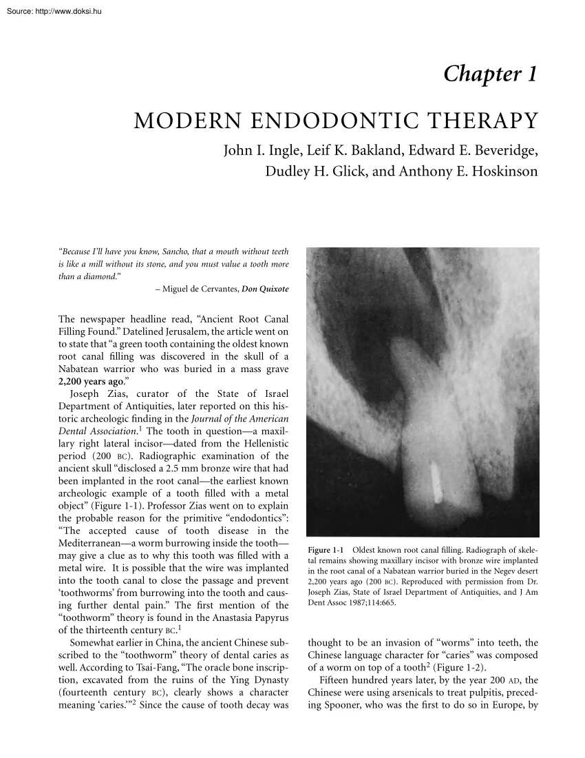
Chapter 1 MODERN ENDODONTIC THERAPY John I. Ingle, Leif K Bakland, Edward E Beveridge, Dudley H. Glick, and Anthony E Hoskinson “Because I’ll have you know, Sancho, that a mouth without teeth is like a mill without its stone, and you must value a tooth more than a diamond.” – Miguel de Cervantes, Don Quixote The newspaper headline read, “Ancient Root Canal Filling Found.” Datelined Jerusalem, the article went on to state that “a green tooth containing the oldest known root canal filling was discovered in the skull of a Nabatean warrior who was buried in a mass grave 2,200 years ago.” Joseph Zias, curator of the State of Israel Department of Antiquities, later reported on this historic archeologic finding in the Journal of the American Dental Association.1 The tooth in questiona maxillary right lateral incisordated from the Hellenistic period (200 BC). Radiographic examination of the ancient skull “disclosed a 2.5 mm bronze wire that had been implanted in the root
canalthe earliest known archeologic example of a tooth filled with a metal object” (Figure 1-1). Professor Zias went on to explain the probable reason for the primitive “endodontics”: “The accepted cause of tooth disease in the Mediterraneana worm burrowing inside the tooth may give a clue as to why this tooth was filled with a metal wire. It is possible that the wire was implanted into the tooth canal to close the passage and prevent ‘toothworms’ from burrowing into the tooth and causing further dental pain.” The first mention of the “toothworm” theory is found in the Anastasia Papyrus of the thirteenth century BC.1 Somewhat earlier in China, the ancient Chinese subscribed to the “toothworm” theory of dental caries as well. According to Tsai-Fang, “The oracle bone inscription, excavated from the ruins of the Ying Dynasty (fourteenth century BC), clearly shows a character meaning ‘caries.’”2 Since the cause of tooth decay was Figure 1-1 Oldest known root
canal filling. Radiograph of skeletal remains showing maxillary incisor with bronze wire implanted in the root canal of a Nabatean warrior buried in the Negev desert 2,200 years ago (200 BC). Reproduced with permission from Dr Joseph Zias, State of Israel Department of Antiquities, and J Am Dent Assoc 1987;114:665. thought to be an invasion of “worms” into teeth, the Chinese language character for “caries” was composed of a worm on top of a tooth2 (Figure 1-2). Fifteen hundred years later, by the year 200 AD, the Chinese were using arsenicals to treat pulpitis, preceding Spooner, who was the first to do so in Europe, by 2 Endodontics A B Figure 1-2 A, A piece of “oracle bone” inscribed with Chinese character meaning “caries” (fourteenth century BC). B, Chinese characters for “worm” and “tooth” combined to form the word “caries.” Reproduced with permission from Dr Tsai-fang Tsao, Faculty of Stomatology, Peking Medical College; Int Endodont J
1984;17:163; and L.F Zhens Diseases of the mouth and teeth 4th ed. Beijing (PRC): People’s Health Publishing House; 1957 p 2–5 1,600 years.2 The Chinese also used amalgam to fill cavities in the teeth as early as 659 AD2 This ancient history, preceding dentistry in North America and even Europe by thousands of years, was a harbinger of things to come. Jumping ahead to more modern times, Dr. Louis I Grossman (Figure 1-3), dean of endodontists in America, if not the world, pointed out that, by 1750, “Pierre Fauchard, the noted French dentist (1678-1761), had dispelled the ‘toothworm’ legend and was recommending the removal of diseased pulps as well.”3 Dr Grossman also chronicled the historical events impacting on root canal therapy since the American Revolution.4 In his usual orderly manner, Dr. Grossman divided the 200 years between 1776 and 1976 into four 50-year periods.4 During the first period, 1776 to 1826, he noted that treatment was crudeabscessed teeth were treated
with leeches or toasted fig poultices, and pulps were cauterized with red-hot wires. Nevertheless, it was during this same period that root canals were being filled from apex to crown with gold foil. The second half-century, 1826 to 1876, was marked by the founding of the first dental journal and the first dental school, the introduction of general anesthesia, rubber dam, gutta-percha root canal points, and the barbed broach, as well as three- and four-sided tapering broaches for cleaning and enlarging root canals, intracanal antiseptics, and oxyphosphate of zinc cement. At the same time, however, pulps were still being removed by driving wooden pegs into the canal, and crowns of the teeth were also being “snipped” off at the gingival level to cure toothache. Arsenicals were still being used to devitalize pulps. The third half-century, 1876 to 1926, was highlighted by the discovery and development of the x-ray, the advent of local anesthetics, and the acceptance of antisepsis as a
part of endodontic therapy. In 1891, for example, Otto Walkhoff introduced camphorated monoclorophenol (CMCP) as an intracanal medicament. It was this same Dr Walkhoff who took the first dental radiograph in 1895.3 Beginning about 1912, dentistry in general and endodontics in particular were set back by the wide acceptance of the theory of focal infection. Wholesale extraction of both vital and pulpless teeth took place. The professions were not to recover their senses until well after World War II. The final 50-year period, 1926 to 1976, saw improvements in radiographs, anesthetics, and procedures as well as the introduction of new methods and agents. Calcium hydroxide made its appearance, as did ethylenediaminetetraacetic acid (EDTA) for chelation. Many root canal medications appeared, and arsenic finally disappeared from the dental pharmacopeia. This same period saw the publication of the first major text devoted to endodontics, Dr. Grossman’s Root Canal Therapy, as well as the
introduction of standardized instruments and cavity preparation.5–7 The period also witnessed the rise and decline of the silver root canal Figure 1-3 The late Louis I. Grossman, DDS, Dr med dent, dean of American endodontists. His substantial contributions over more than half of the twentieth century enormously improved the practice, science, and standing of endodontics. Modern Endodontic Therapy point. A more sensible attitude toward endodontic surgery developed During this period, the American Association of Endodontists (AAE) was formed, followed by the American Board of Endodontics. Continuing education in endodontics widely disseminated information, skills, and techniques to an eager profession. The prevention of pulp disease began to play a more important role in dental practice. In part because of fluoridation, there was a decline in dental caries. Research into the causes and biology of dental trauma led to improved awareness and treatment of dental injuries.
Antibiotics greatly improved the profession’s ability to control infection, while new anesthetics and injection techniques increased control over pain. The high-speed air rotor handpiece added to patient comfort and the speed and ease of operation, as did prepackaged sterilized supplies. The mandatory use of masks, gloves, and better sterilizing methods rapidly emerged with the spread of human immunodeficiency virus/acquired immune deficiency syndrome (HIV/AIDS) and hepatitis. The widespread use of auxiliaries expanded dental services. It is now more than two decades since Grossman’s historic report, a period in which new instruments and techniques for cleaning and shaping as well as filling root canals have been introduced. Some of them are still in the development stage. All in all, the new decade, if not the new millennium, should prove exciting and profitable for the profession and patients alike. RECENT ATTITUDES TOWARD DENTISTRY AND ENDODONTIC THERAPY Increasingly, the term
“root canal” has become fashionable and generally known. In conversation, people proudly proclaim that they have had a “root canal.” The stigma of fear and pain is fast disappearing. Another impressive factor in the acceptance of endodontics is television. Countless advertisements emphasize a beautiful smilenot just toothpaste advertisements, but commercials in every field, from Buicks to beer. At the same time, the constant barrage of denture adhesive and cleanser advertisements produces a chilling effect. The public sees the problems that develop from the loss of teeth. Obvious missing teeth are anathema. There is no question that the public’s acceptance of endodontic treatment is on the rise. In 1969, for example, the American Dental Association (ADA) estimated that 6 million root canal fillings were done each year. By 1990, their estimate had risen to 13,870,000. One might add that the ADA also estimated that another 690,000 “endodontic surgeries and root amputations”
3 were done in l990.8 By the year 2000, it was estimated that 30 million teeth were root-filled annually.9 This upward trend was also documented by the Public Affairs Committee of the AAE. Reporting on surveys of the general public made by the Opinion Research Institute in 1984 and 1986, the Committee noted that 28% of 1,000 telephone respondents reported that they had had root canal therapy by 1986, an increase of 5% over 23% in 1984.10 Also, in 1986, 62% said they would choose root canal therapy over extraction, an increase of 10% over 52% in 1984. More than half the respondents (53%) believed that an endodontically treated tooth would last a lifetime.10 On the other hand, the perceptions of younger people (under 25 years) in this survey were disappointing in that 70% described root canal therapy as “painful” and 58% thought it would be less expensive to extract the tooth and place a bridge.10 Clearly, the profession has a mission in educating this age group to reverse their
image of endodontics and the value of a treated pulpless tooth. A rate of use of endodontic services similar to the rate in the United States (28%) was also reported from Norway, where 27% of an older age group (66 to 75 years) had had root canal therapy, as had 12% of a younger age group (26 to 35 years). Incidentally, 100% of the root-filled teeth in the younger group were still present 10 to 17 years later, a remarkable achievement.11 The growth in endodontic services is also reflected in the sale of endodontic equipment, supplies, and instruments. In 1984, endodontics was a $20 million market, growing at a rate of 4% a year.12 By 1997, 13 years later, the endodontic market, through dental dealer retail stores alone, was $72 million, up from $65.6 million in 1996, a growth of nearly 10% One must add to these sales another 10% to account for mail-order/telephone sales, a grand total in 1997 of nearly $80 million. Worldwide sales are probably double this figure!13 There is no question
that the greatest share of endodontic procedures is carried out by America’s general practitioners. On the other hand, the specialty of endodontics is growing as well. In 1986, for example, only 5% of those patients who had root canal therapy were treated by a specialist.10 By 1990, this percentage had grown to 28.5%8 In 1989, there were 2,500 endodontic specialists in the United States.9 By 2,000, the figure was around 3,300 endodontists,14 and these endodontists were completing 39% of all of the root canal therapy and endodontic surgery in the United States.8 In spite of these encouraging figures for the immediate future of endodontics, one has to question what 4 Endodontics the distant future will bring. The rate of dental caries is declining precipitously. In two separate reports, the US National Institute of Dental Research (NIDR) proudly announced in 1988 that half of all children in the United States aged 5 to 17 years had no decay in their permanent teeth. None!15 In
contrast, in the early 1970s, only 28% of the permanent teeth of American children were caries free. By 1980, this figure had risen to 36.6%, and by 1986–1987, 499% were caries free. Furthermore, there was a 50% improvement in 17 year olds, a most encouraging sign for dentists, who were once faced with repairing the ravaged mouths of adolescents in the 1950s through the 1970s. The national decayed-missing-filled surfaces (DMFS) rate had dropped to 3.1 for all US schoolchildren and, even more importantly, “82 percent of the DMF surfaces are filled, about 13% decayed and 4% are missing.”15 A comparable radiographic survey in 1980 on 1,059 US Air Force basic trainees 18 to 20 years old found that 10% had “no restorations, no decay and no missing teeth.” Moreover, another 10% had at least one root canal filling.16 A comparison between these 1980 recruits and Navy recruits in 1956 proved that missing teeth per recruit had dropped from 2.4 to 075 in 24 years, a reduction of 31%.16
As far as older adults are concerned, the NIDR reported a remarkable decline in edentulism as well, particularly in the middle-aged, a group in which “total tooth loss has been practically eliminated.”17 The elderly (age 65 and older), however, “are still in serious trouble,” root caries and periodontal disease being the primary offenders17 All of these encouraging figures suggest greater preventive measures and higher use of dental services by the public. Part of the improvement can be credited to a healthier economy and lifestyle, part to the national water supply and dentifrice fluoridation programs, part to the dental profession’s efforts, and part to dental insurance. Bailit et al have shown that third-party payment has increased dental use and improved oral health.18 By 1995, the ADA estimated that 63% of all US citizens were covered by a private insurance program and another 5.3% by public assistance A remaining 31.4% were not covered by any insurance program19 In
providing these burgeoning services, the dental profession has fared well financially. Over the past 30 years, the net income of dentists has more than doubled in constant 1967 dollars.20 Moreover, between 1986 and 1995, the net income of dentists rose 30.7%, from $102,953 to $134,590.19 Dental expenditures by the public have increased as well, from $3.4 billion in 1967 to $10 billion in 1977, to $25.3 billion in 1987 and $475 billion in 199621 In 1996, the average person spent $172.70 for dental services, up from $108 (adjusted to 1996 dollars) in 1967 This amounts to a 60% increase in outlay for dental care in 30 years.21 After all of this expenditure and care, one is hardpressed to explain why 15.1 million workdays are lost annually because of dental pain.22 ENDODONTIC CASE PRESENTATION All of these improvements notwithstanding, many patients still must be convinced that root canal therapy is an intelligent, practical solution to an age-old problemthe loss of teeth. The “case for
endodontic treatment” must be presented to the patient in a straightforward manner The patient with the correct “oral image” will be anxious to proceed with therapy. “Is this tooth worth saving, doctor?” This sentiment is voiced more often than not by the patient who has been informed that his or her tooth will require endodontic therapy. Superficially, this appears to be a simple question that requires a direct, uncomplicated answer. It should not be interpreted as hostile or as a challenge to the treatment recommendations presented for the retention of the tooth. Psychologically, however, this initial question is a prelude to a Pandora’s box of additional queries that disclose doubts, fears, apprehensions, and economic considerations: for example, “Is it painful?” “Will this tooth have to be extracted later?” “How long will this tooth last?” “Is it a dead tooth?” “Will it turn black?” and “How much will it cost?” Following the first question, the
dentist should anticipate such a series of questions. These may be avoided, however, by including the answers to anticipated questions in the presentation. In turn, the dentist will gain a decided psychological advantage. By this apparent insight into his or her problems, the patient is assured that the dentist is cognizant of the very questions the patient was about to raise or possibly was too reticent to ask. Most of the patient’s fear and doubts can be allayed by giving a concise answer to each question. The dentist should be able to explain procedures intelligently as ideas are exchanged with the patient. To do this, one must be endodontically oriented. That is, one must believe in the value of endodontic therapy. By believing in such treatment, one cannot help but influence the patient favorably. The dentist will soon gain the confidence of the patient who realizes that professional recommendations emanate from an honest desire to preserve the mouth’s functional efficiency.23
Modern Endodontic Therapy To answer patients’ questions, the ADA produced an inexpensive pamphlet entitled “Your Teeth Can be Savedby Endodontic.”24 The AAE also publishes a number of pamphlets25 for patients: the “Your Guide To”* series: “Endodontic Treatment, Cracked Teeth, Endodontic Retreatment, and Endodontic Surgery.”* Dr. Joel Burns has beautifully illustrated a booklet entitled Why Root Canal Therapy?26 Although this approach is a little impersonal, it is a tangible reference, particularly when the patient returns home and tries to explain to an interested spouse what endodontic therapy involves. Based on previous experiences in the office, the average patient has sufficient confidence in the dentist’s ability to help. He or she is ready to accept the professional knowledge and advice offered but likes to have some part in evaluating the reasonableness of treatment. The professional person and the staff must spend the time and thought necessary to understand
the patient’s initial resistance, which is often based on false assumptions and beliefs in matters dealing with pulpless teeth. However, once the patient is secure in the thought that this is the correct treatment, most of the fears and apprehensions related to unfamiliarity with endodontic therapy will be dissipated. A dental appointment is still associated with fear in the minds of many people27–29 (Figure 1-4). The mere thought of treating the “nerve” of a tooth implies pain. Patients require reassurance, supported by all available psychological and therapeutic methods of relaxation and pain control. The patient must be reassured that endodontic therapy need not be painful and usually requires no more than a local anesthetic. All too often, we hear negative remarks about root canal therapy: “Trying to do anything positive in Tacoma is akin to getting a root canal without Novocain.” Or “Whew! What you just heard was a collective sigh of relief following 7 months of
agonizing root canal”remarks made following President Clinton’s “confession” on television. In contrast to these commonly heard excoriations, LeClaire et al. reported that 43.9% of endodontic patients reported a decrease in fearfulness after having root canal therapy. Furthermore, 96.3% said that “they would have root canal therapy again to save a tooth.”29 It should be explained to the concerned patient that root canal therapy is a specialized form of dental pro- *Available from the American Association of Endodontists Information Services, 211 East Chicago Ave., Suite 1100, Chicago, IL 60611-2691. 5 A B Figure 1-4 A, Rash occurred in this terrified patient, who was merely sitting in the dental chair. B, The patient’s apprehension was allayed by sympathetic management, allowing successful completion of endodontic therapy in a four-canal molar. (Courtesy of Dr Norbert Hertl.) cedure designed to retain a tooth safely and comfortably. The tooth, when properly treated
and restored, can be retained as long as any other tooth. It is not a “dead tooth” as long as the roots of the tooth are embedded in healthy surrounding tissues. Although teeth do not turn “black” following root canal therapy, a slight change in color owing to reduced translucency may occur. Discoloration associated with pulp necrosis and leakage around restorations can be managed successfully (see Chapter 16). Most often, retention of the tooth and bleaching, veneering, or crowning (Figure 15) are preferable to extraction and replacement with a prosthetic appliance.10 There is little doubt that economic considerations play an important role (and for some a supreme role) in the final decision. Some patients “think financially,” and even though they are able to afford treatment, they allow financial considerations to govern decisions that should logically be made on a physiologic basis only. It 6 Endodontics is necessary to point out to these people the financial
advantage of retaining a tooth by endodontic therapy rather than by extraction and prosthetic replacement. The properly informed patient is quick to recognize that the fee for a bridge is more than that for root canal therapy and proper restoration.10 In addition, it should be mentioned in all honesty that any vital tooth prepared for a crown could become a possible candidate for future endodontic therapy. Also, the patient who says “Pull it out”’ should be informed of the problems that arise if a space is left unfilled (ie, tilting, reduced masticatory efficiency, future periodontal problems, and cosmetic effects). Another commonly heard statement by the patient is, “It’s only a back tooth, anyway,” or “If it were a front tooth I would save it, but no one sees it in back.” This patient thinks cosmetically. The disadvantages of the loss of any tooth, let alone a posterior one so essential for mastication, must be explained. A B Figure 1-5 Fractured premolar restored
by endodontics and post-and-core crown. A, Tooth immediately following fracture B, Restoration and periradicular healing at 3-year recall. Note the spectacular fill of arborization (arrows) at the apex. (Courtesy of Dr. Clifford J Ruddle) Fortunately, today’s patient is becoming more sophisticated, too “tooth conscious” to permit indiscriminate extraction without asking whether there is an alternative. Extraction contributes to a crippling aberration from the normal dentition There is no doubt that a normally functioning, endodontically treated, and well-restored tooth is vastly superior to the best prosthetic or implant replacement. INDICATIONS The indications for endodontic therapy are legion. Every tooth, from central incisor to third molar, is a potential candidate for treatment. Far too often, the expedient measure of extracting a pulpless tooth is a short-sighted attempt at solving a dental problem. Endodontic therapy, on the other hand, extends to the dentist and the
patient the opportunity to save teeth. The concept of retaining every possible tooth, and even the healthy roots of periodontally involved teeth, is based on the even distribution of the forces of mastication. The final success of any extensive restorative procedure depends on the root-surface area attached through the periodontal ligaments to the alveolar bone. Like the proverbial horseshoe nail, root-filled teeth may often be the salvation of an otherwise hopeless case. To carry this concept one step further, recognized today is the importance of retaining even endodontically treated roots, over which may be constructed a full denture, the so-called overdenture.30 On some occasions, attachments may be added to these roots to provide additional retention for the denture above. At other times, the treated roots are merely left in place on the assumption that the alveolar process will be retained around roots, and there will not be the usual ridge resorption so commonly seen under full
or even partial dentures. Most dentists would agree that the retained and restored individual tooth is better than a bridge replacement and that a bridge is better than a removable partial denture, which, in turn, is superior to a full denture. Although recent success with dental implants is impressive, the long-term outcome is not known, and, functionally, the patient’s own tooth is superior. Treatment in every case should adhere to the standards set by the dentist for himself or herself and his or her family. Modern dentistry incorporates endodontics as an integral part of restorative and prosthetic treatment. Most any tooth with pulpal involvement, provided that it has adequate periodontal support, can be a candidate for root canal treatment. Severely broken down teeth, and potential and actual abutment teeth, can be candidates for the tooth-saving procedures of endodontics. Modern Endodontic Therapy One of the greatest services rendered by the profession is the retention of
the first permanent molar (Figure 1-6). In contrast, the long-range consequences of breaking the continuity of either arch are also well known (Figure 1-7). Root canal therapy often provides the only opportunity for saving first molars with pulp involvement. In addition to saving molars for children, saving posterior teeth for adults is also highly desirable. Retaining a root-filled terminal molar, for example, means saving two teeththe molar’s opposite tooth as well (Figure 1-8, A). Moreover, root canal treatment may save an abutment tooth of an existing fixed prosthesis. The gain is doubled if the salvaged abutment is also the terminal posterior tooth in the arch and has a viable opponent (Figure 1-8, B). Another candidate for endodontic therapy is the adolescent who arrives in the office with a grossly damaged dentition and is faced with multiple extractions and dentures (Figure 1-9). Many of these children are mortified 7 by their appearance. It is gratifying to see the
blossoming personality when an esthetic improvement has been achieved. The end result in these cases would not be possible without root canal therapy (Figure 1-10) Intentional Endodontics Occasionally, intentional endodontics of teeth with perfectly vital pulps may be necessary. Examples of situations requiring intentional endodontics include hypererupted teeth or drifted teeth that must be reduced so drastically that the pulp is certain to be involved.31 On other occasions, a pulp is intentionally removed and the canal filled so that a post and core may be placed for increased crown retention. In these cases, the endodontic treatment may be completed before tooth reduction is started. Over and above these quite obvious indications for intentional endodontics, it has been recommended that pulpectomy and root canal filling be done for vital teeth badly discolored by tetracycline ingestion. B A C Figure 1-6 A, Pulpless first molar following failure of pulpotomy. Note two periradicular
lesions and complete loss of intraradicular bone Draining sinus tract opposite furca is also present. B, Completion of endodontic therapy without surgery C, Two-year recall radiograph Complete healing was evident in 6 months. New carious lesions (arrows) now involve each interproximal surface 8 Endodontics Figure 1-7 Extrusion, recession (arrow) tipping, malocclusion, rotation, and gingival cemental caries are only a few of the long-range consequences following early extraction of a permanent first molar. Following root canal therapy, internal bleaching may be carried out.32 Considerations Prior to Endodontic Therapy Although it is true that root canal treatment can be performed on virtually any tooth in the mouth, there are some important considerations that must be evaluated prior to recommending root canal treatment. Some of these were delineated by Beveridge (personal communication, June 1971): 1. Is the tooth needed or important? Does it have an opponent? Could it some day
serve as an abutment for prosthesis? 2. Is the tooth salvageable, or is it so badly destroyed that it cannot be restored? 3. Is the entire dentition so completely broken down that it would be virtually impossible to restore? 4. Is the tooth serving esthetically, or would the patient be better served by its extraction and a more cosmetic replacement? 5. Is the tooth so severely involved periodontally that it would be lost soon for this reason? 6. Is the practitioner capable of performing the needed endodontic procedures? In regard to the last point, today in the United States and many other countries, endodontic specialists are available to whom patients may be referred. A decision to refer is preferable before a mishap, such as perforation of the root canal, occurs. If a mishap does occur A B Figure 1-8 A, Terminal molar retained by endodontic therapy saves opposing molar as well. (Courtesy of Dr L Stephen Buchanan.) B, Fixed partial denture possible only because abutment teeth are
retained by root canal therapy (Courtesy of Dr Norbert Hertl.) Modern Endodontic Therapy A 9 B Figure 1-9 A, Caries-decimated dentition in a 14-year-old girl. Personality problems had developed in this youngster related to her feeling embarrassed about her appearance. B, Provisional restoration following endodontic therapy has restored the cosmetic appearance and confidence so necessary for the adolescent during treatment, the patient must be given the option of seeing a specialist before the decision to extract the tooth is made. The well-trained dentist should have no fear of the pulpally involved tooth. If a carious exposure is noted during cavity preparation, the patient is informed of the problem and the recommended treatment, and, if consented to, the endodontic therapy is started while A the tooth is anesthetized. The prepared dentist can begin pulpectomy immediately, using sterile instruments packaged and stored for just such an emergency. Age and Health as
Considerations Age need not be a determinant in endodontic therapy. Simple and complex dental procedures are routinely performed on deciduous teeth in young children and B Figure 1-10 A, Obvious pulp involvement of incisors shown in Figure 1-9. B, Root canal treatment of these incisors makes possible dowel restoration followed by cosmetic provisional plastic crowns. 10 Endodontics on permanent teeth in patients well into their nineties. The same holds true for endodontic procedures. It should be noted, however, that complete removal of the pulp in young immature teeth should be avoided if possible. Procedures for pulp preservation are more desirable and are fully discussed in chapter 15. Health consideration must be evaluated for endodontics as it would for any other dental procedure. Most often, root canal therapy will be preferable to extraction. In severe cases of heart disease, diabetes, or radiation necrosis,33 for example, root canal treatment is far less traumatic than
extraction. Even for terminal cases of cancer, leukemia, or AIDS, endodontics is preferred over extraction. Pregnancy, particularly in the second trimester, is usually a safe time for treatment. In all of these situations, however, endodontic surgery is likely to be as traumatic as extraction. Status of the Oral Condition Pulpally involved teeth may simultaneously have periodontal lesions and be associated with other dental problems such as rampant decay, orthodontic malalignment, root resorption, and/or a history of traumatic injuries. Often the treatment of such teeth requires a team effort of dental specialists along with the patient’s general dentist. The presence of periodontal lesions must be evaluated with respect to the correct diagnosis: Is the lesion of periodontal or endodontic origin, or is it a combined situation? The answer to that question will determine the treatment approach and the outcome; generally, lesions of endodontic origin will respond satisfactorily to
endodontic treatment alone34 (Figure 1-11), whereas those of periodontal origin will not be affected simply by endodontic procedures (Figure 1-12). Combined A B Figure 1-11 A, Mandibular molar with furcal bone loss (arrow) owing to endodontic infection and no periodontal disease. B, Root canal treatment completed without any periodontal intervention. C, One-year control shows recovery of furcal lesion by endodontic treatment alone. C Modern Endodontic Therapy A C 11 B D Figure 1-12 A, Retraction of surgical flap reveals the extent of periodontal lesion completely involving buccal roots of second molar abutment of full-arch periodontal prosthesis. Root canal therapy of a healthy, palatal root is completed before surgery B, Total amputation of buccal roots reveals extent of cavernous periodontal lesion. C, Extensive bone loss, seen in A and B, is apparent in a radiograph taken at the time of treatment (the root outline was retouched for clarity). D, Osseous repair, 1 year
following buccal root amputation A solidly supported palatal root serves as an adequate terminal abutment for a full-arch prosthesis Endodontic therapy was completed in 1959 and has remained successful. (Courtesy of Dr Dudley H Glick) lesionsthose that develop as a result of both pulpal infection and periodontal diseaserespond to a combined treatment approach in which endodontic intervention precedes, or is done simultaneously with, periodontal treatment35 (Figure 1-13). Even teeth with apparently hopeless root support can be saved by endodontic treatment and root amputation (Figure 1-14). Today, many pulpless teeth, once condemned to extraction, are saved by root canal therapy: teeth with large periradicular lesions or apical cysts36–39 (Figure 115), teeth with perforations or internal or external resorption (Figure 1-16), teeth badly broken down by caries or horizontal fracture (Figure 1-17), pulpless teeth with tortuous or apparently obstructed canals or broken instruments
within,40 teeth with flaring open apices (Figure 1-18), teeth that are hopelessly discolored (Figure 1-19), and even teeth that are wholly or partially luxated. All of these conditions can usually be overcome by endodontic, orthodontic, periodontic, or surgical procedures. In some cases, the prognosis may be somewhat guarded. But in the majority of cases, the patient and dentist are pleased with the outcome, especially if the final result is an arch fully restored. 12 A Endodontics B Figure 1-13 A, Maxillary premolar with both periodontal bone loss (open arrow) and an apical lesion (small closed arrows) from pulpal infection. B, Root canal treatment was done along with periodontal pocket maintenance C, One-year control shows apical bony response to the endodontic procedure; the periodontal condition is unchanged. C Figure 1-14 Amputation of periodontally involved distobuccal root allows retention of well-restored maxillary first molar. Root canal therapy of two remaining roots
is necessary. Buccal-lingual narrowing of the occlusal table reduces the forces of mastication on these roots. The vulva-like soft tissue defect should be corrected with gingivoplasty. Modern Endodontic Therapy Figure 1-15 Classic apical cyst (left) apparent in pretreatment radiograph. Total repair of cystic cavity in 6-month recall film is signaled by complete lamina dura that has developed periradicularly Biopsy confirmed the initial diagnosis of an apical cyst A B C D Figure 1-16 A, Extensive defect by internal-external resorption is demonstrated by an explorer in a 67-year-old man. B, Retraction of the rectangular flap reveals a pathologic defect involving over half the tooth. Under no circumstances should root canal therapy be attempted from this lateral approach. C, Silver point root canal filling cemented to place before restoration of resorptive defect. D, Restoration of area of resorption with zinc-free amalgam. Case is completed by suturing flap into position E,
Five-year postoperative photograph (patient, age 72) reveals gingival repair and toleration of subgingival amalgam filling. E 13 14 Endodontics A Figure 1-17 Four maxillary incisors with coronal fractures into pulp. Radiograph is necessary to determine whether root fracture has occurred and the stage of root development and apical closure. Immediate pulpectomy and root canal filling are indicated for all four incisors. B Figure 1-19 A, Intense discoloration of a pulpless maxillary central incisor. B, Successful bleaching with Superoxol (30% H2O2) The incisor has been restored to its normal color. Figure 1-18 Left, Flaring apex of incompletely formed root follows pulpal death caused by impact injury at early age. Right, Obturation of the “blunderbuss” canal is accomplished by retrofilling from surgical approach. Reproduced with permission from Ingle JI. Dent Digest 1956;62:410. Modern Endodontic Therapy ONE-APPOINTMENT THERAPY Single-appointment root canal therapy has
become a common practice. When questioned, however, most dentists reply that they reserve one-appointment treatment for vital pulp and immediate periradicular surgery cases. In 1982, only 128% of dentists queried thought that necrotic teeth would be successfully treated in one appointment.41 Endodontists have been treating patients in one-appointment visits for some time. At one time, 86% of the directors of postgraduate endodontic programs, when surveyed, reported that nonsurgical one-visit treatment was part of their program.42 What are the advantages and disadvantages of single-visit endodontics? Advantages: 1. Immediate familiarity with the internal anatomy, canal shape, and contour facilitates obturation 2. No risk of bacterial leakage beyond a temporary coronal seal between appointments 3. Reduction of clinic time 4. Patient convenienceno additional appointment 5. Less cost Perceived Disadvantages: 1. No easy access to the apical canal if there is a flare-up 2. Clinician fatigue
with extended one-appointment operating time 3. Patient fatigue and discomfort with extended operating time 4. No opportunity to place an intracanal disinfectant (other than allowing NaOCl to disinfect during treatment) What has held back one-appointment endodontics? The major consideration has been concern about postoperative pain and failure. Postoperative Pain The fear that patients will probably develop postoperative pain and that the canal has been irretrievably sealed has probably been the greatest deterrent to single-visit therapy. Yet the literature shows no real difference in pain experienced by patients treated with multiple appointments41–57 In spite of this evidence, however, 40% of the endodontic course directors surveyed were of the opinion that necrotic cases treated in one visit have more flare-ups.41 Galberry did not find this to be true in Louisiana,49 nor did Nakamuta and Nagasawa in Japan, who had only a 7.5% pain incidence after treating 106 infected cases in
single 15 appointments.50 Moreover, the symptoms the patients experienced were mild and needed no drugs or emergency treatment. Oliet reported that only 3% of his sample of 264 patients receiving single-appointment treatment had severe pain, compared with 2.4% of the 123 patients treated in two visits.48 Wolch’s records of over 2,000 cases treated at a single appointment showed that less than 1% of patients indicated any severe reaction.44 Pekruhn reported no statistically significant difference between his two groups.47 Mulhern et al reported no significant difference in the incidence of pain between 30 single-rooted teeth with necrotic pulps treated in one appointment and 30 similar teeth treated in three appointments.51 At the University of Oklahoma, however, Roane and his associates found a “two to one higher frequency of pain following treatment completed in multiple visits when compared to those completed in one visit.”52 More recent reports from Brazil and Fava from the
Netherlands found no difference in the incidence of pain between one- and two-visit cases,53–56 and Trope reported no flare-ups in one-appointment cases with no apical lesions.57 Re-treatment of failed cases with apical periodontitis made the difference, however. These cases suffered a 13.6% flare-up rate57 One might expect pain from any case, as reported by Harrison et al. from Baylor University.58 Of 229 patients treated twice, 55.5% had no interappointment pain, 288% had slight pain, and 15.7% had moderate to severe pain Eleazer and Eleazer compared the flare-up rate between one and two appointments in treating necrotic canal molars. In the two-visit cohort, there was a 16% flare-up rate, whereas in the one-visit group, there was only a 3% flare-up experience, which proved to be significant.59 In 1996, Ørstavik et al also reported fewer flareups following single-appointment therapy60 In light of these studies, pain does not appear to be a valid reason to avoid single-appointment
root canal therapy. Success versus Failure If pain is not a deterrent, how about fear of failure? Pekruhn has published a definitive evaluation of single-visit endodontics.61 From the clinics of the Arabian-American Oil Company, he reported a 1-year recall of 925 root-filled teeth of 1,140 possible cases. His failure rate was 5.2%, very comparable to many multiple-visit studies. Pekruhn was surprised to learn that his rate of failure was higher (15.3%) in teeth with periradicular lesions that had had no prior access opening. If this type of case had been previously opened, the incidence of failure dropped to 6.5% The 16 Endodontics highest failure rate (16.6%) was in endodontic re-treatment cases Symptomatic cases were twice as likely to fail as were asymptomatic cases (10.6% versus 50%) A Japanese study followed one-visit cases for as long as 40 months and reported an 86% success rate.50 Oliet again found no statistical significance between his two groups.48 The majority of the
postgraduate directors of endodontics felt that the chance of successful healing was equal for either type of therapy.42 The original investigators in this field, Fox et al.,43 Wolch,44 Soltanoff,45 and Ether et al.,46 were convinced that single-visit root canal therapy could be just as successful as multiple-visit therapy. None, however, treated the acutely infected or abscess case with a single visit. In more recent times, and in marked contrast to these positive reports, Sjögren and his associates in Sweden sounded a word of caution.62 At a single appointment, they cleaned and obturated 55 singlerooted teeth with apical periodontitis. All of the teeth were initially infected. After cleaning and irrigating with sodium hypochlorite and just before obturation, they cultured the canals. Using advanced anaerobic bacteriologic techniques, they found that 22 (40%) of the 55 canals tested positive and the other 33 (60%) tested negative. Periapical healing was then followed for 5 years.
Complete periapical healing occurred in 94% of the 33 cases that yielded negative cultures! But in those 22 cases in which the canals tested positive prior to root canal filling, “the success rate of healing had fallen to just 68%,” a statistically significant difference.62 In other words, if a canal is still infected before filling at a single dental appointment, there may be a 26% greater chance of failure than if the canal is free of bacteria. Their conclusions emphasized the importance of eliminating bacteria from the canal system before obturation and that this objective could not be achieved reliably without an effective intracanal medicament. This is one limited study, but it was done carefully and provides the recent evidence correlating the presence of bacteria to longer-term outcomes. Ørstavik et al. faced up to this problem and studied 23 teeth with apical periodontitis, all but one infected initially. At the end of each sitting, apical dentin samples were cultured
anaerobically. No chemical irrigants were used during cleaning and shaping, and at the end of the first appointment, 14 of the 23 canals were still infected.63 At an earlier time, Ingle and Zeldow, using aerobic culturing, found much the same.64,65 Ørstavik et al then sealed calcium hydroxide in the canal. In 1 week, at the start of the second appointment, only one root canal had sufficient numbers of bacteria “for quantifica- tion”the calcium hydroxide was that effective! They also found “a tendency for teeth causing symptoms to harbour more bacteria than symptomless teeth.”63 In a follow-up study, Trope et al. treated teeth with apical periodontitis, with and without calcium hydroxide, in one or two visits. They reached a number of conclusions: (1) “[C]alcium hydroxide disinfection after chemomechanical cleaning will result in negative cultures in most cases”; (2) “[I]nstrumentation and irrigation alone decrease the number of bacteria in the canal 1000-fold, however
the canals cannot be rendered free of bacteria by this method alone”; and (3) “[T]he additional disinfecting action of calcium hydroxide before obturation resulted in a 10% increase in healing rates. This difference should be considered clinically important.”66 In another 52-week comparative study in North Carolina, of the “periapical healing of infected roots [in dogs] obturated in one step or with prior calcium hydroxide disinfection,” the researchers concluded that “Ca(OH)2 disinfection before obturation of infected root canals results in significantly less periapical inflammation than obturation alone.”67 One has to ask, therefore, wouldn’t it be better to extend one more appointment, properly medicate the canal between appointments, and improve the patient’s chances of filling a bacteria-free canal? Unfortunately, there is a widely held but anecdotal opinion that current chemomechanical cleaning techniques are superior, predictably removing the entire bacterial
flora. If this is so, single-visit treatment of necrotic pulp cases would definitely be indicated. However, the research has yet to be published to corroborate these opinions. Until then, it may be more prudent to use an intracanal medicament such as calcium hydroxide, within a multiple-visit regimen, for cases in which a mature bacterial flora is present within the canal system prior to treatment. Although single appointments would be very appropriate in cases with vital pulps, on the other hand, for teeth with necrotic pulps and periapical periodontitis, and for failed cases requiring retreatment, there may be a risk of lower success rates in the long term. To date, the evidence for recommending either one- or multiplevisit endodontics is not consistent. The prudent practitioner needs to make decisions carefully as new evidence becomes available Wolch said it best: “In the treatment of any disease, a cure can only be effected if the cause is removed. Since endodontic diseases
originate from an infected or affected pulp, it is axiomatic that the root canal must be thoroughly and carefully debrided and obturated” (personal communication, 1983). Modern Endodontic Therapy “ENDODONTICS AND THE LAW”68 If today’s patients are becoming more sophisticated about their dental wants, they are also becoming more sophisticated about their legal rights. As Milgrom and Ingle have noted, the dentist can no longer consider himself immune to malpractice litigation by hiding behind a doctrine of “local community standards.”69 Local community standards today are those standards set by the specialists in the community, in this case the board-certified endodontists, not the general practitioner. More and more often, specialists are willing to testify in court, supporting patients who, in their view, have been treated below the standard of care. Along with authors who have alluded to the subject,70–72 the AAE has issued guidelines that could well establish a
national standard of care. Titled “Appropriateness of Care and Quality Assurance Guidelines,” it is now in its third edition and may be obtained from the AAE.73 Cohen and Schwartz have pointed out that a meritorious claim by a patient is “any departure from the minimum quality of endodontic care that reasonably prudent practitioners would perform under the same or A 17 similar circumstances.”68 “Any departure”’ is rather broad and includes failure to properly diagnose; failure to perform comprehensive diagnostic tests; failure to properly document and record all findings and treatment; treatment of the wrong tooth; use of paraformaldehyde/steroid pastes such as N2, RC2B, Endomethazone, and SPAD; root perforations; failure to receive informed consent; failure of yet-to-be-approved endodontic implants; failure because of instruments broken in the canal; and failure to use a rubber dam.74 From this list, “failure to use a rubber dam” is unconscionable and may result
in the most disastrous consequences, namely the swallowing or inhalation of an endodontic instrument (Figure 1-20). Instrument breakage or, as it is euphemistically referred to, “instrument separation” is a “disquieting event.” One must ask, “Did the file break because of overzealous use. or was it defectively manufactured?”74 The unbroken end of the file should be saved in a coin envelope and placed in the patient’s treatment record. If defective manufacturing can be proved, liability shifts to the manufacturer In either event, the patient must be promptly informed.74 B Figure 1-20 Two examples of swallowed endodontic instruments because the rubber dam was not used. A, Radiograph taken 15 minutes after an endodontic broach (arrow ) was swallowed. Reproduced with permission from Heling B, Heling I Oral Surg 1977;43:464 B, Abdominal radiograph showing a broach in the duodenum (arrow). The broach was surgically removed 1 month later Reproduced with permission from
Goultschin J, Heling B. Oral Surg 1971;32:621 18 Endodontics A major standard of care controversy has also erupted over the issue of overfilling or overextending the root canal filling versus filling “short.” One would be hard-pressed in court to defend gross overfilling, sometimes even to the point of filling the mandibular canal (Figure 1-21). On the other hand, a “puff ” of cement from the apical constriction has become acceptable. Filling just short of the radiographic apex, at the apical constriction, 0.5 to 10 mm, is backed by a host of positive reports. By the same token, an inadequate root canal filling is hardly defensible as rising to the standard of care, even though the filling might appear to extend to the apex. Grossly underfilled canals, 3.0 to 60 mm short, are also hard to defend, particularly if an associated periradicular lesion is radiographically apparent. One must realize, however, that some root canals are so thoroughly calcified (obliterated) that
penetration to the apex is virtually impossible. Facing this problem, Swedish scientists analyzed 70 cases of “obliterated” canals over a recall period of 2 to 12 years.75 The overall success rate for the partially filled canals was 89%. If in the initial radiograph there was an intact periradicular contour, the success rate was an amazing 97.9% If a preoperative periradicular radiolucency was present, however, the success rate dropped to a disappointing 62.5%75 In the incompletely filled failure cases, it was theorized that canals were present but so narrow that they could not be negotiated by the smallest instruments, Figure 1-21 Massive overextension of RC2B into the inferior alveolar canal. The patient suffered permanent paresthesia A lawsuit was settled out of court against the dentist and in favor of the 26year-old female secretary in Pennsylvania. (Courtesy of Edwin J Zinman, DDS, JD.) but were still large enough for the passage of bacteria and their toxins.75 Buchanan has
shown that with care and persistence, many so-called obliterated canals can be negotiated (personal communication, 1989). In the light of the low success rate (62.5%) of unfilled “obliterated” canals with apical radiolucencies, the dentist must seriously consider a surgical approach and retrofillings. This would be well within the standard of care if done expertly Paresthesia is another patient complaint following endodontic treatment. Lip numbness (“the injection didn’t wear off ”)76 is usually caused by gross overfilling, nearly always when root canal sealers or cements impinge on the inferior alveolar nerve. This is particularly true when neurotoxic filling materials are used (eg, N2, RC2B, Endomethazone, SPAD). Ørstavik et al. surveyed the literature for reported cases of paresthesia related to endodontic treatment.76 They found 24 published cases; 86% of patients were female, and usually a paste-type filling had been used. Although 5 cases “healed in four months to
two years, 14 showed no indication of the paraesthesia healing.from 3 months up to 18 years” The remaining cases were resolved by surgical removal of the offending material. Ørstavik et al reported the twenty-fifth case, paresthesia following overfilling with Endomethazone. The condition still persisted 3 years later and “the possibility of regeneration of the nerve must be considered negligible.”76 Others have reported the same or similar causes of nerve damage and paresthesia.77–80 In California, endodontics became number one in terms of the frequency of malpractice claims filed.68 Nationally, “endodontic claims are the second most frequent producer of claims and dollar losses with oral surgery being number one.”72 There is obviously “an increase in the number of malpractice claims involving endodontics, primarily against general dentists.”73 Many of these tragedies, for dentist and patient alike, could have been avoided had the patient been referred to a dentist
more skilled in endodontics. “When in doubt, refer it out.”74 Just such a tragic casea failure to timely or properly refer a patientinvolved five dentists enmeshed in a recent malpractice suit: one general dentist, three endodontists, and a prosthodontist. None of the four specialists was board certified, although all were educationally qualified. The patient was first seen by the general dentist, who took full-mouth radiographs, did an oral examination, and established a treatment plan that said nothing about an unusual bony lesion in the left mandible. The patient was not satisfied with the gener- Modern Endodontic Therapy alist, asked for her radiographs, and transferred to a prosthodontist, who also used the original films for his examination. He established that a number of crowns and a bridge should be done and that he would start on tooth #19, which had had root canal therapy that failed. So, quite properly, he referred the patient to an endodontist, who, for some
unexplained reason, retreated only two of the three canals. Up to this time, all three dentists had failed to notice the unusual bone trabeculation and apparent lesion that extended from the mesial of #19 and around the roots of #20 and #21 to the distal of #22, nor had they noted the buccal swelling in the region! If they had done so, they should have referred the patient to an oral surgeon, a competent radiologist, or an oral pathologist. The prosthodontist continued treatment, and, finally, when the patient complained, noted the swelling in the vestibule opposite the radiographic lesion. So he sent her back to the endodontist, who was not in his office, so his associate saw her. The associate stated that the patient had an abscess and that root canal therapy would have to be done on both teeth, #20 and #21. She was very displeased with this second endodontist and so went to a third, who stated that she had an abscess and proceeded to do root canal therapy on tooth #21, right in the
middle of the lesion, which, by this time, had grown almost to the midline. The patient was very concerned about the swelling, but the endodontist assured her that it was an abscess that was about to “fistulate,” even though there were no other signs of inflammationno redness, no pain, no loss of functiononly swelling. He did not suggest that she be referred to an oral surgeon, nor did he aspirate the buccal swelling for exudate. He stated that they should “watch and wait” to see if the root canal therapy improved the situation. When it did not and the buccal swelling increased, the patient finally went to an oral surgeon. The case was diagnosed as an ameloblastoma, and the mandible had to be amputated from first molar to first molar. The case against the five dentists was settled out of court for nearly one million dollars This case is a sad example of dentists so eager to treat the patient that they did not thoroughly examine the evidence that was present, ignored the signs
and symptoms, and neglected to refer the patient to someone better trained or more competent. REFERRALS Just when should an endodontic patient be referred? Dietz has listed four general categories in which referral should be considered81: 19 1. The complex case involving multiple, dilacerated, obstructed, or curved canals; malpositioned and malformed teeth; and complex root morphology. To this one might add unusual radiographic lesions that do not appear to be “standard” periradicular lesions. 2. Emergencies in which a patient needs immediate treatment for toothaches, broken crowns, clinical exposures, infection, or traumatically injured teeth. 3. Medically compromised patients with cardiovascular conditions, diabetes, and blood disorders 4. Mentally compromised patients, those with a true mental disorder and those who have problems with dentistry. Then there is “the dentist who is too busy to perform the procedures.”81 To this list, Harman has added, “If the general
dentist believes that a good and proper diagnosis goes beyond his or her abilities, then the dentist should refer the patient.”82 Nash has estimated that 85 to 90% of all endodontic referrals come from other dentists.83 The remainder are self-referrals, walk-ins, and patient or physician referrals. The endodontist would much rather receive the patient at the beginning of treatment than become a “retreat-odontist,” retrieving his fellow dentist’s “chestnuts from the fire.” INFORMED CONSENT Weichman has pointed out the importance of the doctrine of informed consent, as well as other steps that must be taken by the dentist to maintain good patient relations.84 According to the doctrine of informed consent, a dentist must (1) describe the proposed treatment so that it is fully understood by the patient, (2) explain all of the risks attendant to such treatment, and (3) discuss alternative procedures or treatments that might apply to the patient’s particular problem.85 To this
should be added (4) the risks associated with doing nothing! The courts have decided that a patient can give a valid or an informed consent for treatment only after receiving all of this information. If a dentist does not obtain an informed consent, he or she is guilty of professional negligence and is liable for any injury resulting from socalled unauthorized treatment. One way of handling this is to list the options in the patient’s chart and have the patient sign. “Inform before you perform”68 Weichman points out that, at a minimum, the dentist must tell the patient what he or she intends to accomplish and what any follow-up treatment, such as final 20 Endodontics restoration, might entail; the dentist must list other ways of treating the condition, as well as their advantages and disadvantages, such as extraction versus root canal therapy, and, above all, must discuss possible complicationswhat might go wrong or the fact that the treatment could lose its effectiveness
after a few months.84 In spite of this detailed recitation, just informing the patient is not enough, as a famous court decision has made quite clear: “The test for determining whether a potential peril must be divulged to the patient is its materiality to the patient’s decision.” For the patient to give informed consent, he or she must understand what the dentist is stating. In other words, technical terms are to be avoided. For example, use “numbness” rather than “paresthesia.” Also, the explanation must be in the language the patient understands (eg, Spanish rather than English). It should be pointed out that in some states, “guaranteeing” the outcome of professional services is against the law. Another type of informed consent is parental consent. A minor should never be treated without the written consent of a parent Again, “age of consent” varies by state. One may also encounter the “emancipated minor,” who may give consent. The definition of
“emancipated minor” also varies by state. Weichman goes on to list the other aspect of practicing defensive dentistry, maintaining good patient relations. He recommends showing concern for the patient’s welfare by (1) establishing good anesthesia, (2) anticipating problems such as unavoidable pain and forewarning the patient, (3) telephoning patients after treatment to inquire about their comfort, (4) placing high priority on emergencies, (5) consulting with other professionals to provide the best possible care for each patient, and (6) providing competent “coverage” in the event that the dentist is unavailable.84 Selbst has added another caveat. He shows “data suggesting an increased incidence of complications associated with retreatment cases, particularly the retreatment of paste fills” He recommends that special care be taken to advise the re-treatment patient of this increased jeopardy.85 on opening the chamber, as well as the results of all testing before
treatment; any possible complications foreseen or encountered, such as curved roots, obliterated canals, postoperative problems, and associated periodontal problems; a list of allergies and illnesses; any prescription written or medications given, including anesthetics injected; and full disclosure of any procedural accidents occurring during treatment, such as broken instruments or fractured roots.84 Hourigan emphasized that, at the very least, records should show the following: • • • • • Diagnosis (Dx) Treatment (Tx) (eg, “carpules”what, how many) Medications (Rx) (what, how much; write out) Follow-up (Fx) Complications (Cx) (broken instruments, perforations, patient’s reaction to anesthetic, etc)86 When records are filled out, abbreviations may be used, but the dentist must know what they stand for. If someone other than the dentist writes on the patient’s record, the writer must initial the writing. An office record of initials and the names they stand for
should be kept for possible future use. The AAE has suggested an informed consent form that will cover most situations (Figure 1-22). However, the Association has stated that “a written consent form cannot be used as a substitute for the doctor’s discussion with each individual patient.”87 Patient Records The importance of maintaining good patient records, not just financial ones, is also emphasized by Weichman.84 These records should consist, at a minimum, of good, well-processed radiographs; a health history signed by the patient; the patient’s complaints, from “chief complaint” to any variance at subsequent appointments; any objective findings made during treatment, such as the state of the pulp’s vitality found Figure 1-22 Informed consent form for endodontic procedures recommended by the American Association of Endodontists (may be copied and enlarged). Modern Endodontic Therapy Others have written extensively about informed consent.88–92 Bailey and Curley
have both noted that informed consent was an outgrowth of assault and battery lawthe unauthorized “offensive touching without consent.”88,89 In 1960, Kansas was the first state to formalize informed consent applied to dentists The practitioner must bear in mind that informed consent is the “rule of law rather than just a standard of practice.”89 Bailey has pointed out the wide variance among states in applying or interpreting the law. In Alaska and Washington state, for example, informed consent is not mandatory in severe emergencies.88 The Council on Insurance of the ADA made note of the fact that the issue of informed consent will be tried in court as a civil action and that guilt will be based on the “preponderance of evidence,” which is easier to prove than “beyond a reasonable doubt,” used in criminal cases.90 Paladino et al. have warned of the indefensibility of using the Sargenti endodontic technique (N2 or RC2B), informed consent or no informed consent: “A
general dentist who performs a Sargenti root canal is going to have as an expert witness testifying against him virtually every endodontist in town.”91 Further, “any patient who comes to [a lawyer] with a Sargenti treated tooth has a prima facie case of negligence” against the dentist. “There is no way that a dentist can justify performing that procedure.”91 Weichman has stated that the statute of limitations does not begin until the patient discovers (or should have discovered) such problems as a broken instrument or a poorly filled canal.84 He also points out the futility of adding to or changing records at a later date, noting the dishonesty of the procedure and the dentist’s culpability when proved a fraud in court. A serious problem in patient management that has developed in this age of specialization revolves around responsibility. Who among the many professionals caring for the patient shall assume responsibility? “Who should be captain of the ship?” asked
Beveridge. “Let it become a mutual objective that no patient shall move from one practitioner to another without someone in command. Every patient deserves to have a clearly understood, readily identified, ‘captain of his dental ship,’” he stated. Ideally, the dentist most responsible should be the general practitioner who has referred the patient to the endodontist, periodontist, or oral surgeon. His office should be the “clearinghouse” for central records and coordination of treatment Howard has also emphasized the importance of the general dentist being the “captain of the ship.”92 21 It would be easy to become discouraged about providing medical and dental care after reviewing the number of malpractice suits in recent decades. The fact of the matter is that heightened patient awareness of their rights, and the standard of care to be expected, forces the health care provider to be prudent and careful in caring for patients and makes the patient take more
responsibility for his or her medical and dental health. REFERENCES 1. Zias J, Numeroff K Operative dentistry in the second century BCE. J Am Dent Assoc 1987;114:665 2. Tsai-Fang T Endodontic treatment in China Int Endodont J 1984;17:163. 3. Grossman LI Pioneers in endodontics JOE 1987;13:409 4. Grossman LI Endodontics 1776-1976: a bicentennial history against the background of general dentistry. J Am Dent Assoc 1976;93:78. 5. Pucci FM Conductos radiculares Vol II Buenos Aires: Editorial Medico-Quirurgica; 1945. 6. Ingle JI, Levine M The need for uniformity of endodontic instruments, equipment, and filling materials. In: Grossman LI, editor. Transactions of the Second International Congress on Endodontics. Philadelphia: 1958. p 133–45 7. Ingle JI A standardized endodontic technique utilizing newly designed instruments and filling materials. Oral Surg 1961;14:83. 8. American Dental Association 1990 Services rendered report (estimates). 9. American Association of Endodontists
recertification document, 1989 10. Burns R Surveys document more people choosing root canal therapy over extractions. Report of the Public Affairs Committee of the American Association of Endodontists. Public education report. April 1987 11. Molven O, et al Prevalence and distribution of root-filled teeth in former dental school patients: follow-up after 1017 years. Int Endodont J 1985;18:247 12. Torrey Report American Dental Trade Association, 1984 13. Dental products marketing strategic survey-1997: Strategic Dental Marketing. 14. AAE Internet report, 1999 15. National Institute of Dental Research Dental caries continues downward trend in children. J Am Dent Assoc 1988;117:625 16. Burgess JO A panoramic radiographic analysis of Air Force basic trainees. Oral Surg 1985;60:113 17. National Institute of Dental Research Survey of adult dental health. J Am Dent Assoc 1987;114:829 18. Bailit H, et al Does more generous dental insurance coverage improve oral health? J Am Dent Assoc
1985;110:701. 19. ADA 1996 Survey of dental practice 20. Waldman BH A favorable prognosis for dentistry Dent Econom 1984;74:51. 21. US Health Care Financing Administration and the Bureau of Labor Statistics, 1997. 22. Louis Harris Associates Nuprin pain report Newsweek 1985;Dec 2. 23. Gale EN, et al Effect of dentist’s behavior on patient’s attitudes J Am Dent Assoc 1984;109:444 22 Endodontics 24. What is root canal treatment? American Dental Association pamphlet No. W-117 25. American Association of Endodontists, survey ADA News 1985; Apr 15;7. 26. Burns JM Why root canal therapy? Chicago: Quintessence; 1986. 27. Milgrom P, et al The prevalence and practice management consequences of dental fear in a major U.S city J Am Dent Assoc 1988;116:641. 28. Gatchel RJ The prevalence of dental fear and avoidance: expanded adult and recent adolescent surveys. J Am Dent Assoc 1989;118:591. 29. LeClaire AJ, et al Endodontic fear survey JOE 1988;14:560 30. Lord J, Teel S The overdenture:
patient selection, use of copings J Prosthet Dent 1974;32:41 31. Bohannan HM, Abrams L Intentional vital extirpation in periodontal prosthesis. J Prosthet Dent 1961;11:781 32. Abou-Rass M The elimination of tetracycline discoloration by intentional endodontics and internal bleaching. JOE 1982;8:101. 33. Hayward JR, Kerr DA, Jesse RH, Ingle JI The management of teeth related to the treatment of oral cancer. CA Cancer J Clin 1969;19:98. 34. Hiatt WH Regeneration of the periodontium after endodontic therapy and flap operation Oral Surg 1959;12:1471 35. Prichard J The intrabony technique as a predictable procedure J Periodontol 1957;28:202 36. Sommer RF, Ostrander FD, Crowley MC Clinical endodontics 2nd ed Philadelphia: WB Saunders; 1961 37. Grossman LI, Rossman SR Correlation of clinical diagnosis and histopathologic findings in 101 pulpless teeth with areas of rarefaction [abstract]. J Dent Res 1955;34:692 38. Priebe WA, Lazansky JP, Wuehrmann AH The value of roentgenographic film in the
differential diagnosis of periradicular lesions. Oral Surg 1954;7:979 39. Bhaskar SN Synopsis of oral pathology 7th ed St Louis: CV Mosby; 1986. 40. Crump MC, Natkin E Relationship of broken root canal instruments to endodontic case prognosis: a clinical investigation. J Am Dent Assoc 1970;80:1341 41. Calhoun RL, Landers RR One-appointment endodontic therapy: a nationwide survey of endodontists JOE 1982;8:35 42. Landers RR, Calhoun RL One-appointment endodontic therapy: an opinion survey JOE 1980;6:799 43. Fox JL, Atkinson JS, Dinin PA Incidence of pain following one-visit endodontic treatment. Oral Surg 1970;30:123 44. Wolch I The one-appointment endodontic technique J Can Dent Assoc 1975;41:613. 45. Soltanoff W Comparative study of the single visit and multiple visit endodontic procedure JOE 1978;4:278 46. Ether S, et al A comparison of one and two visit endodontics J Farmacia Odontol New Orleans, Louisiana State University 1978;8:215. 47. Pekruhn RB Single-visit endodontic therapy:
a preliminary clinical study. J Am Dent Assoc 1981;103:875 48. Oliet S Single-visit endodontic therapy: a preliminary clinical study. J Am Dent Assoc 1981;103:873 49. Galberry JH Incidence of post-operative pain in one appointment and multi-appointment endodontic treatment: a pilot study [thesis]. Louisiana State University; 1983 50. Nakamuta H, Nagasawa H Study on endodontic treatment of infected root canals in one visit. Personal communication, 1983. 51. Mulhern JM, Patterson SS, Newton CW, Ringel AM Incidence of postoperative pain after one appointment endodontic treatment of asymptomatic pulpal necrosis in single-rooted teeth. JOE 1982;8:370 52. Roane JB, Dryden JA, Grimes EW Incidence of post-operative pain after single- and multiple-visit endodontic procedures. Oral Surg 1983;55:68 53. Genet J Factors determining the incidence of post-operative pain in endodontic therapy. JOE 1986;12:126 54. Fava L A comparison of one versus two appointment endodontic therapy in teeth with
non-vital pulps. Int Endodont J 1979;22:179. 55. Fava L One appointment root canal treatment: incidence of post-operative pain using a modified double flared technique. Int Endodont J 1991;24:258 56. Fava L A clinical evaluation of one and two-appointment root canal therapy using calcium hydroxide. Int Endodont J 1994;27. 57. Trope M Flare-up rate of single-visit endodontics Int J Endodont 1991;24:24. 58. Harrison JW, Baumgartner JC, Svec TA Incidence of pain associated with clinical factors during and after root canal therapy. Part I Interappointment pain JOE 1983;9:384 59. Eleazer PD, Eleazer KR Flare-up rate in pulpally necrotic molars in one-visit versus two-visit endodontic treatment. JOE 1998;24:614. 60. Ørstavik O, et al Sensory and affective characteristics of pain following treatment of chronic apical periodontitis [abstract]. J Dent Res 1996;75:373 61. Pekruhn R The incidence in failure following single-visit endodontic therapy. JOE 1986;12:68 62. Sjogren U, et al Influence
of infection at the time of root filling on the outcome of the endodontic treatment of teeth with apical periodontitis. Int Endodont J 1997;30:297 63 Ørstavik D, et al. Effects of apical reaming and calcium hydroxide dressing on bacterial infection during treatment of apical periodontitis. Int Endodont J 1991;24:1 64. Ingle JI, Zeldow BJ An evaluation of mechanical instrumentation and the negative culture in endodontic therapy J Am Dent Assoc 1958;57:471. 65. Zeldow BJ, Ingle JI Correlation of the positive culture to the prognosis of endodontically treated teeth. J Am Dent Assoc 1963;66:23. 66. Trope M, Delano EO, Orstavik D Endodontic treatment of teeth with apical periodontitis: single vs. multivisit treatment JOE 1999;25:345 67. Katebzadeh N, Hupp J, Trope M Histological periapical repair after obturation of infected canals in dogs. JOE 1999;25:364 68 Cohen S, Schwartz S. Endodontics and the law Calif Dent Assoc J 1985;13:97. 69. Milgrom P, Ingle JI Consent procedures as a quality
control J Oral Surg 1975;33:115. 70. Association reports Code on dental procedures and nomenclature J Am Dent Assoc 1989;118:369 71. Quality assurance guidelines Chicago: American Association of Endodontists; 1988. 72. Harman B A roundtable on referrals Dent Econ 1987;77:44 73. Appropriateness of care and quality assurance guidelines 3rd ed. Chicago: American Association of Endodontics; 1998 74. Cohen S, Schwartz S Endodontic complications and the law JOE 1987;13:191. Modern Endodontic Therapy 75. Åkerblom A, Hasselgren G The prognosis for endodontic treatment of obliterated root canals. JOE 1988;14:565 76. Ørstavik D, et al Paraesthesia following endodontic treatment: survey of the literature and report of a case Int Endodont J 1983;16:167. 77. Rowe AHR Damage to the inferior alveolar nerve during or following endodontic treatment. Br Dent J 1983;153:306 78. Cohenca C, Rotstein I Mental nerve paresthesia associated with a non vital tooth. Endod Dent Traumatol 1996;12:298 79.
Reeh ES Messer HH Long term paresthesia following inadvertent forcing sodium hypochlorite through perforation in incisor. Endod Dent Traumatol 1989;5:200 80. Joffe E Complications during root canal therapy following accidental extrusion of sodium hypochlorite through the apical foramen. Gen Dent 1991;39:460 81. Dietz G A roundtable on referrals Dent Econ 1987;77:51 82. Harman B Op cit, p 72 83. Nash K Endodontic referrals Calif Dent Assoc J 1987;15:47 23 84. Weichman JA Malpractice prevention and defense Calif Dent Assoc J 1975;3:58. 85. Selbst AG Understanding informed consent and its relationship to the incidence of adverse treatment events in conventional endodontic therapy JOE 1990;16:387 86. Hourigan MJ Oral surgery for the general practitioner Palm Springs Seminars, Mar., 1989 87. American Association of Endodontists Informed consent Communique 1986;3:4. 88. Bailey B Informed consent in dentistry J Am Dent Assoc 1985;110:709. 89. Curley A Informed consent, past, present and
future [bulletin] Sacramento (CA): The Dentists Insurance Co.; 1989 p 3 90. Association Reports, Council on Insurance Informed consent: a risk management view. J Am Dent Assoc 1987;115:630 91. Paladino T, Linoff K, Zinman E Informed consent and record keeping. AGD Impact 1986;14:1 92. Howard W A roundtable on referrals Dent Econ 1987;77:50 Chapter 2 HISTOLOGY AND PHYSIOLOGY OF THE DENTAL PULP David H. Pashley, Richard E Walton, and Harold C Slavkin As the principal source of pain within the mouth and as the major site of attention in endodontic treatment, the pulp warrants direct inspection. By its very location deep within the tooth, it defies visualization, other than its appearance as radiolucent lines on radiographs. Occasionally, when required to deal with an accidentally fractured cusp, the dentist is afforded a glimpse of the normal pulp. A pink, coherent soft tissue is noted, obviously dependent on its normal hard dentin shell for protection and hence, once exposed,
extremely sensitive to contact and to temperature changes. When pulp tissue is removed en masse from a tooth in the course of, say, vital pulpectomy, the dentist gains a new perspective of the pulp. Here is connective tissue obviously rich in fluid and highly vascular. After exposure to air, the appearance and volume of the tissue change as the fluid evaporates. Another dimension of the physical characteristics of pulp tissue can be demonstrated by grasping a freshly extirpated vital pulp between thumb and forefinger in both hands and attempting to pull the pulp apart. Surprisingly, this tiny strand has much the feel of dental floss: it is tough, fibrous, and inelastic. This is a reflection of an important structural component of the pulp, namely collagen FUNCTION The pulp lives for the dentin and the dentin lives by the grace of the pulp. Few marriages in nature are marked by a greater interrelationship. Thus it is with the pulp and the four functions that it serves: namely, the
formation and the nutrition of dentin and the innervation and defense of the tooth.1 Formation of the dentin is the primary task of the pulp in both sequence and importance. From the mesodermal aggregation known as the dental papilla arises the specialized cell layer of odontoblasts adjacent and internal to the inner layer of the ectodermal enamel organ. Ectoderm interacts with mesoderm, and the odonto- blasts initiate the process of dentin formation.2 Once under way, dentin production continues rapidly until the main form of the tooth crown and root is created. Then the process slows, eventually to a complete halt. Nutrition of the dentin is a function of the odontoblast cells and the underlying blood vessels. Nutrients exchange across the capillaries into the pulp interstitial fluid, which, in turn, travels into the dentin through the network of tubules created by the odontoblasts to contain their processes. Innervation of the pulp and dentin is linked by the fluid and by its
movement between the dentinal tubules and peripheral receptors, and thus to the sensory nerves of the pulp proper.3 Defense of the tooth and the pulp itself has been speculated to occur by the creation of new dentin in the face of irritants. The pulp may provide this defense by intent or by accident; the fact is that formation of layers of dentin may indeed decrease ingress of irritants or may prevent or delay carious penetration. The pulp galvanizes odontoblasts into action or produces new odontoblasts to form needed hard tissue. The defense of the pulp has several characteristics. First, dentin formation is localized. Dentin is produced at a rate faster than that seen at sites of nonstimulated primary or secondary dentin formation. Microscopically, this dentin is often different from secondary dentin and has earned several designations: irritation dentin, reparative dentin, irregular secondary dentin, osteodentin, and tertiary dentin. The type and amount of dentin created during the
defensive response appear to depend on numerous factors. How damaging is the assault? Is it chemical, ther- *Supported, in part, by grant DE 06427 from the National Institute of Dental and Craniofacial Research. 26 Endodontics mal, or bacterial? How long has the irritant been applied? How deep was the lesion? How much surface area was involved? What is the status of the pulp at the time of response? A second defensive reaction, inflammation within the pulp at the site of injury, should not be ignored. This phenomenon will be explored in more detail in chapter 4. INDUCTION AND DEVELOPMENT OF DENTIN AND CEMENTUM Human tooth development spans an extremely long period of time, starting with the induction of the primary dentition during the second month of embryogenesis until completion of the permanent dentition toward the end of adolescence. The primary dentition is induced during the fifth week of gestation, and biomineralization begins during the fourteenth week of gestation. In
tandem, the first permanent teeth have reached the bud stage, and they begin biomineralization just prior to birth. The first primary teeth begin to erupt in children at 6 months of age, and the first permanent teeth erupt at 5 to 6 years of age. The third molars are the last teeth to be formed, and their crown development is completed between 12 and 16 years of age. Therefore, the induction and development of the human dentition persists during embryonic, fetal, neonatal, and postnatal childhood stages of development. Detailed descriptions of the histology and timing of human tooth development can be readily found in a number of excellent textbooks.4 Inductive tissue interactions, specifically epithelium-mesenchyme interactions, have been extensively investigated and characterized throughout the various stages of tooth crown and root morphogenesis.5–8 The developing tooth system has become a well-characterized model for defining the molecular mechanisms required for reciprocal
signaling during epitheliummesenchyme interactions in crown and root morphogenesis and cytodifferentiation.9–13 It is now evident that the initial inductive signals for tooth formation are synthesized and secreted from specific sites defined as odontogenic placodes within the oral ectoderm that cover the maxillary and mandibular processes.13 The oral ectodermally derived inductive signals are received by multiple cognate receptors located on the cell surfaces of a specific subpopulation of cranial neural crest–derived ectomesenchymal cells during the initial induction process and the subsequent dental lamina, bud, and stages of tooth development8–13 (Figure 2-1). During the transition from bud to cap stages, the ectomesenchyme provides several cell lineages, including one that becomes dental papilla mesenchyme and others that become progenitor cells for the subsequent development of the periodontium.8,14,15 At this time, multiple and reciprocal signals are secreted by the dental
papilla mesenchyme, and these signals bind to the extracellular matrix and transmembrane cell receptors along the adjacent enamel organ epithelium. A discrete structure within the enamel organ, termed the “enamel knot,” synthesizes and secretes a number of additional signals that also participate in the determination for the patterns of morphogenesis within the maxillary and mandibular dentitions: incisiform, caninform, and molariform.12,13 Figure 2-1 Early dental pulp, or dental papilla, exhibiting a cellular mass at the center of this tooth bud in the early bell stage. Nerve fiber bundles are evident in cross-section as dark bodies apical to dental papilla yet are absent in the papilla itself. Human fetus, 19 weeks. Palmgren nerve stain Reproduced with permission from Arwil T, Häggströms I. Innervation of the teeth Transactions Royal School of Dentistry (Stockholm), 3:1958. Histology and Physiology of the Dental Pulp The human genome was basically completed by the year
200016; essentially all of the 100,000 regulatory and structural genes within the human lexicon have been identified, sequenced, and mapped to specific chromosomal locations. Further, combinations of genes encoded within the human genomic deoxyribonucleic acid (DNA) are now known to be expressed during development that controls morphogenesis. A number of these genes have been identified and characterized as being expressed in the odontogenic placode, dental lamina, and the bud, cap, bell, and crown stages of tooth development in either the epithelial or mesenchymal cells or both. Significantly, these genes are expressed in various combinations during induction processes associated with many developing epidermal organ systems including the salivary gland, sebaceous glands, mammary glands, tooth, hair, and skin morphogenesis.5 The specificity of induction reflects the particular combinations of signaling molecules, their cognate cell surface receptors, various intracellular signal
pathways, and a large number of transcriptional factors that regulate gene expression. These combinations further are modified according to temporal and spatial information during development; the combination used to induce the dental lamina is different than that required 27 for cap stage development and subsequent odontoblast cytodifferentiation. The hierarchy of these molecular mechanisms associated with the initiation and subsequent early stages of tooth development is shown in Figure 2-2. Specific mutations or alternations in one or more of these molecules result in clinical phenotypes including cleft lip and/or palate and a range of dental abnormalities with enamel, dentin, cementum, periodontal ligament, or alveolar bone disorders; for example, hypodontia or missing teeth as seen in X-linked anhydrotic ectodermal dysplasia, which is caused by a mutation in the gene EDA; familial tooth agenesis, which is caused by a mutation in the gene MSX1; and X-linked amelogenesis
imperfecta, which is caused by a mutation in the gene amelogenin.17,18 Recently, a mutation in the gene CBF-alpha1, a transcription factor, was found to cause cleidocranial dysplasia with hyperdontia.19 The development of dentin is intriguing for several reasons. First, signals within the inner enamel epithelia of the enamel organ induce adjacent cranial neural crest–derived ectomesenchymal cells to become progenitor odontoblasts. Mutations in either the signaling molecules, cognate receptors, intracellular signal pathways, transcription factors, or extracellular matrix molecules can result in severe dental anomalies including Figure 2-2 Molecular controls for the initiation and early stages of tooth morphogenesis. The initiation or site selection for tooth development is controlled by oral ectoderm-derived epithelial fibroblast growth factor (FGF) and bone morphogenetic protein (BMP) through their signaling pathways. The actions of these signaling pathways or circuits are dependent
on the stage and position of development and the combination of transcription factors that are either induced () or repressed ( ) This establishes the odontogenic placode Thereafter, epithelium regulates the adjacent mesenchyme during the bud stage and the mesenchyme then promotes differentiation and influences the adjacent inner enamel epithelium through feedback loops in the cap, bell, and crown stages of tooth development The reader is encouraged to see the following references for detailed analyses of these morphoregulatory molecules.2,7–10,14,16 28 Endodontics tooth agenesis, hypodontia, and oligodontia and defects in dentin deposition or biomineralization, termed dentinogenesis imperfecta. Several dentin extracellular matrix proteinsdentin sialoprotein, dentin matrix protein, and dentin phosphoproteinare produced by alternative splicing from one single gene product. Mutations in any one of these encoded sequences, or in type I collagen, result in dentinogenesis
imperfecta. Mutations that are limited to type I collagen produce osteogenesis imperfecta with a form of dentinogenesis imperfecta. Following crown morphogenesis, cells from the cervical loop give rise to Hertwig’s epithelial root sheath (HERS) cells, which, in turn, induce adjacent dental papilla mesenchymal cells to engage in root formation.6–8 Progressive cell proliferation and migrations eventually outline the shape and size of the forming roots. In this developmental process, two interpretations are currently being considered First, evidence is available to support the hypothesis that HERS cells transdifferentiate into cementoblasts and secrete acellular cementum matrix.8 Second, evidence is also available to support the hypothesis that peripheral ectomesenchymal cells penetrate through the HERS and become cementoblasts and secrete acellular cementum.6,7 Of course, both processes may take place at different times and positions of cementogenesis The understanding of the
induction and development of cementum has also progressed in recent years. Hertwig’s epithelial root sheath cells provide signals that induce the differentiation of odontoblasts and the formation of the first peripheral layer of dentin. It has been known for almost 100 years that a small percentage of the human population expresses enamel pearls along the root surfaces of permanent teeth. These enamel-like aberrations in cementogenesis are intriguing and could offer new insights and strategies to regenerate acellular cementum. Recent advances have indicated that molecules presumed to be uniquely restricted to inner enamel epithelium and ameloblasts associated with enamel formation are also expressed in HERS.6–8 Specifically, ameloblastin has recently been identified in both ameloblasts and HERS.20 This association between enamel and cementum has been previously discussed with respect to coronal cementum.21 These and other studies have suggested that enamel matrix contains molecular
constitutents, presumably with growth factor bioactivities, that can induce acellular regeneration when active on denuded human dentin root surfaces. Recently, an enamel matrix preparation has been demonstrated to induce acellular cementum regenera- tion on root surfaces associated with advanced periodontitis.22 The available evidence strongly supports the hypothesis that enamel organ epithelium-derived cell phenotypes control coronal enamel deposition and the initiation of acellular cementum formation.23,24 ANATOMY The living pulp, as we have seen, creates and shapes its own locale in the center of the tooth. The pulp, under normal conditions, tends to form dentin evenly, faciolingually and mesiodistally.25 The pulp therefore tends to lie in the center of the tooth and shapes itself to a miniaturization of the tooth. This residence of the pulp is called the pulp cavity, and one speaks of its two main parts as the pulp chamber and the root canal. The clinical implications of pulp form
and variation are extensively covered in chapter 10 Indeed, the key word in understanding the gross anatomy of the pulp is “variation.” Equally evident in any study of the pulp is the reduction in size of the chamber and canals with age. Such reduction in size thus becomes a new variation. In addition to changes in pulp size and shape with aging, external stimuli also exert an effect. Caries, attrition, abrasion, erosion, impact trauma, and clinical procedures are some of the major irritants that may cause formation of irritation dentin. The clinician must appreciate the resultant alterations in internal anatomy that accompany disease and damage of the pulp and dentin. Pulp Chamber At the time of eruption, the pulp chamber of a tooth reflects the external form of the enamel.1 Anatomy is less sharply defined, but the cusp form is present. Often the pulp suggests its original perimeter (and threatens its future) by leaving a filament of itself, the pulp horn, within the coronal
dentin. A specific stimulus such as caries leads to the formation of irritation dentin on the roof or wall of the chamber adjacent to the stimulus. Of course, with time, the chamber undergoes steady reduction in size as secondary, and irritation dentin is produced on all surfaces (Figure 2-3). Root Canal An unbroken train of connective tissue passes from the periodontal ligament through the apical root canal(s) to the pulp chamber. Each root is served by at least one such pulp corridor. Actually, the root canal is subject to the same pulp-induced changes as the chamber. Its diameter becomes narrowed, rapidly at first as the foramen takes shape in the posteruptive months but with increasing slowness once the apex is defined. The canal diameter Histology and Physiology of the Dental Pulp 29 tends to decrease slightly with age; irritants such as periodontal disease may cause further constriction. According to Orban, the shape of the canal, “to a large degree, conforms to the shape
of the root. A few canals are round and tapering, but many are elliptical, broad and thin.”26 A curve at the end of the root means almost invariably that the canal follows this curve. Meyer stated that “roots that are round and cone-shaped usually contain only one canal, but roots that are elliptical and have flat or concave surfaces more frequently have more canals than one”27 (Figure 2-4). The foramen can change in shape and location because of functional influences on the tooth28 (eg, tongue pressure, occlusal pressure, mesial drift). The pattern that develops is the reverse of the changes in the alveolar bone around the tooth. Cementum resorption occurs on the wall of the foramen farthest from the force and apposition on the wall nearest. The net result is a deviation of the foramen away from the true apex (Figure 2-5). Figure 2-3 Schematic diagram of a mandibular molar showing hard tissue apposition with time and/or irritation. Black arrows indicate physiologic secondary
cementum and dentin apposition; white arrows indicate formation of dentin in response to irritants. Pulp space undergoes continual reduction in size and volume. Note that the floor of the chamber is a region of maximum secondary dentin formation. Figure 2-4 Canal size and shape are a reflection of the external root surface. The upper diagrams show the possible canal configurations within each shaped root The maxillary molar root sections illustrate that all sizes and shapes of canals may be seen in a single tooth. Figure 2-5 Features often noted in normal yet aging tooth: deviation of apical foramen as a result of mesial migration of the tooth (top arrow). Selective resorption and apposition of cementum have changed the position of the apex, causing narrowing of the apical third of the root canal by increments of secondary dentin and flaring of apical foramen from its smallest diameter at the dentinocemental junction (bottom arrow) to its greatest diameter at the cemental surface.
Note the abundance of collagen fibers in the radicular pulp. Reproduced with permission from Matsumiya S Atlas of oral pathology. Tokyo Dental College Press; 1955 30 Endodontics Foramina The anatomy of the root apex is partially determined by the number and location of apical blood vessels present at the time of formation of the apex. When the tooth is young and just erupting, the foramen is open. Islands of dentin may appear within the mainstream of connective tissue when the root sheath has induced them, but these islands are widely separated. Progressively, the main channel narrows. The primary vessels and nerves, though never directly threatened with strangulation, have a restricted passage. Increments of cementum apposition contribute to this continuous modeling. The possibilities of vascular branching are so varied at the apex that prediction of the number of foramina in a given tooth becomes impossible (Figure 2-6). It is known that the incidence of multiple foramina is
high.29,30 The majority of single-rooted teeth have a single canal that terminates in a single foramen. Less often, they possess an apical delta, which terminates in a major channel and one or more collateral exits. Occasionally, the delta has several channels of equal magnitude. Root canals of the multirooted teeth, on the other hand, tend to have a more complex apical anatomy. Multiple foramina are the rule rather than the exception. When accessory foramina are found in one root of a multirooted tooth, the other roots usually have a similar condition.29 Moreover, because the individual roots of such teeth often contain two or even three canals, a new factor is introduced. These canals can merge, but they need not and often do not merge before making their exit. Indeed, each may leave the root independently. Branching of the emergent canals within the apical area is a common finding because the preexisting vessels are linked.30 It is also important to recall that cementum forms in
abundance at the root apex. Because of the apposition of new layers of cementum, in response to eruption, the foramen anatomy is by no means constant. As stated above, the center of the foramen tends to deviate increasingly from the apical center.31 Also, many root canals have two apical diameters. The minor diameter at the level of the dentinocemental junction can be as small as one half that of the major diameter at the external surface of the root. Cementum deposition tends to produce an apical funnel of increasing divergence. Contributing to this is secondary dentin formation that narrows the dentinal orifices of the canal (see Figure 2-5). Stereomicroscopic analysis of some 700 posterior root apices showed that at least half of the major foramina take eccentric positions that deviate as far as 2 mm from the apex.29Accessory foramina, on average, were found to be located twice the distance of the major foramina from the vertex (Figure 2-7).29 Accessory Canals Figure 2-6 Diverse
ramifications of apical pulp space anatomy. These models were made by drawing pulps from serial histologic sections and “stacking” the drawings. Many regions are obviously inaccessible to conventional débridement methods. Adapted with permission from Meyer W. Dtsch Zahnaerztl Z 1970;25:1064 Communication of pulp and periodontal ligament is not limited to the apical region. Accessory canals are found at every level. Vascular perfusion studies have demonstrated vividly how numerous and persistent these tributaries are32 Many, in time, become sealed off by cementum and/or dentin; however, many remain viable. The majority appear to be encountered in the apical half of the root. These generally pass directly from the root canal to the periodontal ligament (Figure 2-8). A common area in which accessory canals appear is the furcation area of molar teeth (Figure 2-9). Burch and Hulen33 and Vertucci and Anthony34 found that molars frequently presented openings in the furcation areas.
However, the studies did not determine how many of these represented patent (continuous) accessory canals all the way from the pulp to the periodontal ligament. Morphologic and scanning electron microscopic studies consistently show the presence of patent accessory canals or depressions that were assumed to be the openings to such canals.34–37 In other studies, dyes were injected or drawn by vacuum into the furcation of molars. Approximately one half of Histology and Physiology of the Dental Pulp 31 tributes to a close symbiosis. The way in which the normal pulp relates to its immediate environment can be best explained by a review of its own morphology and that of the tissues with which it is confluent, namely, the dentin and the periodontal ligament. In general, the pulp demonstrates a homogeneity in its blend of cells, intercellular substance, fiber elements, vessels, and nerves. Peripheral Pulp Zone. On the periphery of the pulp, adjacent to the calcified dentin, structural
layers are apparent. These are usually evident in a medium-power photomicrograph (Figure 2-10). Next to the predentin lies the palisade of columnar odontoblast cells. Central to the odontoblasts is the subodontoblastic layer, termed the cell-free zone of Weil40 A Figure 2-7 Accessory foramen (arrow) in mandibular anterior tooth. Accessory foramina are usually located within the apical 2 to 3 mm of root. (Orban collection) Reproduced with permission from Sicher H. Oral histology and embryology 5th ed St Louis: CV Mosby; 1962. the teeth studied demonstrate patent accessory canals from pulp space to furcation.38 HISTOLOGY Regions Classically, the pulp is described as having two defined regions, central and peripheral.39 Typically, however, studying sections of pulp under the microscope reveals that the classic description is not consistent; levels of activity dictate regional morphology. However, it is helpful to know the textbook description before studying the variations. The pulp is
in intimate contact with the dentin and survives only through the protection of its hard outer covering. As the price of this protection, the pulp con- B Figure 2-8 A, Necrotic pulp in maxillary second premolar in which irritants egress through lateral canals to the periodontium, creating inflammatory lesions (arrows). Although often invisible on pretreatment radiographs, the presence of lateral canals may be confirmed following obturation. B, Bony lesions healed several months following root canal treatment. Accessory canals are important, not so much for the irritants they contain but because of their communication to the PDL. (Courtesy of Dr Manuel I Weisman) 32 Endodontics A B Figure 2-9 Inset demonstrates method of sectioning molars for viewing furcations. A, Foramina of accessory canal indicated by arrow (×20 original magnification). B, Foramen seen in A, magnified ×1,000 Canal is about 35 microns across, flaring to 60 microns at surface Note two peripheral foramina on
the rim. Reproduced with permission from Koenigs JG, Brilliant JD, Foreman DW Oral Surg 1974;38:773 Plexuses of capillaries and small nerve fibers ramify in this subodontoblastic layer. Deep to the odontoblastic layer is the cell-rich zone, which blends in turn with the dominant stroma of the pulp. The cell-rich zone contains fibroblasts and undifferentiated cells, which sustain the population of odontoblasts by proliferation and differentiation.41 These zones vary in their prominence from tooth to tooth and from area to area in the pulp of the same tooth. The cell-free and cell-rich zones are usually indistinct or absent in the embryonic pulp and usually appear when dentin formation is active. The zones tend to become increasingly prominent as the pulp ages. Both of these zones are less constant and less prominent near the root apex. Central Pulp Zone. The main body of the pulp occupies the area circumscribed by cell-rich zones. It contains the principal support system for the
peripheral pulp, which includes the large vessels and nerves (Figure 2-11) from which branches extend to supply the critical outer pulp layers. The principal cells are fibroblasts; the principal extracellular components are ground substance and collagen. The environment of the pulp is unique in that it is surrounded by an unyielding tissue and fed and drained by vessels that pass in and out at a distant site. However, it is classified as an areolar, fibrous connective tissue, containing cellular and extracellular elements that are found in other similar tissues. These elements will be discussed in more detail Structural Elements, Cellular Reserve Cells. The pulp contains a pool of reserve cells, descendants of undifferentiated cells in the primitive dental papilla. These multipotential cells are likely a fibroblast type that retains the capability of dedifferentiating and then redifferentiating on demand into many of the mature cell types. Beneath the odontoblasts, in the cell-rich
zone, are concentrations of such cells. However, Frank demonstrated by radioautography that these cells produce little collagen, which is circumstantial evidence that they are not mature fibroblasts.42 Baume has reviewed ultrastructural studies that suggest cytoplasmic connections between the odontoblasts and these subjacent mesenchymal cells.43 Through such connections, on odontoblast injury or death, signals may be provided to these less differentiated cells that may cause them to divide and differentiate into odontoblasts or odontoblast-like cells, as required.43 Also important are the reserve cells scattered throughout the pulp, usually in juxtaposition to blood vessels. These retain the capacity, on stimulation, to divide and differentiate into other mature cell types. For example, mast cells and odontoclasts (tooth resorbers) arise in the presence of inflammation. Significant are the unique cells that differentiate to form the calcified tissue that develops under a pulp cap or
pulpotomy when calcium hydroxide is placed in direct contact with the pulp. These unique cells are also frequently observed along the calcified tissue forming at the base of tubules involved with caries, restorations, attrition, or abrasion. This calcified tissue is not a true Histology and Physiology of the Dental Pulp A 33 B D E C Figure 2-10 A, Medium-power photomicrograph from human pulp specimen showing dentin (D), predentin (P), odontoblast layer (O), cell-free zone (CF), cell-rich zone (CR), and central pulp (CP). B, Region similar to area bracketed in A Cell-free zone contains large numbers of small nerves and capillaries not visible at this magnification Underlying CR does not have high concentration of cells but contains more cells than does central pulp. (A and B courtesy of Drs Dennis Weber and Michael Gaynor) C, Diagram of peripheral pulp and its principal elements D, Scanning electron micrograph of dentin-pulp junction Note corkscrew fibers between odontoblasts
(arrow) Reproduced with permission from Jean A, Kerebel JB, Kerebel LM. Oral Surg 1986;61:592 E, Scanning electron micrograph of pulpal surface of odontoblast layer Thread-like structures are probably terminal raveling of nerves (Courtesy of Drs R White and M Goldman) 34 Endodontics Figure 2-11 Cross-section from the central pulp showing major support systems, including arterioles (A) with a muscular wall, thin-walled lymphatics (L), venules (V), and nerve bundles (NB) containing myelinated and unmyelinated nerves. Reproduced with permission from Walton R, Leonard L, Sharawy M, Gangarosa L. Oral Surg 1979;48:545. dentin, just as the cells that produce it are not true odontoblasts. However, like the odontoblast, these cells trace their origins to undifferentiated cells. Fibroblasts. Most of the cells of the pulp are fibroblasts. These cells exhibit wide variation in their degree of differentiation.44 Baume refers to them as mesenchymal cells, pulpoblasts, or pulpocytes in their
progressive levels of maturation.43 These distinctions are made, in part, because of the ability of these cells to form calcified tissues, something regular connective tissue fibroblasts apparently cannot accomplish. Pulpal fibroblasts are spindle-shaped cells with ovoid nuclei (Figure 2-12). They synthesize and secrete the bulk of the extracellular components, that is, collagen and ground substance. The classic autoradiographic studies of Weinstock and Leblond, using 3H-proline, demonstrated the process of collagen synthesis and secretion by the fibroblast.45 Not only are fibroblasts the principal producers of collagen, they also eliminate excess collagen or participate in collagen turnover in the pulp by resorption of collagen fibers. This has been demonstrated to occur intracellularly by the action of lysosomal enzymes, which literally digest the collagen components.46 Defense Cells. Histiocytes and Macrophages. Undifferentiated mesenchymal cells (see Figure 2-12) around blood
vessels (pericytes) can differentiate into fixed or wandering histiocytes under appropriate stimulation.47 Wandering histiocytes (macrophages) may also arise from monocytes that have migrated from vessels. These cells are highly phagocytic and can remove bacteria, foreign bodies (endodontic paste, zinc oxide, etc), dead cells, or other debris.48 Pulpal macrophages and dendritic cells thought to function like Langerhans’ cells have been identified in normal rat pulp.49 These cells seem to be associated with pulpal immunosurveillance. Polymorphonuclear Leukocytes. The most common form of leukocyte in pulpal inflammation is the neutrophil, although eosinophils and basophils are occasionally detected. It is important to know that although neutrophils are not normally present in intact healthy pulps, with injury and cell death they rapidly migrate into the areas from nearby capillaries and venules.50 They are the major cell type in microabscess formation and are very effective at
destroying and phagocytizing bacteria or dead cells. Unfortunately, their participation often injures adjacent cells and may contribute to the development of wider zones of inflammation. Figure 2-12 Pulpal fibroblasts showing spindle-shaped cytoplasm. Plump nuclei (dark arrow) usually indicate active collagen formation. Condensed nucleus is in a “quiet” cell, often termed a fibrocyte (open arrow). Pericytes (P) lie in close apposition to vessels and differentiate into other typical pulp cell types on demand Histology and Physiology of the Dental Pulp Lymphocytes and Plasma Cells. These inflammatory cell types generally appear following invasion into the area of injury by neutrophils. These cells are not normally present in healthy pulp tissue but are associated with injury and resultant immune responses attempts to destroy, damage, or neutralize foreign substance(s). Their presence would therefore indicate the presence of a persistent irritant. Mast Cells. Interestingly, mast
cells are seldom in large numbers in normal, healthy pulps51 but are commonly found in inflamed pulps.52,53 The granules of these cells contain histamine, a potent inflammatory mediator, and heparin. These cells release these granules or degranulate into the surrounding tissue fluid during inflammation.52 Since these cells are generally found near blood vessels, degranulation of mast cells releases histamine close to vascular smooth muscle, causing vasodilation. This increases vessel permeability, allowing fluids and leukocytes to escape. Odontoblasts. The principal cell of the dentinforming layer, the odontoblast, is the first cell type encountered as the pulp is approached from the dentin (Figure 2-13). These cells arise from peripheral mesenchymal cells of the dental papilla during tooth development (see Structural Elements) and differentiate by acquiring the characteristic morphology of glycoprotein synthesis and secretion54 (Figure 2-14). Glycoprotein forms the predentin matrix,
which is rendered mineralizable by the odontoblast, a unique cell producing a unique tissue, dentin. Synthesizing and secretory activities render the odontoblast highly polarized, with synthesis occurring in the cell body and secretion from the odontoblastic process. The cell body contains organelles that represent different stages of secretion of collagen, glycoproteins, and calcium salts.55 Matrix secretion precedes mineralization with these two events separated in time and space by the predentin. As happens in bone, the initial mineral seeding of predentin at the dentinoenamel junction is by formation of “matrix vesicles.”56,57 Classic studies by Weinstock and colleagues,45,58,59 using an autoradiographic technique, have demonstrated the functional sequence of matrix production and secretion. This material has recently been reviewed by Holland.60 In histologic sections viewed under a light microscope, odontoblasts appear to vary from tall, pseudostratified columnar cells in the
coronal pulp (see Figure 2-10) to a single row of cuboidal cells in radicular pulp to a flattened, almost squamous shape near the apex.61,62 These squamous cells often form an irregular, atubular dentin. 35 Figure 2-13 Pseudostratified appearance of odontoblasts. Dark horizontal line (arrow) delineates cell body from the odontoblastic processes and predentin and was once termed “pulpodentinal membrane.” Ultrastructural examination has shown this to be a terminal cell web that forms a support and attachment area between adjoining cells. Reproduced with permission from Walton R, Leonard L, Sharawy M, Gangarosa L. Oral Surg 1979;48:545 Scanning electron microscopy has provided a better view of the external morphology of the odontoblasts (see Figure 2-10, C and D). The large nucleus is located in the base of the cell, giving it a pear-shaped appearance.63 From an exquisite scanning electron microscopic study, French dental scientists have demonstrated that odontoblast cell bodies
“appear tightly packed in the pulp horn and successively pear shaped, spindle shaped, club shaped, or globular from the crown to the apex”64 (Figure 2-15). During dentin formation in the crown, the odontoblasts are pushed inward to form the periphery of a pulp chamber, the circumference of which is increasingly smaller than the original circumference at the dentinoenamel junction. This explains why the cells are packed and palisaded into a pseudostratified appear- 36 Endodontics A B C D E Figure 2-15 Diagram summarizing shape variation of odontoblast cell bodies. A, Pulp horn (pear shaped); B, coronal midpulp level (spindle shape); C, coronal midroot level (elongated club shape); D, mid-third of root (short club shape); E, apical third of root (globules). Reproduced with permission from Marion D, et al Oral Surg 1991;72:473. Figure 2-14 Odontoblasts, as viewed under the electron microscope, show organelles essential for protein synthesis, a plump nucleus filled with
euchromatin, and cytoplasm rich with rough endoplasmic reticulum (R) and a well-developed Golgi apparatus (G). (Courtesy of Dr Dale Eisenmann) ance of coronal odontoblasts. Conversely, because the space is not so compressed in the radicular pulp, the odontoblasts maintain a columnar, cuboidal, or (in the apical region) squamous shape. Also, the resulting cell and tubule density is much higher in the pulp chamber than in the root pulp.65 This increased tubule density in the chamber may explain the greater sensitivity and permeability of the dentin of the crown. The cell body manufactures the matrix material; the material is transported to and secreted from the odontoblastic process. Classically, the odontoblastic process has been described as extending from the cell body to the dentinoenamel junction, a distance of 2 to 3 mm (ie, 2,000 to 3,000 µm). This concept was based on the observations of many light microscopists using a variety of special procedures and stains. 66 When dentin
was examined by electron microscopy, the odontoblastic process was determined to be limited to the inner third of dentin, with the outer two-thirds of the tubule devoid of processes or of nerves but filled with extracellular fluid.67–70 More recent investigations indicate that odontoblastic processes may indeed extend to the dentinoenamel junction.71,72 However, tubular structures in dentinal tubules are not necessarily odontoblastic processes73,74 Unequivocal identification can be done only by identifying a trilaminar plasma membrane around the putative process using transmission electron microscopy.75,76 The extent of the odontoblastic process remains controversial77 Therefore, modern interpretations of pulpal injury following conservative cavity preparation may not be attributable to amputation of odontoblastic processes but to desiccation, heat, and osmotic effects. Further, dentin sensitivity may not be related to direct stimulation of either odontoblastic processes or nerves in
peripheral dentin since the tubules may be devoid of such structures in the periphery of dentin. After initial dentin formation, the odontoblast, via its process, can still modify dentin structure by producing peritubular dentin. This is a hypermineralized cuff with little organic matrix within the tubule, decreasing the diameter of the tubule.78–80 When irritated, the odontoblast can accelerate peritubular dentin forma- Histology and Physiology of the Dental Pulp 37 tion to the point of complete occlusion of the tubule (Figure 2-16).81–83 When tubule occlusions extend over a large area, this is referred to as sclerotic dentin, commonly found in teeth with cervical erosion.44 Alternatively, irritated odontoblasts can secrete collagen,84 amorphous material, or large crystals into the tubule lumen; these occlusions result in decreases in dentin permeability to irritating substances.81–83 Although these secretions have been described as a defensive reaction by the odontoblast
to protect itself and the underlying pulp, this “protection” has never been proved. Extracellular. The dental pulp has most of its volume primarily composed of fibers and ground substance These form the body and integrity of the pulp organ. Fibers. The morphology of collagen fibers, a principal constituent in the pulp, has been described at the level of both light and electron microscopy. At the ultrastructural level, typical 640 angstrom banding or electron-dense periodicity provides positive proof of collagen fiber identity.85 These fibers form a loose, reticular network to support other structural elements of the pulp. Collagen is synthesized and secreted by odontoblasts and fibroblasts. However, the type of collagen secreted by odontoblasts to subsequently mineralize differs from the collagen produced by pulpal fibroblasts, which normally does not calcify. They also differ not in basic structure but in the degree of cross-linking, and in slight variation in hydroxylysine
content.86 Tropocollagen is immature collagen fibers that remain thin and stain black with silver nitrate, described in light microscopy as argyrophilic or reticular fibrils. If tropocollagen molecules aggregate into larger fibers, they no longer stain with silver and are generally termed “collagen fibers.” If several collagen fibers aggregate (cross-link) and grow more dense, they are termed “collagen bundles.” Collagen generally becomes more coarse (ie, develops more bundles) as the patient ages. Age also seems to permit ectopic calcification of pulp connective tissue, ranging from the development of random calcifications to diffuse calcifications87 to denticle (pulp stone) formation. Elastin, the only other fibrous connective tissue protein, is found only in the walls of pulp arterioles. Collagen has been described as having a unique arrangement in the peripheral pulp; these bundles of collagen are termed von Korff ’s fibers. Most textbooks describe von Korff ’s fibers
as being corkscrew-like and originating between odontoblasts to pass into the dentin matrix (Figure 2-17). The tight packing of odontoblasts, predentin, capillaries, and nerves produces very narrow spaces between and around odontoblasts that can retain heavy metal stain (precipitates). Ten Cate’s electron microscopic studies of the distribution of these precipitates demonstrated their presence in narrow intracellular tissue spaces.88 He claimed that they represented artifactual “stains” that were not attached to collagen fibers.88 However, more recent scanning electron microscopic studies (see Figure 2-10, Figure 2-16 Mineralization of dentinal tubules in an aging human tooth. Electron micrograph of transparent root dentin from a 45-year-old person. The more densely mineralized peritubular dentin is white and the tubule itself is black. Two tubules are visible in cross-section. The tubule on the left is almost completely occluded The progressive nature of mineralization is
evident in the tubule on the right. (Courtesy of Dr John Nalbandian) Figure 2-17 Peripheral pulp. The corkscrew-like structures (von Korff ’s fibers) reportedly are collagen fibers that originate in the pulp, pass between odontoblasts, and are incorporated into predentin. That these are collagen fibers is disputed Reproduced with permission from Bernick S.155 38 Endodontics D) of the predentin-pulpal border demonstrating screw-like fibrous material have stimulated new speculation that von Korff ’s fibers are real structures.63 Ground Substance. This structureless mass, gel-like in consistency, makes up the bulk of the pulp organ. It occupies the space between formed elements The ground substance resembles that of other areolar, fibrous connective tissues, consisting primarily of complexes of proteins and carbohydrates and water. More specifically, these complexes are composed of combinations of glycosaminoglycans, that is, hyaluronic acid, chondroitin sulfate, and other
glycoproteins.89,90 The ground substance surrounds and supports structures and is the medium through which metabolites and waste products are transported to and from cells and vessels. Aging of the pulp alters the ground substance,91 although there is no substantive proof that these alterations significantly inhibit pulp functions. Supportive Elements. Pulpal Blood Supply. Numerous investigators have described the blood supply of the dental pulp.92–95 Because the pulp itself is small, pulp blood vessels do not reach a large size. At the apex and extending through the central pulp, one or more arterioles branch into smaller terminal arterioles or metarterioles that are directed peripherally (Figure 2-18, A). Before the arterioles break up into capillary beds, arteriovenous anastomoses often arise to connect the arteriole directly to a venule.96 These arteriole-venule shunts are identified by the presence of irregularly oriented myoepithelium-like cells surrounding them and by the
cuboidal nature of the cells lining their lumen.97 The classic description of microcirculatory beds includes capillaries that branch off arterioles at right angles (Figure 2-18, B). Unfortunately, no such structures have been found in human pulps. Instead, arterioles branch into terminal arterioles, which, in turn, give rise to capillaries (Figure 2-19). Capillary density is highest in the subodontoblastic region with loops passing between odontoblasts (Figure 2-20).98–100 In the subodontoblastic region, capillaries with fenestrations occur frequently and regularly in both primary and permanent teeth (Figure 221). The fenestrae (“windows”) are spanned by a thin diaphragm of plasma membrane. The frequency of fenestration falls off rapidly when examining central capillaries, being as low as 4% in the coronal pulp.98,101 Capillaries empty into small venules that connect with fewer and successively larger venules. At the apex, multiple venules exit the pulp. These venules connect
with vessels that drain the periodontal ligament or adjacent alveolar bone. The vessels of the pulp have A B Figure 2-18 A, Perfused pulp of dog premolar showing the size and location of vessels. Larger central vessels are arterioles and venules. Peripherally directed are smaller metarterioles that branch into the rich network of looping capillaries of peripheral pulp. B, Schematic diagram of classic microcirculatory vascular bed that would represent a region of central and peripheral vessels as seen in A. Arterioles are invested by a continuous layer of smooth muscle cells. Metarterioles have discontinuous clusters of smooth muscle with capillaries branching directly off metarterioles. Precapillary smooth muscle sphincters are strategically located to control capillary blood flow. True capillaries lack smooth muscle Arteriovenous shunts represent direct connections between arterioles and venules. Their muscles are innervated by sympathetic nerve fibers. A reproduced with permission
from Seltzer S, Bender IB The dental pulp 2nd ed. Philadelphia: JB Lippincott; 1975 p 106 Histology and Physiology of the Dental Pulp A 39 C B Figure 2-19 Corrosion resin casts of the pulp vessels of a dog premolar. A, Higher magnification of the region enclosed by the square labeled A in C demonstrates many hairpin capillary loops within the pulp horn (×300 original magnification). B, Higher magnification of the area enclosed by the square labeled B in C Arterioles (a) arborize from artery to form a capillary (c) network on the surface of the radicular pulp. Venule (V) is about 50 micrometers in diameter Arterioles are about 10 micrometers in diameter Superficial capillary network demonstrates cross-fence shape. C, Montage of the entire pulp made from multiple, overlapping scanning electron micrographs (×50 original magnification). The three projections in the coronal region represent three pulp horns, apical foramen (af). (Courtesy of Drs. K Takahashi, Y Kishi, and S Kim)
thinner muscular walls (tunica media) than vessels of comparable diameter in other parts of the body. Undoubtedly, this is an adaptation to the surrounding protective and unyielding walls.102 Kim and his associates have obtained evidence that suggests that most vasodilating agents induce only a transient, brief increase in pulpal blood flow followed by a decrease in blood flow owing to collapse of local venules.103 Apparently, the vasodilation either directly impinges on venules or permits transudation of fluid across capillaries that indirectly compresses the thin-walled venules in the low-compliance system of the pulp chamber. The above-described general vascular architecture is found in each tooth root. Alternate blood supply is available to multicanaled teeth, with the resulting rich anastomoses in the chamber. The occasional vessels that communicate via accessory canals have not been 40 Endodontics Figure 2-20 Electron micrograph of subodontoblastic capillary looping
between odontoblasts. Portions of several red blood cells are seen within the lumen. Reproduced with permission from Avery J100 A demonstrated to contribute significantly as a source of collateral circulation. Lymphatics. The presence of pulpal lymphatics is disputed.104,105 However, lymphatics have been identified in the pulp at the ultrastructural98 and histologic levels by the absence of red blood cells in their lumina, the lack of overlapping of endothelial margins, and the absence of a basal lamina.87,98,106–108 They arise as lymphatic capillaries in the peripheral pulp zone (Figure 222) and join other lymph capillaries to form collecting vessels.87 These vessels unite with progressively larger lymphatic channels that pass through the apex with the other vasculature. Numerous authors, using both histologic and functional methods, have described extensive anastomoses between lymph vessels of the pulp, periodontal ligament, and alveolar bone.108–114 Functional Implications. The
presence of arteriovenous shunts32,96 in the pulp provides the opportunity for blood to shunt115,116 past capillary beds since these arteriole-venule connections are “upstream” from the capillaries. Alternatively, the arteriole-venule shunts could remain nearly closed (in a constricted state), and most of the blood would pass peripherally in the pulp to perfuse capillaries and the cells that they support.115 It has been suggested that the distribution of blood flow might change during pulp inflammation.117 Increased dilation of arteriole-venule shunts may produce “hyperemia,” in which more blood vessels than normal are open and filled with blood cells; this may indicate more rapid blood flow or represent partial stasis. Further, this dilation of arteriole-venule shunts may “steal” blood from capillary beds, causing accumulation of waste products. B Figure 2-21 A, Electron micrograph of cross-section of the capillary loop passing between and closely surrounded by
odontoblasts. Inset shows higher magnification of a portion of endothelial wall of the capillary demonstrating fenestrations (arrow). B, Subodontoblastic capillary with large nucleus of endothelial cell impinging on lumen. Plasticity of red blood cell (black) allows it to adapt to irregular contours of lumen Prominent basement membrane encircles the periphery of the cell. (A courtesy of Dr K Josephsen, Denmark; B courtesy of Dr. Robert Rapp) Capillary fenestration may indicate that these capillaries are more permeable to large molecules or that they allow more rapid fluid movement across the endothelium.118 However, studies on pulp capillaries Histology and Physiology of the Dental Pulp Figure 2-22 Lymphatic capillary arising and collecting from within the odontoblast-subodontoblast region of a human pulp. Arrows delineate margins of the vessel, draining toward the central pulp. Reproduced with permission from Bernick S.87 suggest a lower than normal permeability to large
molecules. On the other hand, a higher rate of fluid movement has not been ruled out Increased transudation of plasma and polymorphonuclear neutrophil leukocytes from the circulation occurs most often in venules50 rather than capillaries. The structural identification of lymph capillaries87,98,107 complements the functional studies of Walton and Langeland,108 who demonstrated that substances placed in the pulp chamber can be found in regional lymph nodes. The open endothelial margins and incomplete basal lamina permit entry of large molecules and even bacteria. The fact that materials placed on pulps can migrate to lymph nodes119 indicates the possibility of immunologic reactions to substances that enter the pulp.120 Bernick described the appearance of lymphatics in the inflamed pulp and surmised that their function is to remove the excess fluid and debris that accompanies inflammation.87 Unfortunately for the pulp, the lymphatics may collapse as pulp pressure rises, thus inhibiting
removal of 41 irritants and fluid. Clearly, more investigation is required before one can understand how lymphatic function is disturbed in pulp inflammatory conditions. The anastomoses of pulpal, periodontal, and alveolar lymphatics may be important routes for the spread of pulpal inflammation into adjacent tissues during the removal of irritants and fluid from the pulp. These structural interrelationships have not received the attention they deserve. Finally, the extent and degree of anastomoses of apical venules with those of the periodontal ligament and alveolar bone need investigation.92 Vessels may provide a route for local anesthetic movement during intraosseous or periodontal ligament injections121 rather than the fluid “dissecting” through perivascular tissue spaces.122 These same pathways have been implicated as routes of spread of inflammation from pulp to periodontal ligament and/or bone and vice versa. Nerves. Several nerve bundles, each containing numerous
unmyelinated and myelinated nerves, pass into each root via the apical foramen. The majority are unmyelinated nerves,123–125 most of which are part of the sympathetic division of the autonomic nervous system; these have been shown to cause reductions in pulp blood flow when stimulated.126,127 The remaining nerves are myelinated sensory nerves of the trigeminal system (Figure 2-23). The myelinated nerve fibers branch extensively beneath the cell-rich zone to form the so-called plexus of Raschkow (Figure 2-24). From here, many fibers lose their myelin sheath and pass through the cell-free zone to terminate as receptors or as free nerve endings near odontoblasts (Figure 2-25); others pass between odontoblasts to travel a short distance up the dentinal tubules adjacent to odontoblastic processes.128 The nerve endings terminate far short of the dentinoenamel junction; rather, endings are found only in tubules of the inner dentin and predentin, on or between odontoblasts.129 Byers has
found that intradental nerves pass approximately 100 µm into the tubules, regardless of the dentin thickness, in a wide variety of animal species.130 Perhaps nerve fibers cannot be nourished beyond a 100 µm diffusion distance. Some sensory axons exhibit terminal aborizations that innervate up to 100 dentinal tubules.130 Significantly, sensory nerves of the pulp respond to noxious stimuli with pain sensation only, regardless of the stimulus. This pain is produced whether the stimulus is applied to dentin or the pulp. Cavity preparation in the unanesthetized tooth is painful at any depth of dentin. How can this occur if there are no sensory nerves in the outer two-thirds of dentin? The answer probably lies in the hydrodynamic 42 Endodontics Figure 2-23 Schematic drawing showing sensory nerve location in pulp and dentin. Percentage of innervated tubules at regions A through D is indicated (left). Px = plexus of Raschkow; cfz = cell-free zone; O = odontoblasts; p = predentin
Reproduced with permission from Byers M144 Figure 2-24 Branching of nerve bundles as they approach the subodontoblastic region (plexus of Raschkow). (Courtesy of Dr James Avery.) Figure 2-25 Nerve terminal (arrow) located between adjacent odontoblasts near calcifying dentin. Large number of vesicles is characteristic of nerve terminal. Terminal is closely applied to the adjacent odontoblastic process; this does not, however, represent a synapse. (Courtesy of Dr James Avery) Histology and Physiology of the Dental Pulp theory in which fluid movement within tubules stimulates distant sensory nerve endings (see Byers et al and Avery128–131 for reviews). Highly organized junctions have been demonstrated between some nerve fibers and odontoblasts.132–136 Although they do not appear to be typical synaptic junctions, their existence must be functional. It is unclear whether the activity is sensory or motor. An additional function of sympathetic nerves is the possible regulation of
the rate of tooth eruption. Sympathetic nerve activity influences local blood flow and tissue pressure by opening or closing arteriovenous shunts as well as arteriolar blood flow; this may secondarily affect eruptive pressure.137 Activation of sympathetic fibers not only reduces pulpal blood flow138 but also decreases the excitability of intradental nerves.139 Thus, there is a very intimate relationship between pulpal nerves and their excitability and local blood flow.140 Numbers and concentrations of nerves vary with the stage of tooth development and also with location. Fearnhead and others have reported that very few nerves appear in the human pulp prior to tooth eruption.41,141,142 After eruption, the highest number of nerves is found in the pulp horns (about 40% of the tubules are “innervated”). The number of nerves per tubule drops off to about 4.8% in the more lateral parts of the coronal dentin to less than 1% in the cervical region (see Figure 2-23), with only an
occasional nerve in radicular dentin.143 Patterns of branching nerves seen with the light microscope would confirm numbers of nerves at different levels. There is little branching off the main nerve bundles until the coronal pulp. Regions of sensitivity also correlate in that coronal pulp and dentin are more painful to stimuli than are radicular pulp and dentin. The same stimuli applied to dentin were described as “sharp” when applied to coronal dentin but “dull” when applied to radicular dentin.144 Restorative procedures in rat teeth cause sprouting of pulpal and intradental nerves that may modify both dentin sensitivity145 and local inflammatory reactions.146 Interestingly, removal of the pulp by extraction of the tooth or by pulpectomy and, presumably, pulpotomy results in the successive degeneration of the cell bodies located in the spinal nucleus of the trigeminal nerve, the main sensory ganglion, and the peripheral nerve leading to the tooth in the socket.147 Bernick
observed the effects of caries and restorations on underlying nerves in the pulp.148 He found a degeneration of the subodontoblastic plexus of nerves associated with the production of irritation dentin He concluded that “the ter- 43 minal nerves in the injured pulp are sensitive to the noxious products of caries and the restorative procedures. An apparent decrease in sensitivity results in restored teeth.” The lack of sensitivity that accompanies the caries process may be attributable, at least partially, to degeneration of underlying nerves. Calcifications. Basically, there are two distinct types of pulpal calcifications: formed structures commonly known as pulp stones (denticles) and tiny crystalline masses generally termed diffuse (linear) calcifications (Figure 2-26). Pulp stones seem to be found predominantly in the coronal pulp, whereas the calcifications found in radicular pulp seem to be of the diffuse variety.149 Calcifications are common in the dental pulp, with a
tendency to increase with age and irritation. It has been speculated that these calcifications may aggravate or even incite inflammation of pulp or may elicit pain by pressing on structures; however, these speculations have not been proved and are improbable. Although these calcifications are not pathologic, their presence under certain conditions may be an aid in diagnosis of pulpal disease. Moreover, their bulk and position may interfere with endodontic treatment. Pulp Stones. These discrete calcific masses appear with frequency in mature teeth.150 Although there is increased incidence with age, they are not uncommon Figure 2-26 Uninflamed pulp. Typical pattern of calcifications Larger pulp stones in the chamber blend into linear diffuse calcifications in the canal. Reproduced with permission from Bernick S155 44 Endodontics in young teeth. It has also been demonstrated that their occurrence and size often increase with external irritation.151 Pulp stones also may arise
spontaneously; their presence has been identified on radiographs (Figure 227) and, on histologic examination, even in impacted teeth.152 Interestingly, there appears to be a predisposition for pulp stone formation in certain individuals, possibly a familial trait. Pulp stones have been classified as two types, true or false. However, recent careful histologic examination has discounted the true pulp stone. Supposedly, true pulp stones are islands of dentin, demonstrating tubules and formative odontoblasts on their surface. However, serial sectioning has shown that these are not islands but peninsulasextrusions from dentin walls.152,153 Therefore, the term “denticle,” which would imply dentin structure, is a misnomer. The term “pulp stone” is more correct, particularly because the “false” pulp stone so closely resembles gallstones and kidney or ureter stones. Pulp stones, like other types of stones, are formed from clearly concentric or diffuse layers of calcified tissue on
a matrix that seems to consist primarily of collagen.154 Their structure may help explain their origin; it has been shown that potential nidi of pulp stones may occur in the sheaths associated with blood vessels (Figure 2-28) and nerves.155 Other potential nidi are calcifications of thrombi in vessels or calcification of clumps of necrotic cells.153 Whatever the nidus, growth is by incremental layering of a matrix that quickly acquires mineral salts. Figure 2-27 Second molar demonstrates chamber nearly filled with pulp stones (arrow). Stones may have arisen spontaneously, or their presence may indicate chronic irritation and inflammation from caries and/or deep restoration. (Courtesy of Dr G Norman Smith.) Figure 2-28 Calcification of the sheath surrounding this small vessel may form a nidus for growth of a larger calcified structure, a pulp stone. Reproduced with permission from Bernick S J Dent Res 1967;46:544. Pulp stones are also classified according to location. “Free”
stones are those that are islands, “attached” stones are free pulp stones that have become fused with the continuously growing dentin, and “embedded” stones are formerly attached stones that have now become surrounded by dentin. Pulp stones may be important to the clinician who attempts access preparation or to negotiate canals. Either free or attached denticles may attain large size and occupy considerable volume of the coronal pulp (Figure 2-29). Their presence may alter the internal anatomy and confuse the operator by obscuring, but not totally blocking, the orifice of the canal. Attached denticles may deflect or engage the tip of exploring instruments in the canals, thus preventing their easy passage down the canal.156 Pulp stones of sufficient size are readily visible on radiographs, although the majority are too small to be seen except on histologic examination. The large, discrete masses, occasionally appearing to nearly fill the chamber (Figure 2-30), are likely to be
those of natural occurrence. The chamber that appears to have a diffuse and obscure outline may represent a pulp that has been subjected to a persistent irritant and has responded by forming large numbers of irregular pulp stones. This finding is a diagnostic aid and indicates a pulp exposed to a persistent chronic irritant. Diffuse Calcifications. Also known as linear calcifications because of their longitudinal orientation, Histology and Physiology of the Dental Pulp 45 PULP CHANGES WITH AGE Teeth age, not only with the passage of time but also under the stimulus of function and irritation. Therefore, age is a chronologic occurrence, but even more importantly, an “aged” tooth may represent a premature response to the abuses of caries, extensive restorative procedures, and inflicted trauma. Since the pulp reacts to its environment and is in intimate contact with dentin, it responds to abuses by altering the anatomy of its internal structures and surrounding hard tissue.
Dimensional Figure 2-29 Large pulp stone may have been formed by growth and fusion of smaller stones such as those below it. Examination of other serial sections may show that this apparently “free” pulp stone is actually attached to dentin walls. Reproduced with permission from Bernick S.155 these are common pulp findings. They may appear in any area of the pulp but predominate in the radicular region.157 Their form is that of tiny calcified spicules,156 usually aligned close to blood vessels and nerves or to collagen bundles (see Figure 2-26). Because of their size and dispersion, they are not visible in radiographs and are seen only on histologic specimens. Like pulp stones, diffuse calcifications also tend to increase with age and with irritation but otherwise have no known clinical significance. Figure 2-30 Large pulp stones may fill and nearly obscure the entire chamber. In some areas, the margins of the stone have fused with the dentin walls. Reproduced with permission
from Bernick S155 With time and/or injury, the pulp volume decreases by forming additional calcified tissues on the walls (see Figure 2-30). Ordinarily, with time, formation of dentin continues, with the greatest increase on the floor of the chamber of posterior teeth158 (Figure 231) and on the incisal of anterior teeth. In such teeth, the location of the pulp chamber and/or root canals may be difficult. In anterior teeth, the clinician may have to search cervically to locate a remnant of the chamber. In molars, dentin formation may have rendered the chamber almost disk-like; while searching, it is easy to inadvertently pass a bur through the flattened chamber (Figure 2-32). If the preparation is continued, the next hemorrhage encountered will arise Figure 2-31 Pattern of dentin formation in “aged” posterior tooth from a 60-year-old patient. Typically irregular hard tissue apposition is greatest on the floor, decreasing chamber depth (Courtesy of Dr. Sol Bernick) 46
Endodontics from the furcation, not from the chamber. Careful examination of radiographs to identify chamber size and location, followed by measurements of the occlusochamber distance, will prevent this mishap. Irritation dentin formation will also alter internal anatomy. Therefore, when the dentin has been violated by caries or by attrition, one should expect increased amounts of hard tissue in the underlying pulp. Irritation dentin may occasionally be extensive enough to obscure or fill large areas of the chamber. A Structural Although exacting quantitative studies have not been published, there is agreement that the number of cells decreases and the fibrous component increases with aging of the pulp (Figure 2-33).159 The increased fibrosis with time is not from continued formation of collagen but rather may be attributable to a persistence of connective tissue sheaths in an increasingly narrowed pulp space.155,160 Bernick observed a decrease in the number of blood vessels155 and
nerves161 supplying the aging pulp, noting that many of the arteries demonstrated arteriosclerotic changes similar to those seen in other tissues161 (Figure 2-34). These changes involve decreases in lumen size with intimal thickening and hyperplasia of elastic fibers in the media. Also common is calcification of arterioles and precapillaries.161 Although these structural changes are described, it is not clear whether the vascular and neural changes alter the function of the older pulp. Figure 2-32 Disk-like chamber (arrow) is a result of hard tissue apposition on the floor and roof. The center of the chamber is difficult to locate. An access preparation should be started by locating the large orifice of the distal canal first. (Courtesy of Dr G Norman Smith) B Figure 2-33 Age changes in cross-sections of human pulp. A, Young pulp with characteristic cellularity and relatively small scattered fibrous components of central pulp. B, Acellularity and large fiber bundles are common
findings in mature pulps. (Courtesy of Dr. Dennis Weber) Not only do cells decrease in number, notably fibroblasts and odontoblasts, but the remaining cells are likely to appear relatively inactive. These ordinarily active cells demonstrate fewer organelles associated with synthesis and secretion.1 Figure 2-34 Cross-section of small arterioles in the apical third of pulp from an older person. The lumina are decreased in size, showing thickening of the tunica intima and hyperplasia of the tunica mediachanges characteristic of arteriosclerosis. Reproduced with permission from Bernick S. J Dent Res 1967;46:544 Histology and Physiology of the Dental Pulp “Regressive” Changes The term “regressive” is defined as a condition of decreased functional capability or of returning to a more primitive state. Older pulps have been described as regressive and as having a decreased ability to combat and recover from injury. This has been surmised because older pulps have fewer cells, a
less extensive vasculature, and increased fibrous elements. In fact, there have never been experiments proving that aged pulps are more susceptible to irritants or less able to recover. Until these have been conclusively demonstrated, the term “regression” is not appropriate, and the dentist should not assume that pulps in older individuals are less likely to respond favorably than are younger pulps. PULPAL RESPONSE TO INFLAMMATION Pulp structures and functions are altered, often radically, by injury and resulting inflammation. As a part of the inflammatory response, neutrophilic leukocytes are chemotactically attracted to the site. Bacteria or dying pulp cells are phagocytosed, causing release of potent lysosomal enzymes. These enzymes may attack surrounding normal tissue, resulting in additional damage For instance, by-products of the hydrolysis of collagen and fibrin may act as kinins, producing vasodilation and increased vascular permeability.162,163 Escaping fluid tends to
accumulate in the pulp interstitial space, but because the space is confined, the pressure within the pulp chamber rises. This elevated tissue pressure produces profound, deleterious effects on the local microcirculation. When local tissue pressure exceeds local venous pressure, the local veins tend to collapse, increasing their resistance; hence blood will flow away from this area of high tissue pressure as it seeks areas of lower resistance. This process of blood diversion can be illustrated by applying slight pressure to the end of a fingernail. As the pressure increases, the nail bed blanches as blood is squeezed out of the local vessels, and new blood is prevented from flowing through this area of elevated tissue pressure. Persistent pressure continues to compromise circulation. The consequences of reduced local blood flow are minor in normal tissue but disastrous in inflamed tissue because the compromised circulation allows the accumulation of irritants such as injurious enzymes,
chemotoxic factors, and bacterial toxins. This event may lead to the development of the “compartment syndrome,”164–166 a condition in which elevated tissue pressure in a confined space alters structure and severely depresses function of tissues within that space. Depressed function often leads to cell death, which, in turn, produces inflammation resulting in 47 fluid escape and increased pressure within the compartment. The increased tissue pressure collapses veins, thereby increasing the resistance to blood flow through capillaries. Blood is then shunted from areas of high tissue pressure to more “normal” areas. Thus, a vicious cycle is produced in which inflamed regions tend to become more inflamed because they tend to limit their own local nutrient blood flow (Figure 2-35). This is not to say that the pulp “strangulation” theory is valid. As shown by Van Hassel,167 and more recently by Nahri168 and Tönder and Kvinnsland,169 pressures are not readily transmitted
throughout the pulp. Therefore, inflammation and increased pressure in the coronal pulp will not collapse veins in the apical region. Pulps physiologically have multiple compartments throughout. It is as if small volumes of pulp tissue are enclosed in separate connective tissue sheaths, each of which can contain local elevations in tissue pressure. Although no histologic evidence exists to support this notion, these functional compartments may break down individually to become necrotic and may coalesce to form microabscesses. The recent micropuncture work by Tönder and Kvinnsland demonstrated that there are highly localized elevations in interstitial tissue pressure in inflamed pulp.169 This is thought by some to be caused by the release of vasoactive neuropeptides such as substance P and calcitonin gene–related peptide, both found in pulp nerve fibers.170 During pulpal inflammation, there is an increase in the number of calcitonin gene–related peptide–containing nerves in areas
previously devoid of nerves. The release of these peptides seems to promote and sustain inflammation, prompting some to call it neurogenic inflammation.171 PULPODENTINAL PHYSIOLOGY As long as dentin is covered peripherally by enamel on coronal surfaces and cementum on radicular surfaces, the dental pulp will generally remain healthy for life, unless the apical blood supply is disrupted by excessive orthodontic forces or severe impact trauma. Most pathologic pulp conditions begin with the removal of one or both of these protective barriers via caries, fractures, or abrasion. The result is the communication of pulp soft tissue with the oral cavity via dentinal tubules, as has been demonstrated by dye penetration studies172 and radioactive tracer experiments.173 It is apparent that substances easily permeate dentin, permitting thermal, osmotic, and chemical insults to act on the pulpal constituents. The initial stages involve stimulation or irritation of odontoblasts and may proceed to
inflammation and often to tissue destruction. 48 Endodontics Figure 2-35 Vicious cycle of pulpal inflammation, which begins with irritation (top), leads to a localized response, and may progress to a lesion of increasing severity and eventual irreversible pulpitis. To understand how these steps may lead to pulp damage, the pulpodentinal complex will be examined in its separate forms. Dentin Structure Dentin is a calcified connective tissue penetrated by millions of tubules; their density varies from 40,000 to 70,000 tubules per square mm.174,175 Tubules are from 1 µm in diameter at the dentinoenamel junction to 3 µm at their pulpal surface and contain fluid that has a composition similar to extracellular fluid.176 If the fluid becomes contaminated, for example, with carious bacterial endotoxins and exotoxins, then it develops a reservoir of injurious agents that can permeate through dentin to the pulp to initiate inflammation.177 It is useful to understand the important
variables that control dentin permeability. Dentin Permeability. Dentinal tubules in the coronal dentin converge from the dentinoenamel junction to the pulp chamber.178 This tends to concentrate or focus permeating substances into a smaller area at their terminus in the pulp. The surface area occupied by tubules at different levels indicates the effect of tubule density and diameter. One can calculate from Garberoglio and Brännström’s175 observations that the area of dentin occupied by tubules is only 1% at the dentinoenamel junction and increases to 45% at the pulp chamber. The clinical implications of this are enormous. As dentin becomes exposed to increasing depths by restorative procedures, attrition, or disease, the remaining dentin becomes increasingly permeable.179,180 Thus, dentin removal, although necessary, renders the pulp more susceptible to chemical or bacterial irritation. This functional consequence of tubule area is also responsible for the decrease in dentin
microhardness closer to the pulp181,182; as tubule density increases, the amount of calcified matrix between the tubules decreases. This relative softness of the dentin lining the pulp chamber somewhat facilitates canal enlargement during endodontic treatment.183 Overall dentin permeability is directly proportional to the total surface area of exposed dentin. Obviously, a leaking restoration over a full crown preparation provides more diffusional surface for bacterial products than would a small occlusal restoration.184 Restorations requiring extensive and deep removal of dentin (ie, preparation for a full crown) would open more and larger tubules and increase the rate of injurious substances diffusing from the surface to the pulpthus the importance of “remaining dentin thickness.”185,186 The Histology and Physiology of the Dental Pulp 49 Figure 2-36 Schematic representation of the interface of dentin and restorative material. The globular constituents of the smear layer have
been exaggerated out of proportion for emphasis. Reproduced with permission from Pashley DH.193 Oper Dent 1984;3:13 permeability of the root is 10 to 20 times less than that of a similar thickness of coronal dentin.187 This may account for the lack of pulpal reactions to periodontal therapy that removes cementum and exposes root dentin to the oral cavity. Recent evidence indicates that dentin permeability is not constant after cavity preparation. In dogs, dentin permeability fell over 75% in the first 6 hours following cavity preparation.188 Although there were no histologic correlates of the decreased permeability, dogs depleted of their plasma fibrinogen did not decrease their dentin permeability following cavity preparation.189 The authors speculated that the irritation to pulpal blood vessels caused by cavity preparation increased the leakage of plasma proteins from pulpal vessels out into the dentinal tubules, where they absorb to the dentin, decreasing permeability. Future study
of this phenomenon is required to determine if it occurs in humans The character of the dentin surface can also modify dentin permeability. Two extremes are possible: tubules that are completely open, as seen in freshly fractured190 or acid-etched dentin,191 and tubules that are closed either anatomically66 or with microcrystalline debris.192 This debris creates the “smear layer” (Figure 2-36), which forms on dentin surfaces whenever they are cut with either hand or rotary instruments.193 The smear layer prevents bacterial penetration194,195 but permits a wide range of molecules to readily permeate dentin. Small molecules permeate much faster than large molecules. Smear layers are often slowly dissolved over months to years as oral fluids percolate around microleakage channels between restorative materials and the tooth.196 Removal of the “smear layer” by acid etching or chelation increases dentin permeability197 because the microcrystalline debris no longer restricts
diffusion of irritants and also permits bacteria to penetrate into dentin.198 There is considerable debate as to whether smear layers created in the root canal during biomechanical preparation (Figure 2-37) should be removed.199 Its removal may increase the quality of the seal between endodontic filling materials and root dentin. It may also increase the bond strength of resin posts.200 Pulp Metabolism. The rate at which pulpal cells are metabolizing can be quantitated by measuring their rate of oxygen consumption, CO2 liberation, or lactic acid production.201 Fisher and colleagues reported that zinc oxide–eugenol (ZOE) cement, eugenol, calcium hydroxide, silver amalgam, and procaine all depressed pulp oxygen consumption.202 Shalla and Fisher demonstrated that lowering the medium pH of pulp 50 Endodontics Figure 2-37 Scanning electron micrograph of root canal dentin treated with 5% NaOCl and 17% EDTA (ethylenediaminetetraacetic acid) to remove pulpal tissue and smear layer. A
number 8 file drawn over the clean surface in the middle of the field creates a smear layer (×2,000 original magnification). Reproduced with permission from Goldman M et al.199 cells below 6.8 caused a progressive decrease in oxygen consumption.203 This undoubtedly occurs during the development of pulp abscesses. Even though pulp respiration may decrease in an acid environment, Fisher and Walters have shown that bovine pulp has a very active ability to produce energy through anaerobic glycolysis.204 Oxygen consumption has also been measured in dental pulp tissue using an oxygen electrode205 A more sensitive technique has recently been applied to studying pulp respiration.206–208 Pulp tissue was placed in 14C-labeled substrates such as succinate and measured the rate of appearance of 14CO2 from the reaction vessel. Using this technique, reduced pulp metabolism was demonstrated when ZOE cement, Dycal, Cavitec, and Sargenti’s formula/N2 were used.206 It was also reported that the
application of orthodontic force to human premolars for 3 days led to a 27% reduction in pulp respiration.207 The depressant effects of eugenol on pulp respiration were reported as well.208 Similar results were recently reported by Hume.209,210 Pulpal irritation generally causes elevated tissue levels of cyclooxygenase products. Eugenol in ZOE cement has been shown to block this reaction.211 Pulp Reaction to Permeating Substances. What happens when permeating substances reach the pulp chamber? Although bacteria may not actually pass through dentin, their by-products212,213 have been shown to cause severe pulp reaction.177,212,214 The broad spectrum of pulp reaction, from no inflamma- tion to abscess formation, may be related to the concentration of these injurious substances in the pulp. Although exposed dentin may permit substances to permeate, their concentrations may not reach levels high enough to trigger the cascade of events associated with inflammation. This would indicate that
the interstitial fluid concentration of these substances can be maintained at vanishingly low concentrations. As long as the rate of pulp blood flow is normal, the microcirculation is very efficient at removing substances diffusing across dentin to the pulp chamber.184 There is enough blood flowing through the pulp each minute to completely replace between 40 and 100% of the blood volume of the pulp.215 Since blood is confined to the vasculature, which comprises only about 7% of the total pulpal volume,215,216 the blood volume of the pulp is replaced 5 to 14 times each minute. If pulpal blood flow is reduced,217–230 there will be a resultant rise in the interstitial fluid concentration of substances that permeated across dentin.184 The increased concentration of injurious agents may degranulate mast cells,49,50 release histamine231 or substance P,232–236 produce bradykinin,237 or activate plasma proteins.238,239 All of these effects would initiate inflammation. The endogenous
mediators of inflammation produce arteriolar vasodilation, elevated capillary hydrostatic pressure, increased leakage of plasma proteins into the pulp interstitium,240 and increased pulp tissue pressure.167,169,241 These events, by causing collapse of local venules, lead to a further reduction in pulp blood flow,242 with an even higher interstitial concentration of irritants; thus, a vicious cycle243 is created that may terminate in pulp death (see Figure 2-35). Techniques for accurately measuring pulp tissue pressures were developed in the 1960s.244–248 These methods all involve drilling carefully through the enamel and dentin to tap into the pulp chamber. Recently, several indirect methods of measuring pulp pressure through intact dentin have been devised. One group measured the pressure in a chamber cemented on cat dentin necessary to prevent outward movement of fluid as 15 cm H2O.249 Ciucchi et al, using the same technique in humans, reported a normal pulp pressure of 14 cm H2O
(10.4 mm Hg), far below systemic blood pressure but close to pulp capillary pressure (Figure 238).250 Recent direct measurements of pulpal interstitial fluid pressures by micropuncture have given pressures of 6 to 10 mm Hg However, they were done on pulps exposed to the atmosphere.169 Pulp blood flow has been measured by numerous authors using many different techniques.251–254 Recently, Gazelius and his associates reported the use of Histology and Physiology of the Dental Pulp a laser Doppler blood flowmeter that was sensitive enough to measure changes in pulpal blood flow in intact human teeth.253 This method has begun to be used in pulp biology research.138 Blood flow in the pulp falls in direct proportion to any increase in pulp tissue pressure. Van Hassel,167 Stenvik and colleagues,241 and Tönder and Kvinnsland169 reported that pulp tissue pressure is elevated in pulpitis but that the elevation is localized within specific regions of the pulp, being normal in noninflamed
areas. The localized reduction in pulp blood flow, however, allows the accumulation of mediators of inflammation, which, in turn, causes a spread in the elevation of tissue pressure, reducing pulp blood flow to a larger volume of pulp, etc.167,243 The elevated pulp tissue pressure causes dull, aching, poorly localized pulp pain, a type of pain that differs from the brief, sharp, well-localized dentinal pain that is postulated to be caused by fluid movement within dentin.3 Accordingly, when teeth with elevated pulp pressures are opened to the pulp, the pain generally subsides rapidly as tissue pressures rapidly fall. Dentin Sensitivity. Clinicians recognize that dentin is exquisitely sensitive to certain stimuli.255,256 It is unlikely that this sensitivity results from direct stimulation of nerves in dentin (Figure 2-39). As previously stated, A B Figure 2-38 Comparison of pulp tissue pressure with systemic blood pressure in dog. A, Systemic blood pressure, shown on a different
sensitivity and scale, demonstrates a mean value of about 120 mm Hg and a pulse pressure of about 60 mm Hg. B, Pulp tissue pressure taken simultaneously with systemic blood pressure and shown on different scale. Pulp tissue pressure demonstrates a mean value of about 30 mm Hg and a pulse pressure of about 10 mm Hg. (Courtesy of Dr. J G Weatherred) 51 nerves cannot be shown in peripheral dentin.143,144 Another speculation is that the odontoblastic process may serve as excitable “nerve endings” that would, in turn, excite nerve fibers shown to exist in deeper dentin, closer to the pulp.143,144,257 The experiments of Anderson and colleagues258 and Brännström259 suggest that neither odontoblastic processes nor excitable nerves within dentin are responsible for dentin’s sensitivity. This led Brännström and colleagues to propose the “hydrodynamic theory” of dentin sensitivity, which sets forth that fluid movement through dentinal tubules, moving in either direction,
stimulates sensory nerves in dentin or pulp.259,260 Further support for the hydrodynamic theory came from electron microscopic examination of animal67,261,262 and human dentin,64,66,67 demonstrating that odontoblastic processes seldom extend more than one-third the distance of the dentinal tubules. Work by LaFleche and colleagues suggested that the process may retract from the periphery during extraction or processing77 Obviously, more investigation will be required before any definitive statement can be made regarding the distribution of the process. The tubules are filled with dentinal fluid that is similar in composition to interstitial fluid.176 The hydrodynamic theory satisfies numerous experimental observations. Although it cannot yet be regarded as fact, it has provided and will continue to provide a very useful perspective for the design of future experiments263–265 (see Figure 2-39). Effect of Posture on Pulpal Pain. Whenever an appendage is elevated above the heart, gravity
acts on blood on the arterial side to reduce the effective pressure and, hence, appendage blood flow. This is why one’s arm rapidly tires when working overhead. The reduced pressure effect occurs in structures in the head that, in normal upright posture, are well above the heart. When the patient lies down, however, the gravitational effect disappears, and there is a significant increase in pulp blood pressure and corresponding rise in tissue pressure over and above that caused by endogenous mediators of inflammation. In this position, an irritated and inflamed pulp becomes more sensitive to many stimuli and may spontaneously begin to fire a message of pain. This is why patients with pulpitis frequently call their dentists after lying down at night. In the supine position, a higher perfusion pressure and, presumably, a higher tissue pressure develop in the patient, which cause more pulp pain. Patients often discover that they are more comfortable if they attempt to sleep sitting up,
which again emphasizes the effects of gravity on pulp blood flow. 52 Endodontics Figure 2-39 Schematic diagram of essentials of three theories of dentin sensitivity. A, Classical theory proposed that stimuli applied to dentin caused direct simulation of nerves in dentin. B, Modified theory proposed that stimuli applied to the odontoblastic process would be transmitted along the odontoblast and passed to the sensory nerves via some sort of synapse. C, Hydrodynamic theory proposed that fluid movement within tubules transmits peripheral stimuli to highly sensitive pulpal nerves. C more accurately represents the actual length of the odontoblastic process relative to the tubules. Nerves are seldom found more than one-third the distance from pulp to surface. Modified with permission from Torneck CD90 Another factor contributing to elevated pulp pressure on reclining is the effect of posture on the activity of the sympathetic nervous system. When a person is upright, the baroreceptors
(the so-called “carotid” sinus), located in the arch of the aorta and the bifurcation of the carotid arteries, maintain a relatively high degree of sympathetic stimulation to organs richly innervated by the sympathetic nervous system. Tönder demonstrated that canine pulps showed large reductions in blood flow when the baroreceptor system was manipulated.226 If the human dental pulp is similar, it would result in slight pulpal vasoconstriction whenever a person is standing or sitting upright. Lying down would reverse the effect with an increase in blood flow and tissue pressure in the pulp. Lying down, then, increases pulp blood flow by removing both the effects of gravity and the effects of baroreceptor nerves, which decrease pulpal vasoconstriction. Thus, the increase in pain from inflamed pulps at night or the transforma- tion of the pain from a dull to a throbbing ache has rational physiologic bases. The lack of documentation in the literature is owing to a lack of
investigation. Systemic Distribution of Substances from Dentin and Pulp. The rate of blood flow115,230,266,267 in the pulp is moderately high and falls between that of organs of low perfusion, such as skeletal muscle, and highly perfused organs, such as the brain or kidney267 (Figure 2-40). Since dentinal fluid (the fluid filling the tubules) is in communication with the vasculature of the pulp,240 in theory, substances placed directly on pulp or dentin diffuse to the interstitial fluid and are quickly absorbed into the bloodstream or into the lymphatics. In vivo evidence indicates that both may occur. Numerous authors have demonstrated that substances placed onto dentin or into pulp chambers are absorbed systemically. These substances include radioactive labeled cortisone,268 tetracycline,269 lead,270,271 formocresol,272–275 glutaraldehyde,276,277 and camphorated monochlorophenol.278 Figure 2-40 Comparison of blood flows among various tissues and organs, adjusted according to
weight. Pulpal blood flow is intermediate between muscle and heart blood flow. Reproduced with permission from Kim S. J Dent Res 1985;64:590 Histology and Physiology of the Dental Pulp This direct communication of dentin to systemic circulation was proved by Pashley,184 who demonstrated that radioactive iodide and albumin placed on dog dentin rapidly gave measurable blood levels of the substances. Systemic absorption of substances following pulp application was shown by Myers and colleagues, who measured the systemic appearance of 131I from pulpotomy sites in monkeys both before and after treatment of the pulp stumps with formocresol.276 Similar studies have recently been completed using 14C-formaldehyde274,275 and 14C-glutaraldehyde.276,277 Barnes and Langeland demonstrated that circulating antibodies were formed against bovine serum albumin and sheep erythrocytes placed on exposed pulps of monkeys.120 Thus, the pulp provides an access route not only to the systemic circulation
but also to the lymphatic system.106 Noyes and Ladd forced fluid into dog pulps and observed its collection in submaxillary lymph nodes.279 Kraintz and coworkers placed radioactive colloidal gold on dog dentin and found that it appeared in the lymphatic drainage.119 Walton and Langeland studied the distribution of zinc oxide and eugenol from pulpotomy sites in monkeys.108 Within days, the particles were distributed throughout the pulp (Figure 2-41) and periodontium and appeared in the submandibular lymph nodes of the animals. Feiglin and Reade deposited radioactive microspheres in rat pulps and found more microspheres in the submandibular lymph nodes in those rats whose pulps had been exposed for 5 days in comparison with those with acute pulp exposure.280 This suggests enhanced lymphatic function during inflammation. The relationship of teeth to the cardiovascular and lymphatic systems is intimate and absolute. Clinicians should remember this when performing dental procedures since
their placement of materials on dentin or pulp may result in widespread distribution of that material or medicament. HISTOLOGY OF THE PERIRADICULAR REGION At the periapex, the connective tissues of root canal, foramen, and periradicular zone form a tissue continuum that is inseparable. This intimate relationship is confirmed by the frequency of disease in the pulp, inciting disease beyond the tooth. When both the pulp and periapex are jointly involved, immediate therapy must often focus on the periradicular region. More commonly, only pulp therapy is necessary. Healing of the periradicular tissue generally occurs spontaneously, demonstrating its capacity to repair. During preparation of the pulp space, the cardinal principles of instrumentation and obturation, aimed at confining every- 53 thing to the canal space, indicate how necessary it is to respect the periradicular connective tissue. The intimate communicative relationship of the structures at the periapex has been shown in
experiments that traced substances placed in the coronal pulp to the periodontium. Markers migrated from the pulp and were observed in all areas of the periodontal ligament, the alveolar and medullary bone, and even in the marginal gingiva.94,108 The periapex is the apical continuation of the periodontal ligament. Actually, the tissue at the immediate apex of the tooth is more akin to the content of the root canal than to the periodontal ligament. The concentration of nerves and vessels coursing into the pulp is such, in fact, that attachment fibers and bone normally associated with the ligament space are generally excluded. The radiographic appearance of the interruption in bone to permit passage of the neurovascular bundle must not be confused with the bone resorption that accompanies periradicular inflammation (Figure 2-42). The connective tissue sheaths of vessel and nerve groups lie close together. It is small wonder that inflammatory change is found concentrated at this zone of
vessel egress; the spread of inflammation occurs via the connective tissue sheaths of vessels as a pathway of spread.113,114 Physiologically, as well as structurally, sharp contrasts set the periodontal ligament apparatus off from pulp tissue: (1) It is, for example, an organ of the finest tactile reception. The lightest contact on the tooth will stimulate its numerous pressor receptors The pulp contains no such receptors. Proprioceptors of the periodontal ligament present the capability of spatial determination It is for this reason that an inflamed periodontium can be more easily localized by the patient than can an inflamed pulp. (2) Collateral blood supply, so lacking within the pulp, is abundant in this area. This rich blood supply is undoubtedly a major factor in the periapex’s ability to resolve inflammatory disease. In contrast, the pulp often succumbs to inflammation because it lacks collateral vessels. (3) The apical periodontium communicates with extensive medullary spaces
of alveolar bone. The fluids of inflammation and resultant pressures apparently diffuse through this region more readily than is possible in the confined pulp space. Histologically, the periapex demonstrates the major features of the remaining periodontium. Collagenous fibers anchor cementum to alveolar bundle bone. The arrangement of bone and fibers is discontinuous where the neurovascular bundle passes through to the pulp. A significant component of the periodontal ligament at all levels is the cords of ectodermal cells derived from the 54 Endodontics A B C Figure 2-41 Experiment in monkeys showing time and spatial pattern of migration of material placed in contact with vital pulp. A, Human mandibular first and second molars after pulpotomy; application of a silver–zinc oxide–eugenol sealer and restoration with amalgam. Arrow indicates region shown histologically in B and C B, Area of pulp canal indicated in A Particles of silver and zinc oxide are visible in
extracellular spaces (E) adjacent to vessels and intracellularly (I) within endothelial or perivascular cells. Material is also contained in venules (V) and in small vessels resembling capillaries, or in lymphatics (L). C, Same field as B; polarized light demonstrates particles seen in B, which are birefringent sealer particles Reproduced with permission from Walton R and Langeland K.108 Histology and Physiology of the Dental Pulp Figure 2-42 Radiographic appearance of bone surrounding apical neurovascular bundle (arrows). Because of configuration, these structures should not be confused with radiolucent apical lesion that is accompanied by loss of lamina dura and has a “hanging drop” appearance. original root sheath, which form a tight network in this narrow zone between tooth and bone. These embryonic remnants, the epithelial rests of Malassez, may serve in a constructive capacity, and several such functions have been postulated.281 However, interest has focused on their
potential to undergo rapid hyperplasia when stimulated by periradicular inflammation. As will be seen in chapter 5, it is these cells that provide the epithelial “seed” for the lining sheet of the apical cysts. Beyond the ligament is the alveolar bone with its associated marrow. The transition from ligament space to marrow is made through the myriad perforations of the alveolar bone proper. This bone, at the periapex as much as on the lateral walls of the socket, is truly a cribriform plate.122 Interstitial connective tissue of the periodontal membrane passes through it, carrying vessels and nerves, to blend with the fatty marrow of the alveolar-supporting bone. The potentials of this periradicular marrow are rich and significant. The reserve and other cells of the marrow contribute to nature’s débridement and repair in the diseased periradicular zone following adequate pulp therapy. REFERENCES 1. Bhaskar SN Orban’s oral histology and embryology 9th ed St Louis; CV Mosby;
1980. 2. Slavkin HC The nature and nurture of epithelial-mesenchymal interactions during tooth morphogenesis J Biol Buccale 1978;6:189. 3. Brännström M, Astrom A The hydrodynamics of the dentin: its possible relationship to dentinal pain. Int Dent J 1972;22:219. 55 4. Ten Cate AR Oral histology, development, structure and function St Louis: CV Mosby Year Book; 1994 5. Thesleff I The teeth In: Thorogood P, editor Embryos, genes and birth defects. New York: John Wiley & Sons; 1997 p 329 6. MacNeil RI, Thomas HF Development of the murine periodontium I Role of basement membrane in formation of a mineralized tissue on the developing root dentin surface. J Periodontol 1993;64:95. 7. MacNeil RI, Thomas HF Development of the murine periodontium II Role of the epithelial root sheath in formation of the periodontal ligament. J Periodontol 1993;64:285 8. Slavkin HC, Chai Y, Hu CC, et al Intrinsic molecular determinants of tooth development from specification to root formation: a review
In: Davidovitch Z, editor The biological mechanisms of tooth eruption, resorption and replacement by implants. Birmingham (AL): EBSCO Media; 1994 p 263 9. Slavkin HC, Diekwisch T Evolution in tooth developmental biology of morphology and molecules. Anat Rec 1996;245:131. 10. Bei M, Maas R FGFs and BMP4 induce both Msx1-independent and Msx1-dependent signaling pathways in early tooth development. Development 1998;125:4325 11. Chen Y, Bei M, Woo I, et al Msx1 controls inductive signaling in mammalian tooth morphogenesis. Development 1996;122:3035. 12. Neubuser A, Peters H, Balling R, Martin GR Antagonistic interactions between FGF and BMP signaling pathways: a mechanism for positioning the sites of tooth formation. Cell 1997;90:247. 13. Tucker AS, Matthews KL, Sharpe PT Transformation of tooth type induced by inhibition of BMP signaling. Science 1998;282:1136. 14. Lumsden AGS Neural crest contribution to tooth development in the mammalian embryo In: Maderson PFA, editor Developmental and
evolutionary aspects of the neural crest. New York: John Wiley and Sons; 1988 p 261 15. Thomas HF, MacNeil RL, Haydenblit R The role of epithelium in the developing and adult murine periodontium. In: Davidovitch Z, editor. The biological mechanisms of tooth eruption, resorption and replacement by implants. Birmingham (AL): EBSCO Media; 1994. p 317 16. Collins F, Patrinos A, Jordan E, et al New goals for the US human genome project: 1998-2003. Science 1998;282:682 17. Nuckolls GH, Shum L, Slavkin HC Ectodermal dysplasia: a synthesis between evolutionary, developmental and molecular biology and human clinical genetics. In: Chuong CM, editor. Molecular basis of epithelial appendage morphogenesis Georgetown (TX): RG Landes; 1998 p15 18. Bachman B Inherited enamel defects In: Chadwick DJ, Cardew G, editors. Dental enamel London: John Wiley and Sons; 1997. p 175 19. Online Mendelian Inheritance of Man, OMIM™ [online] Center for Medical Genetics, Johns Hopkins University (Baltimore,
Maryland) and National Center for Biotechnology Information, National Library of Medicine (Bethesda, Maryland); 1998. http://www3ncbinlmnihgov/omim/ 20. Fong CD, Slaby I, Hammarstrom L Amelin, an enamel related protein transcribed in the epithelial root sheath of rat teeth. J Bone Miner Res 1996;11:892 21. Schroeder HE Development, structure and function of periodontal tissues In: Oksche A, Vollrath L, editors Handbook of microscopic anatomy. Berlin: Springer-Verlag; 1985 p 23 56 Endodontics 22. Hammarström L The role of enamel matrix proteins in the development of cementum and periodontal tissues. In: Chadwick DJ, Cardew G, editors. Dental enamel London: John Wiley and Sons; 1997. p 246 23. Slavkin HC, Bringas P, Bessem C Hertwig’s epithelial root sheath differentiation and initial cementum and bone formation during long term organ culture of mouse mandibular first molars using serumless, chemicallydefined medium. J Periodontal Res 1988;23:28 24. Slavkin HC Towards a cellular
and molecular understanding of periodontics: cementogenesis revisited. J Periodontol 1976;11:331. 25. Green D Morphology of the pulp cavity of permanent teeth Oral Surg 1955;8:743. 26. Coolidge E, Kesel RG A textbook of endodontology 2nd ed Philadelphia: Lea & Febiger; 1956. 27. Meyer W Ist das Foramen apicale stationar? Dtsch Monats F Zahnk 1927;45:1016. 28. Pucci FM, Reig R Conductos radiculares Vol 1 Montevideo (Uruguay): Casa A. Barreiro y Ramos SA; 1944 29. Green D Stereomicroscopic study of 700 root apices of maxillary and mandibular posterior teeth Oral Surg 1960;13:728. 30. Green D A stereomicroscopic study of the root apices of 400 maxillary and mandibular anterior teeth. Oral Surg 1956;9:1224. 31. Kuttler Y Microscopic investigation of root apices J Am Dent Assoc 1955;50:544. 32. Kramer IRH The vascular architecture of the human dental pulp. Arch Oral Biol 1960;2:177 33. Burch JG, Hulen S A study of the presence of accessory foramina and the topography of molar furcations
Oral Surg 1974;38:451. 34. Vertucci FJ, Anthony R A scanning electron microscopic investigation of accessory foramina in the furcation and pulp chamber floor of molar teeth. Oral Surg 1986;62:319 35. Lowman JV, Burke RS, Pellen GV Patent accessory canals: incidence in molar furcation region Oral Surg 1973;36:580 36. Perlich MA, Reader A, Foreman DW A scanning electron microscopic investigation of accessory foramens on the pulpal floor of human molars. JOE 1981;7:402 37. Vertucci FJ, Williams RG Furcation canals in the human mandibular first molar. Oral Surg 1974;38:308 38. Gutmann JL Prevalence, location and patency of accessory canals in the furcation region of permanent molars. J Periodontol 1978;49:21. 39. Cappuccino CC, Sheehan RF The biology of the dental pulp In: Shaw JH, Sweeney EA, Cappuccino CC, Meller SM, editors. Textbook of oral biology Philadelphia: WB Saunders; 1978. 40. Avery JK Structural elements of the young and normal human pulp. Oral Surg 1971;32:113 41. Zach I,
Topal R, Cohen G Pulpal repair following operative procedure: radioautographic demonstration with tritiated thymidine. Oral Surg 1969;28:587 42. Frank RM Etude autoradiographique de la dentinogenèse en microscopie électronique à l’aide de la proline tritiée chez le chat. Arch Oral Biol 1970;15:583 43. Baume LJ The biology of pulp and dentin A historic, terminologicotaxonomic, histologic-biochemical, embryonic and clinical survey. Monographs in oral science Myers HM, Karger S, editors. New York, 1980;8:69–123 44. Han SS The fine structure of cells and intercellular substance in the dental pulp. In: Finn SB, editor Biology of the dental pulp organ Birmingham (AL): University of Alabama Press; 1968. 45. Weinstock M, Leblond CP Synthesis, migration and release of precursor collagen odontoblasts as visualized by radioautography after 3H-proline administration. J Cell Biol 1974;60:92. 46. Torneck CD Intracellular destruction of collagen in the human dental pulp. Arch Oral Biol
1978;23:745 47. Stanley HR The cells of the dental pulp Oral Surg 1962; 15:839. 48. Watts A, Paterson RC Migration of materials and microorganisms in the dental pulp of dogs and rats JOE 1982;8:53 49. Jontell M, Bergenholtz G, Scheynius A, Ambrose W Dendritic cells and macrophages expressing class II antigens in the normal rat incisor pulp. J Dent Res 1988;67:1263 50. Kogushi M, Nakamura S, Kishi Y, et al A study of leukocyte extravasation in early inflammatory changes in the pulp. JOE 1988;14:475. 51. Miller GS, Sternberg RN, Pilrero SJ, Rosenberg PA Histologic identification of mast cells in human dental pulp. Oral Surg 1978;46:559. 52. Zachrisson BU Mast cells in human dental pulp Arch Oral Biol 1971;16:555. 53. Farnoush A Mast cells in human dental pulp JOE 1984;10:250 54. Gartner LP, Siebel W, Hiatt JL, Provenza DV A fine structural analysis of mouse molar odontoblast maturation. Acta Anat 1979;103:16. 55. Garant PR Microanatomy of the oral mineralized tissues In: Shaw JH, Sweeney
EA, Cappuccino CC, Meller SM, editors. Textbook of oral biology Philadelphia: WB Saunders; 1968. p 181 56. Eisenmann DR, Glick PL Ultrastructure of initial crystal formation in dentin J Ultrastruc Res 1972;41:18 57. Katchburian E Membrane-bound bodies as initiators of mineralization of dentine J Anat 1973;116:285 58. Weinstock A Matrix development in mineralizing tissues as shown by radioautography: formation of enamel and dentin. In: Slavkin HC, Bavetta LA, editors Developmental aspects of oral biology. New York: Academic Press; 1972 59. Weinstock M, Leblond CP Radioautographic visualization of a phosphoprotein at the mineralization front in the rat incisor. J Cell Biol 1973;56:838 60. Holland GR The odontoblast process: form and function J Dent Res 1985;64:499. 61. Grosdenovic-Selecki S, Qvist V, Hansen HP Histologic variations in the pulp of intact premolars from young individuals Scand J Dent Res 1973;81:433 62. Seltzer S, Bender IB The dental pulp: biologic considerations in
dental procedures. 2nd ed Philadelphia: JB Lippincott Co.; 1975 p 48 63. Jean A, Kerebel B, Kerebel L-M Scanning electron microscope study of the predentin-pulpal border zone in human dentin. Oral Surg 1986;61:392 64. Marion D, Jean A, Hamel H, et al Scanning electron microscope study of odontoblasts and circum-pulpal dentin in a human tooth. Oral Surg 1991;72:473 65. Forsell-Ahlberg K, Brännström M, Edwall L The diameter and number of dentinal tubules in rat, cat, dog and monkey. A comparative scanning electron microscopic study. Acta Odontol Scand 1975;33:243. Histology and Physiology of the Dental Pulp 66. Avery JK In: Bhaskar SM, editor Orban’s oral histology and embryology. 9th ed St Louis: CV Mosby; 1980 p 108 67. Tsatsas BG, Frank RM Ultrastructure of the dentinal tubular substances near the dentino-enamel junction. Calcif Tissue Res 1972;9:238. 68. Holland GR The extent of odontoblastic process in the cat J Anat 1976;121:133. 69. Brännström M, Garberoglio R The
dentinal tubules and the odontoblast processes. A scanning electron microscopic study. Acta Odontol Scand 1972;30:29 70. Thomas HF The extent of the odontoblastic process in human dentin. J Dent Res 1979;58:2207 71. Maniatopoulos C, Smith DC A scanning electron microscopic study of the odontoblastic process in human coronal dentine. Arch Oral Biol 1984;28:701 72. Sigel MJ, Aubin JE, Ten Cate AR An immunocyto-chemical study of the human odontoblast process using antibodies against tubulin, actin and vimentin. J Dent Res 1985; 64:1348. 73. Thomas HF, Payne RC The ultrastructure of dentinal tubules from erupted human premolar teeth. J Dent Res 1983; 62:532. 74. Thomas HF, Carella P Correlation of scanning and transmission electron microscopy of human dentinal tubules Arch Oral Biol 1984;29:641. 75. Thomas HF The dentin-predentin complex and its permeability: anatomic overview J Dent Res 1985;64:607 76. Weber DF, Zaki AE Scanning and transmission electron microscopy of tubular structures
presumed to be human odontoblast processes. J Dent Res 1986;65:982 77. LaFleche RG, Frank RM, Steuer P The extent of the human odontoblast process as determined by transmission electron microscopy: the hypothesis of a retractable suspensor system. J Biol Buccale 1985;13:293 78. Nalbandian A, Gonzales F, Sognnaes RF Sclerotic changes in root dentin of human teeth as observed by optical, electron and x-ray microscope. J Dent Res 1960;39:598 79. Johansen E, Parks HF Electron-microscopic observations on sound human dentine. Arch Oral Biol 1962;7:185 80. Mjor IA Dentin-predentin complex and its permeability: pathology and treatment overview. J Dent Res 1985;64:621 81. Tronstad L Scanning electron microscopy of attrited dentinal surfaces and subjacent dentin in human teeth. Scand J Dent Res 1973;81:112. 82. Mendis BRRM, Darling AI A scanning electron microscopic and microradiographic study of human coronal dentinal tubules related to occlusal attrition and caries. Arch Oral Biol 1979;24:725.
83. Brännström M, Garberoglio R Occlusion of dentinal tubules under superficial attrited dentin. Swed Dent J 1980;4:87 84. Dai XF, Ten Cate AR, Limeback H The extent and distribution of intratubular collagen fibers in human dentine. Arch Oral Biol 1991;36:775. 85. Griffin CJ, Harris R Ultrastructure of collagen fibrils and fibroblasts of the developing human dental pulp. Arch Oral Biol 1966;11:656. 86. Barbanell RL, Lian JB, Keith DA Structural proteins of the connective tissues. In: Shaw JH, Sweeney EA, Cappuccino CC, Meller SM, editors. Textbook of oral biology Philadelphia: WB Saunders; 1978. p 419 57 87. Bernick S Lymphatic vessels of the human dental pulp J Dent Res 1977;56:70. 88. Ten Cate R A fine structural study of coronal and root dentino-genesis in the mouse; observations on the so-called ‘Von Korff fibers’ and their contribution to mantle dentine. J Anat 1978;125:183. 89. Embery G Glycosaminoglycans of human dental pulp J Biol Buccale 1976;4:229. 90. Torneck CD In:
Ten Cate AR, editor Dentin-pulp complex Oral histology. Toronto: CV Mosby Co, 1980, p 169 91. Zerlotti E Histochemical study of the connective tissue of the dental pulp. Arch Oral Biol 1964;9:149 92. Boling LR Blood vessels of the dental pulp Anat Rec 1942;82:25. 93. Cutright DE, Bhaskar SN A new method of demonstrating microvasculature. Oral Surg 1967;24:422 94. Cutright DE, Bhaskar SN Pulpal vasculature as demonstrated by a new method. Oral Surg 1969;27:678 95. Takahashi K, Kishi V, Kim S A scanning electron microscope study of the blood vessels of dog pulp using corrosion resin casts. JOE 1982;8:131 96. Kim S, et al Arteriovenous distribution of hemodynamic parameters in the rat dental pulp. Microvasc Res 1984;27:28 97. Harris R, Griffin CJ The ultrastructure of small blood vessels of the normal dental pulp. Aust Dent J 1971;16:220 98. Dahl E, Mjor IA The fine structure of the vessels in the human dental pulp. Acta Odontol Scand 1973;31:223 99. Harris R, Griffin CJ The fine
structure of the mature odontoblasts and cell rich zone of the human dental pulp Aust Dent J 1969;14:168. 100. Avery JK Repair potential of the pulp JOE 1981;7:205 101. Rapp R, et al Ultrastructure of fenestrated capillaries in human dental pulps. Arch Oral Biol 1977;22:317 102. Ekblom A, Hansson P A thin-section and freeze fracture study of the pulp blood vessels in feline and human teeth. Arch Oral Biol 1984;29:413. 103. Kim S, Dorscher-Kim J, Liu MT, Trowbridge HO Biphasic pulp blood flow response in the dog as measured with a radiolabelled microsphere injection method. Arch Oral Biol 1988;33:305. 104. Isokava S Uber das lymph System des Zahnes Z Zellforsch 1960;52:140. 105. Kilhara T Das extravaskulare Saftbahn system Okajimas Folia Anat Jpn 1956;28:601. 106. Riedel H, et al Elektronenmikroskopische Untersuchunger zur Frage der Kapillarmorphologie in der menschlichen Zahnpulpa. Arch Oral Biol 1966;11:1049 107. Frank RM, Wiedermann P, Fellingeret E Ultrastructure of lymphatic
capillaries in the human dental pulp. Cell Tissue Res 1977;178:229. 108. Walton RE, Langeland K Migration of materials in the dental pulp of monkeys. JOE 1978;4:167 109. Stein JB A study of the maxillae with regard to their blood and lymph supply. VI, Items of Interest (Dental) 1919;31:81 110. Stein JB A study of the maxillae with regard to their blood and lymph supply. VIII, Items of Interest (Dental) 1919;31:401 111. MacGregor A An experimental investigation of the lymphatic system of the teeth and jaws. Proc R Soc Med 1936;29:1237 112. Dewey KW, Noyes FB A study of the lymphatic vessels of the dental pulp. Dent Cosmos 1917;59:436 113. Ruben MR, et al Visualization of the lymphatic microcirculation of oral tissues: vital retrograde lymphography J Periodontol 1971;42:774. 58 Endodontics 114. Levy BM, Bernick S Studies on the biology of the periodontium of marmosets Lymphatic vessels of the periodontal ligament. J Dent Res 1968;47:1166 115. Path MG, Meyer M Heterogeneity of blood
flow in the canine tooth of the dog. Arch Oral Biol 1980;25:83 116. Kim S, et al Anatomical and functional heterogeneity of microcirculation in the dental pulp. Int J Microcir 1984;3:407. 117. Feiglin B, Reade PC Arteriovenous shunts demonstrated in the apical circulation of rat incisor teeth by the use of radio-labeled microspheres. Oral Surg 1979;47:364 118. Maul GG Structure and formation of pores in fenestrated capillaries. J Ultrastruc Res 1971;36:768 119. Kraintz L, et al Lymphatic drainage of teeth in dogs demonstrated by radioactive colloid gold J Dent Res 1959;38:198 120. Barnes GW, Langeland K Antibody formation in primates following introduction of antigens into the root canal J Dent Res 1966;45:1111. 121. Smith GN, Walton RE Periodontal ligament injection: distribution of the injected solutions Oral Surg 1983;55:232 122. Walton RE Distribution of solutions with the periodontal ligament injection: clinical, anatomical and histological evidence JOE 1986;12:492 123. Johnsen D,
Johns D Quantitation of nerve fibers in the primary and permanent canine and incisor teeth in man Arch Oral Biol 1978;23:825. 124. Johnson DC Innervation of teeth: qualitative, quantitative and developmental assessment. J Dent Res 1985;64:555 125. Hirvonen TJ A quantitative electron microscopic analysis of the axons at the apex of the canine tooth pulp in the dog. Acta Anatomica 1987;128:134. 126. Edwall L, Kindlova M The effect of sympathetic nerve stimulation on the rate of disappearance of tracers from various oral tissues. Acta Odont Scand 1971;29:387 127. Tönder KH, Naess G Nervous control of blood flow in the dental pulp in dogs. Acta Physiol Scand 1978;104:13 128. Byers MR, Närhi MVO, Mecifi KB Acute and chronic reactions of dental sensory nerve fibers to cavities and desiccation in rat molars Anat Rec 1988;221:872 129. Avery JK Anatomic considerations in the mechanism of pain and sensitivity. In: Chasens AI, Kaslick RS, editors The teeth and supporting structures. East
Brunswick (NJ): Fairleigh Dickinson University; 1974. p 16 130. Byers MR Terminal aborization of individual sensory axons in dentin and pulp of rat molars. Brain Res 1985;345:181 131. Taylor PE, Byers MR, Redd PE Sprouting of CGRP nerve fibers in response to dentin injury in rat molars. Brain Res 1988;461:371. 132. Arwill T Studies on the ultrastructure of dental tissue II The predentin-pulp border zone. Odontol Rev 1967;18:191 133. Frank RM Attachment sites between the odontoblast, process and intradentinal nerve fibers. Arch Oral Biol 1968;13:833 134. Arwill T, Edwall L, Lilja J, et al Ultrastructure of nerves in the dentinal-pulp border zone after sensory and autonomic nerve transsection in the cat. Acta Odontol Scand 1973;31:273 135. Dahl E, Mjor IA The structure and distribution of nerves in the pulp-dentin organ. Acta Odontol Scand 1973;31:349 136. Holland GR The effect of nerve section on the incidence and distribution of gap junctions in the odontoblast layer of the cat. Anat
Rec 1987;218:458 137. Van Hassel HJ, McMinn RG Pressure differential favouring tooth eruption in the dog. Arch Oral Biol 1972;17:183 138. Edwall B, et al Neuropeptide Y (NPY) and sympathetic control of blood flow in oral mucosa and dental pulp in the cat. Acta Physiol Scand 1985;125:253 139. Olgart LM The role of local factors in dentin and pulp in intradental pain mechanisms. J Dent Res 1985;64:572 140. Kim S Neurovascular interactions in the dental pulp in health and inflammation. JOE 1990;16:48 141. Fearnhead RW The neurohistology of human dentin Proc R Soc Med 1961;54:877. 142. Bernick S Differences in nerve distribution between erupted and non-erupted human teeth. J Dent Res 1964;43:406 143. Byers MR Development of sensory innervation in dentin J Comp Neurol 1980;191:413. 144. Byers MR Dental sensory receptors Int Rev Neurobiol 1984;25:39. 145. Johnsen DC, Karlsson UL Electron microscopic quantitations of feline primary and permanent incisor innervation. Arch Oral Biol
1974;19:671. 146. Lilja J Sensory differences between crown and root dentin in human teeth. Acta Odontol Scand 1980;38:285 147. Westrum LE, Canfield RB, Black RG Transganglionic degeneration in the spinal trigeminal nucleus following removal of tooth pulps in adult cats. Brain Res 1976;101:137 148. Bernick S Vascular and nerve changes associated with healing of the human pulp. Oral Surg 1971;33:983 149. Foreman PC Micromorphology of mineralized deposits in the pulps of human teeth. Int Endodont J 1984;17:183 150. Moss-Salentijn L, Hendricks-Klyvert M Calcified structures in human dental pulps. JOE 1988;14:184 151. Hall DC Pulpal calcificationsa pathologic process? In: Symons NBB, editors. Dentine and pulp: their structure and function Symposium at the Dental School, University of Dundee. Edinburgh-London: E. & S Livingston; 1968 p 269 152. Langeland K Tissue changes in the dental pulp An experimental histological study Odontol Rev 1957;65:239 153. Johnson PL, Bevelander G
Histogenesis and histochemistry of pulp calcification. J Dent Res 1956;35:714 154. Appleton J, Williams MJR Ultrastructural observations on the calcification of human dental pulp. Calcif Tissue Res 1973;11:222. 155. Bernick S Effects of aging on the human pulp JOE 1975;1:88 156. Langeland K The histopathologic basis in endodontic treatment Dent Clin North Am 1967;491 157. Plackova A, Vah J Ultrastructure of mineralizations in the human pulp. Caries Res 1974;8:172 158. Tidmarsh BG Micromorphology of molar pulp chambers [abstract]. J Dent Res 1979;59:1873 159. Frohlich E Altersveranderungen der Pulpa und des Parodontiums. Dtsch Zahnerztl Z 1970;25:175 160. Stanley HR, Ranney RR Age changes in the human dental pulp: the quantity of collagen. Oral Surg 1962;15:1396 161. Bernick S Effects of aging on the nerve supply to human teeth J Dent Res 1967;46:694. 162. Buczko W, et al Biological effects of degradation products of collagen by bacterial collagenase. Br J Pharmacol 1980;69:551. 163.
Wisniewski K, et al The effects of products of fibrinogen digestion by plasmin (P-FDP) on the central nervous system. Acta Neurobiol Exp 1975;35:275 Histology and Physiology of the Dental Pulp 164. Matsen FA Compartmental syndrome Clin Orthop 1975;113:8. 165. Romanus EM, et al Pressure-induced ischemia Part I An experimental model for intravital microscopic studies in hamster cheek pouch. Eur Surg Res 1977;9:444 166. Reneman RS, et al Muscle blood flow disturbances produced by simultaneously elevated venous and total muscle tissue pressure. Microvasc Res 1980;10:307 167. Van Hassel HJ Physiology of the human dental pulp Oral Surg 1971;32:126. 168. Nahri M Activation of dental pulp nerves of the cat and the dog with hydrostatic pressure. Proc Finn Dent Soc 1978; Suppl V:1. 169. Tönder K, Kvinnsland I Micropuncture measurements of interstitial fluid pressure in normal and inflamed dental pulp in cat. JOE 1983;9:105 170. Kimberly CL, Byers MR Inflammation of rat molar pulp and
periodontium causes increased calcitonin gene-related peptide and axonal sprouting. Anat Res 1988;222:289 171. Khayat B, Byers MR, et al Response of nerve fibers to pulpal inflammation and periapical lesions in rat molars demonstrated by CGRP immunocytochemistry. JOE 1988;14:577 172. Fish EW The physiology of dentine and its reaction to injury and disease. Br Dent J 1928;49:593 173. Pashley DH, Kehl T, Pashley E, Palmer P Comparison of in vitro and in vivo dog dentin permeability. J Dent Res 1981;60:763. 174. Ketterl W Studie uber das Dentin der permanenten Zahne des Menschen. Stoma 1961;14:79 175. Garberoglio R, Brännström M Scanning electron microscopic investigation of human dentinal tubules. Arch Oral Biol 1976;21:355. 176. Coffey CT, Ingram MJ, Bjorndal A Analysis of human dentinal fluid Oral Surg 1970;30:835 177. Bergenholtz, G: Effect of bacterial products on inflammatory reactions in the dental pulp. Scand J Dent Res 1977;85:122 178. Walton R, Outhwaite WC, Pashley DH
Magnificationan interesting optical property of dentin. J Dent Res 1976;55:639. 179. Outhwaite W, Livingston M, Pashley DH Effects of changes in surface area, thickness, temperature and post-extraction time on dentine permeability. Arch Oral Biol 1976;21:599 180. Reeder OW, Walton RE, Livingston MJ, Pashley DH Dentin permeability: determinants of hydraulic conductance. J Dent Res 1978;57:187. 181. Craig RG, Gehring PE, Peyton FA Relation of structure to the microhardness of human dentin. J Dent Res 1959;38:624 182. Fusayama T, Okuse K, Hosoda H Relationship between hardness, discoloration and microbial invasion of carious dentin. J Dent Res 1966;45:1033 183. Pashley DH, Okabe A, Parham P The relationship between dentin microhardness and tubule density. Endod Dent Traumatol 1985;1:176. 184. Pashley DH The influence of dentin permeability and pulpal blood flow on pulpal solute concentration. JOE 1979;5:355 185. Stanley HR The factors of age and tooth size in human pulpal reactions. Oral
Surg 1961;14:498 186. Pashley DH Dentin-predentin complex and its permeability: physiologic overview. J Dent Res 1985;64:613 187. Fogel H, Marshall FJ, Pashley DH Effect of distance from the pulp and thickness on the hydraulic conductance of human radicular dentin. J Dent Res 1988;67:1381 59 188. Pashley DH, Kepler EE, Williams EC, O’Meara JA The effect of dentine permeability of time following cavity preparation in dogs. Arch Oral Biol 1984;29:65 189. Pashley DH, Galloway SE, Stewart FP Effects of fibrinogen in vivo on dentine permeability in the dog. Arch Oral Biol 1984;29:725. 190. Johnson G, Brännström M The sensivity of dentin: changes in relation to conditions at exposed tubules apertures. Acta Odontol Scand 1974;32:29. 191. Pashley DH, Livingston MJ, Reeder OW, Horner J Effects of the degree of tubule occlusion on the permeability of human dentin, in vitro. Arch Oral Biol 1978;23:1127 192. Pashley DH, Tao L, Boyd L, et al Scanning electron microscopy of the substructure of
smear layers in human dentine. Arch Oral Biol 1988;33:265 193. Pashley DH The smear layer: physiological considerations Oper Dent 1984;Suppl 3:13. 194. Olgart L, Brännström M, Johnson G Invasion of bacteria into dentinal tubules. Experiments in vivo and in vitro Acta Odontol Scand 1974;32:61. 195. Michelich VJ, Schuster GS, Pashley DH Bacterial penetration of human dentin, in vitro. J Dent Res 1980;59:1398 196. Brännström M Dentin and pulp in restorative dentistry 1st ed. Nacka (Sweden): Dental Therapeutics AB; 1981 197. Dippel HW, et al Morphology and permeability of the dentinal smear layer J Prosthet Dent 1984;52:657 198. Pashley DH, Michelich V, Kehl T Dentin permeability: effects of smear layer removal. J Prosthet Dent 1981;46:531 199. Goldman LB, Goldman M, Kronman JH, Lin PS The efficacy of several irrigating solutions for endodontics: a scanning electron microscopic study. Oral Surg 1981;52:197 200. Goldman M, DeVitre R, Pier M Effect of the dentin smeared layer on tensile
strength of cemented posts. J Prosthet Dent 1984;52:485. 201. Biesterfeld RC, et al The significance of alterations of pulpal respiration. A review of literature J Oral Pathol 1979;8:129 202. Fisher AK, et al The effects of dental drugs and materials on the rate of oxygen consumption in bovine dental pulp. J Dent Res 1957;36:447. 203. Shalla CI, Fisher AK Influence of hydrogen ion concentrations on oxygen consumption in bovine dental pulp. J Dent Res 1970;49:1154. 204. Fisher AK, Walters VE Anaerobic glycolysis in bovine dental pulp. J Dent Res 1968;47:717 205. Taintor JF, Shalla C Comparison of respiration rates in different zones of rat incisor pulp J Dent 1978;6:63 206. Jones PA, et al Comparative dental material cytotoxicity measured by depression of rat incisor pulp respiration JOE 1979;5:48. 207. Hamersky PA, et al The effect of orthodontic force application on the pulpal tissue respiration rate in the human premolar. Am J Orthod 1980;77:368 208. Vallé GF, et al The effect of
varying liquid-to-powder in zinc oxide and eugenol of pulpal respiration. JOE 1980;6:400 209. Hume WR Effect of eugenol on respiration and division of human pulp, mouse fibroblasts and liver cells in vitro. J Dent Res 1984;63:1262. 210. Hume WR The pharmacologic and toxicological properties of zinc oxide eugenol. J Am Dent Assoc 1986;113:789 60 Endodontics 211. Hashimoto S, et al In vivo and in vitro effects of zinc oxide-eugenol (ZOE) on biosynthesis of cyclo-oxygenase products in rat dental pulp. J Dent Res 1988;67:1092 212. Mjor IA, Tronstad L Experimentally induced pulpitis Oral Surg 1972;34:102. 213. Bergenholtz G Inflammatory response of the dental pulp to bacterial irritation. JOE 1981;7:100 214. Warfringe J, Dahlen G, Bergenholtz G Dental pulp response to bacterial cell wall material. J Dent Res 1985;64:1046 215. Kraintz L, Conroy CW Blood volume measurements of dog teeth. J Dent Res 1960;39:1033 216. Kraintz L, et al Blood volume determination of human dental pulp. J
Dent Res 1980;59:544 217. Pohto M, Scheinin A Effects of local anesthetic solutions on the circulation of the pulp in rat incisor. Bibl Anat 1960;1:46. 218. Scheinin A Flow characteristics of the pulpal vessels J Dent Res 1963;438 Suppl 2:411. 219. Scott D, Scheinin A, Karjalainen S, Edwall L Influence of sympathetic nerve stimulation on flow velocity in pulpal vessels Acta Odontol Scand 1972;30:277 220. Taylor AC Microscopic observation of the living tooth pulp Science 1950;111:40. 221. Meyer M, et al Blood flow in the dental pulp Proc Soc Exp Biol Med 1964;116:1038. 222. Ogilvie RW, et al Physiologic evidence for the presence of vasoconstrictor fibers in the dental pulp. J Dent Res 1966;45:980. 223. Ogilvie RW Direct observations of the cat dental pulp microvascular response to electrical and drug stimuli. Anat Rec 1967;157:379. 224. Edwall L, Kindlova M The effect of sympathetic nerve stimulation on the rate of disappearance of tracers from various oral tissues. Acta Odontol Scand
1971;29:387 225. Edwall L, Scott D Influence of changes in microcirculation on the excitability of the sensory unit in the tooth of the cat. Acta Physiol Scand 1971;85:555. 226. Tönder KH The effect of variations in arterial blood pressure and baroreceptor reflexes on pulpal blood flow in dogs. Arch Oral Biol 1975;20:345. 227. Ahlberg KF, Edwall L Influence of local insults on sympathetic vasoconstrictor control in the feline dental pulp Acta Odontol Scand 1977;35:103. 228. Olgart L, Gaelius B Effects of adrenalin and elypressin (octapressin) on blood flow and sensory nerve activity in the tooth. Acta Odontol Scand 1977;35:69 229. Tönder KH, Naess G Nervous control of blood flow in the dental pulp in dogs. Acta Physiol Scand 1978;104:13 230. Kim S Microcirculation of the dental pulp in health and disease JOE 1985;11:465 231. DelBalso AM, et al The effects of thermal and electrical injury on pulpal histamine levels. Oral Surg 1976;41:110 232. Olgart L, Gazelius B, Brodin E, Nilsson G
Release of substance P-like immunoreactivity from the dental pulp. Acta Physiol Scand 1977;101:510. 233. Brodin E, Gazelius B, Lundberg JM, Olgart L Substance P in trigeminal nerve endings: Occurrence and release. Acta Physiol Scand 1981;111:501. 234. Brodin E, Gazelius B, Panopoulos P, Olgart L Morphine inhibits substance P release from peripheral sensory nerve endings. Acta Physiol Scand 1983;117:567 235. Wakisaka S, et al Immunohistochemical study on regeneration of substance P-like immunoreactivity in rat molar pulp and periodontal ligament following resection of the inferior alveolar nerve. Arch Oral Biol 1987;32:225 236. Edwall L Regulation of pulpal blood flow JOE 1980;6:434 237. Inoki R, et al Elaboration of a bradykinin-like substance in dog’s canine pulp during electrical stimulation and its inhibition by narcotic and non-narcotic analgesics. Naunyn Schmiedebergs’ Arch Pharmacol 1973;279:387. 238. Okamura K, et al Serum proteins and secretory component in human carious
dentin. J Dent Res 1979;58:1127 239. Okamura K, et al Dentinal response against carious invasion: localization of antibodies in odontoblastic body and process. J Dent Res 1980;59:1368 240. Pashley DH, Nelson R, Williams EC, Kepler EE Use of dentine-fluid protein concentrations to measure pulp capillary reflection coefficients in dogs. Arch Oral Biol 1981;26:703 241. Stenvik A, Iversion J, Mjor IA Tissue pressure and histology of normal and inflamed tooth pulps in macaque monkeys. Arch Oral Biol 1972;17:1501. 242. Kim S, Trowbridge H, Kim B, Chien S Effects of bradykinin on pulpal blood flow in dogs. J Dent Res 1982;61:1036 243. Heyeraas KJ Pulpal microvascular and tissue pressure J Dent Res 1985;64:585. 244. Weatherred JG, Kroeger DC, Smith EL Pressure response in dental pulp to inferior alveolar nerve stimulation. Fed Proc 1963;22:756. 245. Wynn W, et al Pressure within the pulp chamber of the dog’s tooth relative to arterial pressure. J Dent Res 1963;42:11 246. Brown AC, Yankowitz
D Tooth pulp tissue pressure and hydraulic permeability. Circ Res 1964; 15:42 247. Beveridge EE, Brown AC The measurement of human dental intrapulpal pressure and its response to clinical variables. Oral Surg 1965;19:655. 248. Brown AC, Beveridge EE The relationship between tooth pressure and systemic arterial pressure. Arch Oral Biol 1966;11:1181. 249. Christiansen RL, Meyer MW, Visscher MD Tonometric measurement of dental pulpal and mandibular marrow blood pressures. J Dent Res 1977;56:635 250. Ciucchi B, Bouillaguets S, Holz J, Pashley DH In vivo estimation of pulp pressure in humans J Dent Res 1993;72:1359 251. Meyer MW, Path MG Blood flow in the dental pulp in dogs determined by hydrogen polarography and radioactive microsphere methods. Arch Oral Biol 1979;24:601 252. Meyer MW Methodologies for studying pulpal hemodynamics JOE 1980;6:466 253. Gazelius B, Olgart L, Edwall B, Edwall L Noninvasive recording of blood flow in human dental pulp Endodont Dent Traumatol 1986;2:219. 254.
Trope M, Jaggi J, Barnett F, Trontad L Vitality testing of teeth with a radiation probe using 133xenon radioisotope. Endodont Dent Traumatol 1986;2:215. 255. Rowe NH, editor Hypersensitive dentin: origin and management Ann Arbor (MI): University of Michigan; 1985 256. Tronstad L, editor Symposium on dentinal hypersensitivity Endodont Dent Traumatol 1986;2:124. 257. Rapp R, Avery JK, Strachan D The distribution of nerves in human primary teeth. Anat Rec 1967;159:89 258. Anderson DJ, et al Sensory mechanisms in mammalian teeth and their supporting structures. Physiol Rev 1970;50:171 Histology and Physiology of the Dental Pulp 259. Brännström M Sensitivity of dentin Oral Surg 1966;21:517 260. Brännström M, et al The hydrodynamics of the dentinal tubule and of pulp fluid. A discussion of its significance in relation to dentinal sensitivity. Caries Res 1967;1:310 261. Garant PR The organization of microtubules within rat odontoblast processes revealed by perfusion fixation with
glutaraldehyde Arch Oral Biol 1972;17:1047 262. Holland GR The dentinal tubules and the odontoblast process in the cat. J Anat 1975;120:169 263. Greenhill JD, Pashley DH The effects of desensitizing agents on the hydraulic conductance of human dentin, in vitro. J Dent Res 1981;60:686. 264. Nahri MVO Dentin sensitivity: a review J Biol Buccale 1985;13:75. 265. Pashley DH Dentine permeability, dentine sensitivity and treatment through tubule occlusion. JOE 1986;12:465 266. Kim S, et al Effects of change in systemic hemodynamic parameters on pulpal hemodynamics. JOE 1980;6:394 267. Kim S Regulation of pulpal blood flow J Dent Res 1985;64:590. 268. deDeus QD, Hans SS The fate of 3H-cortisone applied on the exposed dental pulp. Oral Surg 1967;24:404 269. Page PO, Trump GN, Schaeffer LD Pulpal studies I Passage of 3H-tetracycline into circulatory system through rat molar pulps. Oral Surg 1973;35:555 270. Oswald RJ, Cohen SA Systemic distribution of lead from root canal fillings. JOE
1975;1:59 271 Chong R, Senzer J. Systemic distribution of 201PbO from root canal fillings. JOE 1976;2:301 61 272. Myers DR, Shoaf HK, Dirksen TR, et al Distribution of 14C-formaldehyde after pulpotomy with formocresol. J Am Dent Assoc 1978;96:805. 273. Pashley EL, Myers DR, Pashley DH, Whitford GM Systemic distribution of 14C-formaldehyde from formocresol-treated pulpotomy sites. J Dent Res 1980;59:603 274. Ranley DM Assessment of the systemic distribution and toxicity of formaldehyde following pulpotomy treatment: part one. J Dent Child 1985;52:431 275. Ranley DM, Horn D Assessment of the systemic distribution and toxicity of formaldehyde following pulpotomy treatment: part two. J Dent Child 1987;54:40 276. Myers DR, Pashley DH, Lake FT, et al Systemic absorption of 14C-glutaraldehyde from glutaraldehyde-treated pulpotomy sites. Pediatr Dent 1986;8:134 277. Ranley DM, Horn D, Hubbard JB Assessment of the systemic distribution and toxicity of glutaraldehyde as a pulpotomy agent. J
Pediatr Dent 1989;11:8 278. Fager FK, Messer HH Systemic distribution of camphorated monochlorophenol from cotton pellets sealed in pulp chambers. JOE 1986;12:225 279. Noyes FB, Ladd RL The lymphatics of the dental region Dent Cosmos 1929;71:1041. 280. Feiglin B, Reade PC The distribution of 14C-leucine and 85Sr microspheres from rat incisor root canals. Oral Surg 1979;47:277. 281. Lindskog S, Blomlöf L, Hammarström L Evidence for a role of odontogenic epithelium in maintaining the periodontal space. J Clin Periodontol 1988;15:371 Chapter 3 MICROBIOLOGY OF ENDODONTICS AND ASEPSIS IN ENDODONTIC PRACTICE J. Craig Baumgartner, Leif K Bakland, and Eugene I Sugita Microorganisms cause virtually all pathoses of the pulp and the periradicular tissues. To effectively treat endodontic infections, clinicians must recognize the cause and effect of microbial invasion of the dental pulp space and the surrounding periradicular tissues. Once bacterial invasion of pulp tissues has taken
place, both nonspecific inflammation and specific immunologic response of the host have a profound effect on the progress of the disease. Knowledge of the microorganisms associated with endodontic disease is necessary to develop a basic understanding of the disease process and a sound rationale for effective management of patients with endodontic infections. Although the vast majority of our knowledge deals with bacteria, we are now aware of the potential for endodontic disease to be associated with fungi and viruses.1–4 The topics of this chapter are directed toward the role of microorganisms in the pathogenesis of endodontic disease with recommendations for treatment of endodontic infections. Owing to much recent controversy over the “theory of focal infection,” an update on this issue will be presented first. THEORY OF FOCAL INFECTION REVISITED In 1890, W. D Miller associated the presence of bacteria with pulpal and periapical disease In 1904, F Billings described a “focus
of infection” as a circumscribed area of tissue infected with pathogenic organisms. One of his students was E C Rosenow, who in 1909 described the “Theory of Focal Infection” as a localized or generalized infection caused by bacteria traveling through the bloodstream from a distant focus of infection. In 1910, a British physician, William Hunter, presented a lecture on the role of sepsis and antisepsis in medicine to the faculty of McGill University. He condemned the practice of dentistry in the United States, which emphasized restorations instead of tooth extraction. Hunter stated that the restorations were “a veritable mausoleum of gold over a mass of sepsis.” He believed that this was the cause of Americans’ many illnesses, including pale complexion, chronic dyspepsias, intestinal disorders, anemias, and nervous complaints. Soon pulpless teeth (teeth with necrotic pulps) and endodontically treated teeth were also implicated. Weston Price began a 25-year study on
pulpless and endodontically treated teeth and their association with focal infection. With expansion of the theory, many dentists and physicians became “100 Percenters,” who recommended the extraction of all pulpless and endodontically treated teeth. The dental literature contained numerous testimonials reporting cures of illnesses following tooth extraction These reports were empirical and without adequate follow-up. However, they wrongfully supported the continued extraction of teeth without scientific reason. In many cases, the diseases returned, and the patients had to face the additional difficulty of living with mutilated dentitions In the 1930s, editorials and research refuted the theory of focal infection and called for a return to constructive rather than destructive dental treatment rationale.5,6 The studies by Rosenow and Price were flawed by inadequate controls, the use of massive doses of bacteria, and bacterial contamination of endodontically treated teeth during
tooth extraction. In 1939, Fish recognized four zones of reaction formed in response to viable bacteria implanted in the jaws of guinea pigs.7 He described the bacteria as being confined by polymorphonuclear neutrophil leukocytes to a zone of infection. Outside the zone of infection is the zone of contamination containing inflammatory cells but no bacteria. Next, the zone of irritation contained histocytes and osteoclasts. On the outside was a zone of stimulation with mostly fibroblasts, capillary buds, and osteoblasts. Fish theorized that removal of the nidus of infection would lead to resolution of the 64 Endodontics infection. This theory became the basis for successful root canal treatment Today the medical and dental professions agree that there is no relationship between endodontically treated teeth and the degenerative diseases implicated in the theory of focal infection. However, a recent book entitled Root Canal Cover-up Exposed has resurrected the focal infection theory
based on the poorly designed and outdated studies by Rosenow and Price.8 This body of research has been evaluated and disproved. Unfortunately, uninformed patients may receive this outdated information and believe it to be credible new findings To further confuse the issue, recent epidemiologic studies have found relationships between periodontal disease and coronary heart disease, strokes, and preterm low birth rate.9,10 It must be kept in mind that epidemiologic research can identify relationships but not causation Further research may show that periodontal disease constitutes an oral component of a systemic disorder or has etiologic features in common with medical diseases. They may occur at the same time without necessarily indicating a cause-effect relationship. Endodontic infections can spread to other tissues. An abscess or cellulitis may develop if bacteria invade periradicular tissues and the patient’s immune system is not able to stop the spread of bacteria and bacterial
by-products. This type of infection/inflammation spreads directly from one anatomic space to an adjacent space. This is not an example of the theory of focal infection, whereby bacteria travel through the circulatory system and establish an infection at a distant site. Practitioners are well aware of the relationship between bacteremias caused by dental procedures (especially tooth extraction) and infective endocarditis. This is an example of focal infection that is not related to the classic theory of focal infection. A bacteremia associated with a dental procedure introduces bacteria into the circulation. It does not arise because of the mere presence of an endodontically treated tooth. Studies have shown that the incidence and extent of a bacteremia are related to the amount of bleeding (trauma) produced by a dental procedure.11–14 These studies have shown that nonsurgical endodontic procedures produce a relatively low incidence of bacteremia when compared to tooth extraction.
Simple tooth extraction produces an extensive bacteremia 100% of the time.12,15 Endodontic therapy should be the treatment of choice instead of tooth extraction for patients believed to be susceptible to infective endocarditis following a bacteremia. A recent study found the frequency of bacteremia associated with nonsurgical root canal instrumentation to be from 31 to 54%.16 If the endodontic instrument was confined to inside the root canal 1 mm short of the apical foramen, the incidence of bacteremia was 4 in 13 (31%). If the instruments (sizes 15, 20, and 25) were deliberately used to a level 2 mm beyond the apical foramen, the incidence of bacteremia was 7 in 13 (54%). Ribotyping with restriction enzymes showed identical characteristics for the clinical isolates from the root canals and for the bacteria isolated from the blood. This typing method suggests that the microorganisms recovered from the bloodstream during and after endodontic treatment had the root canal as their
source. However, to show a causal relationship between an oral infection and systemic disease, it is not adequate to show only a potential relationship via a bacteremia. Hard evidence is needed to show that the organism in the nonoral site of infection actually came from the oral cavity. If possible, Koch’s postulates should be fulfilled to establish a causal role of the microorganism from the oral cavity. Successfully completed root canal therapy should not be confused with an untreated infected root canal system or a tooth with a periradicular abscess that may be a source of bacteremias. In addition, numerous bacteremias occur every day as a result of a patient’s normal daily activities Endodontics has survived the theory of focal infection because of recognition by the scientific community that successful root canal treatment is possible without endangering systemic health. ENDODONTIC INFECTIONS Colonization is the establishment of microbes in a host if appropriate biochemical
and physical conditions are available for growth. Normal oral flora is the result of a permanent microbial colonization in a symbiotic relationship with the host. Although the microbes in the normal oral flora participate in many beneficial relationships, they are opportunistic pathogens if they gain access to a normally sterile area of the body such as the dental pulp or periradicular tissues and produce disease. The steps in the development of an endodontic infection include microbial invasion, multiplication, and pathogenic activity. Much of the pathogenic activity is associated with host response Pathogenicity is a term used to describe the capacity of a microbe to produce disease, whereas virulence describes the degree of pathogenicity. Bacteria have a number of virulence factors that may be associated with disease. They include pili (fimbriae), capsules, extracellular vesicles, lipopolysaccharides, enzymes, short-chain fatty acids, polyamines, and low-molecular-weight products
such as ammonia and hydrogen sulfide. Pili may be important for attachment to sur- Microbiology of Endodontics and Asepsis in Endodontic Practice faces and interaction with other bacteria in a polymicrobial infection. Bacteria including gram-negative black-pigmented bacteria (BPB) may have capsules that enable them to avoid or survive phagocytosis.17 Lipopolysaccharides are found on the surface of gram-negative bacteria and have numerous biologic effects when released from the cell in the form of endotoxins. The endotoxin content in canals of symptomatic teeth with apical rarefactions and exudate is higher than that of asymptomatic teeth.18 Endotoxins have been associated with periapical inflammation and activation of complement.19,20 Enzymes are produced by bacteria that may be spreading factors for infections or proteases that neutralize immunoglobulins and complement components.21–24 The enzymes in neutrophils that degenerate and lyse to form purulent exudate also have an
adverse effect on the surrounding tissues. Gram-negative bacteria produce extracellular vesicles (Figure 3-1). They are formed from the outer membrane and have a trilaminar structure similar to the outer membrane of the parent bacteria. These vesicles may contain enzymes or other toxic chemicals It is believed that these vesicles are involved in hemagglutination, hemolysis, bacterial adhesion, and proteolytic activities.25,26 Because these vesicles have the same antigenic determinants on their surface as their parent bacteria, they may protect the bacteria by combining with and neutralizing antibodies that would have reacted with the bacteria. Anaerobic bacteria commonly produce short-chain fatty acids including propionic, butyric, and isobutyric Figure 3-1 Extracellular vesicles are shown between Prevotella intermedia cells (×20,000 original magnification). 65 acids. As virulence factors, these acids may affect neutrophil chemotaxis, degranulation, chemiluminescence, and
phagocytosis Butyric acid has been shown to have the greatest inhibition of T-cell blastogenesis and to stimulate the production of interleukin-1, which is associated with bone resorption.27 Polyamines are biologically active chemicals found in infected canals.28 Bacteria and host cells contain polyamines. Putrescine, cadaverine, spermidine, and spermine are involved in the regulation of cell growth, regeneration of tissues, and modulation of inflammation. The amount of total polyamines and putrescine is higher in the necrotic pulps of teeth that are painful to percussion or with spontaneous pain.28 When a sinus tract was present, a significantly greater amount of cadaverine was detected in the pulp space.28 Although some correlations between some virulence factors and clinical signs and symptoms have been shown, an absolute cause and effect relationship has not been proven. ASSOCIATION OF MICROBES WITH PULPAL DISEASE Antony van Leewenhoek, the inventor of single-lens microscopes, was
the first to observe oral flora.29 His description of the “animalcules” observed with his microscopes included those from dental plaque and from an exposed pulp cavity. The father of oral microbiology is considered to be W D Miller In 1890, he authored a book, Microorganisms of the Human Mouth, which became the basis for dental microbiology in this country. In 1894, Miller became the first researcher to associate the presence of bacteria with pulpal disease.30 The true significance of bacteria in endodontic disease was shown in the classic study by Kakehashi et al in 1965.31 They found that no pathologic changes occurred in the exposed pulps or periradicular tissues in germ-free rats (Figure 3-2, A). In conventional animals, however pulp exposures led to pulpal necrosis and periradicular lesion formation (Figure 3-2, B). In contrast, the germ-free rats healed with dentinal bridging regardless of the severity of the pulpal exposure.31 Thus, the presence or absence of microbial flora
was the major determinant for the destruction or healing of exposed rodent pulps. Invasion of the pulp cavity by bacteria is most often associated with dental caries. Bacteria invade and multiply within the dentinal tubules (Figure 3-3) Dentinal tubules range in size from 1 to 4 µm in diameter, whereas the majority of bacteria are less than 1 µm in diameter. If enamel or cementum is missing, microbes may invade the pulp through the exposed tubules. A 66 Endodontics A B Figure 3-2 Role of bacteria in dentin repair following pulp exposure. A, Germ-free specimen obtained 14 days after surgery, with food and debris in the occlusal exposure. Nuclear detail of surviving pulp tissue (arrow) can be observed beneath the bridge consisting of dentin fragments united by a new matrix B, Intentional exposure of first molar in control rat (with bacteria 28 days postoperatively) Complete pulp necrosis with apical abscess. A reproduced with permission from Kakehashi S, Stanley HR, Fitzgerald
RJ Oral Surg 1965;20:340 B reproduced with permission from Clark JW, Stanley HR Clinical Dentistry Hagerstown (MD): Harper & Row; 1976;4:10 tooth with a vital pulp is resistant to microbial invasion. Movement of bacteria in dentinal tubules is restricted by viable odontoblastic processes, mineralized crystals, and various macromolecules within the tubules. Caries remains the most common portal of entry for bacteria and bacterial by-products into the pulpal space. However, bacteria and their by-products have been shown to have a direct effect on the dental pulp even without direct exposure.32–34 These studies demonstrated inflammatory reactions opposite the exposed dentinal tubules. Although the inflammatory Figure 3-3 Coccal forms of bacteria seen in the cross-section of a fractured dentinal tubule (×15,000 original magnification). reactions could result in pulpal necrosis, the majority of pulps were able to undergo healing and repair.32–34 Following trauma and direct
exposure of the pulp, inflammation, necrosis, and bacterial penetration are no more than 2 mm into the pulp after 2 weeks.35 In contrast, a necrotic pulp is rapidly invaded and colonized. Peritubular dentin and reparative dentin may impede the progress of the microorganisms. However, the “dead tracts” of empty dentinal tubules following dissolution of the odontoblastic processes may leave virtual highways for the microbes’ passage to the pulp cavity. Microbes may reach the pulp via direct exposure of the pulp from restorative procedures or trauma injury and from pathways associated with anomalous tooth development. It is believed that the egress of irritants from an infected root canal system through tubules, lateral or accessory canals, furcation canals, and the apical foramina may directly affect the surrounding attachment apparatus. However, it is debatable whether periodontal disease directly causes pulpal disease36–39 The presence of pulpitis and bacterial penetration into
exposed dentinal tubules following root planing in humans has been demonstrated.40 Langeland et al found that changes in the pulp did occur when periodontal disease was present, but pulpal necrosis occurred only if the apical foramen was involved.38 Recently, Kobayashi et al. compared the bacteria in root canals to those in periodontal pockets.41 The authors believe that bacteria concurrent in both areas suggest that the sulcus or periodontal pocket is the source of Microbiology of Endodontics and Asepsis in Endodontic Practice the bacteria in root canal infections. To differentiate an abscess of periodontal origin from that of endodontic origin, the enumeration of spirochetes has been recommended.42 Abscesses of periodontal origin contained 30 to 58% spirochetes, whereas those of endodontic origin were 0 to 10% spirochetes. Anachoresis is a process by which microbes may be transported in the blood or lymph to an area of inflammation such as a tooth with pulpitis, where they may
establish an infection. The phenomenon of anachoresis has been demonstrated in animal models both to nondental inflamed tissues and inflamed dental pulps.43–45 However, the localization of bloodborne bacteria in instrumented but unfilled canals could not be demonstrated in an animal mode.46,47 Infection of unfilled canals was possible only with overinstrumentation during the bacteremia to allow bleeding into the canals.47 Anachoresis may be the mechanism through which traumatized teeth with intact crowns become infected.48 The process of anachoresis has been especially associated with bacteremias and infective endocarditis. Once the dental pulp becomes necrotic, the root canal system becomes a “privileged sanctuary” for clusters of bacteria, bacterial by-products, and degradation products of both the microorganisms and the pulpal tissue.49–51 PULPAL INFECTION Polymicrobial interactions and nutritional requirements make the cultivation and identification of all organisms from
endodontic infections very difficult. Prior to 1970, very few strains of strict anaerobes were isolated and identified because of inadequate anaerobic culturing methods. The importance of anaerobic bacteria in pulpal and periapical pathoses has been revealed with the development of anaerobic culturing methods and the use of both selective and nonselective culture media. However, even with the most sophisticated culturing methods, there are still many microorganisms that remain uncultivable The bacteria in an infected root canal system are a restricted group compared to the oral flora. Most of the bacteria in an endodontic infection are strict anaerobes. These bacteria grow only in the absence of oxygen but vary in their sensitivity to oxygen. They function at low oxidation-reduction potentials and generally lack the enzymes superoxide dismutase and catalase Microaerophilic bacteria can grow in an environment with oxygen but predominantly derive their energy from anaerobic energy
pathways. Facultative anaerobes grow in the presence or absence of oxygen and usually have the enzymes superoxide dismutase and catalase. Obligate aerobes require oxy- 67 gen for growth and possess both superoxide dismutase and catalase. Most species in endodontic infections have also been isolated from periodontal infections, but the root canal flora is not as complex.41 Using modern techniques, five or more species of bacteria are usually isolated from root canals with contiguous apical rarefactions. The number of colony-forming units (CFUs) in an infected root canal is usually between 102 and 108. A positive correlation exists between an increase in size of the periapical radiolucency and both the number of bacteria species and CFUs present in the root canal.52,53 The dynamics of bacteria in infected root canals have been studied in monkeys.51,54,55 After infecting the monkey root canals with indigenous oral bacteria, the canals were sealed and then sampled for up to 3 years.
Initially, facultative bacteria predominated; however, with increasing time, the facultative bacteria were displaced by anaerobic bacteria.51,54,55 The results indicate that a selective process takes place that allows anaerobic bacteria an increased capability of surviving and multiplying. After almost 3 years (1,080 days), 98% of the cultivable bacteria were strict anaerobes. The root canal system is a selective habitat that allows the growth of certain species of bacteria in preference to others. Tissue fluid and the breakdown products of necrotic pulp provide nutrients rich with polypeptides and amino acids. These nutrients, low oxygen tension, and bacterial by-products determine which bacteria will predominate. Antagonistic relationships between bacteria may occur. Some metabolites (eg, ammonia) may be either a nutrient or a toxin, depending on the concentration. In addition, bacteria may produce bacteriocins, which are antibiotic-like proteins produced by one species of bacteria
to inhibit another species of bacteria. When Sundqvist et al. cultured intact root canals, 91% of the organisms were strict anaerobes.56 When Baumgartner et al. cultured the apical 5 mm of root canals exposed by caries, 67% were found to be strict anaerobes.57 A polymicrobial ecosystem seems to be produced that selects for anaerobic bacteria over time. Gomes et al.58,59 and Sundqvist50,60 used odds ratios to show that some bacteria tend to be associated in endodontic infections. This suggests a symbiotic relationship that may lead to an increase in virulence by the organisms in that ecosystem. Clinicians may consider chemomechanical cleaning and shaping of the root canal system as total disruption of that microbial ecosystem. Although no absolute correlation has been made between any species of bacteria and severity of endodontic infections, several species have been implicated with 68 Endodontics some clinical signs and symptoms. Those species include BPB, Peptostreptococcus,
Peptococcus, Eubacterium, Fusobacterium, and Actinomyces.53,56,58,61–72 Table 3-1 shows the percentage of incidence of bacteria isolated from intact root canals from five combined studies.53,56,73–75 Table 3-2 shows the taxonomic changes that have taken place with the bacteria formerly in the genus Bacteroides. Studies of endodontically treated teeth requiring retreatment have shown a prevalence of facultative bacteria, especially Streptococcus faecalis, instead of strict anaerobes.76–80 In addition, fungi have been shown to be associated with failed root canal treatment.1,80,81 Infection at the time of refilling and the size of the peri- Table 3-1 Bacteria Cultured and Identified from the Root Canals of Teeth with Apical Radiolucencies Bacteria Fusobacterium nucleatum Streptococcus sp Bacteroides sp* Prevotella intermedia Peptostreptococcus micros Eubacterium alactolyticum Peptostreptococcus anaerobius Lactobacillus sp Eubacterium lentum Fusobacterium sp Campylobacter sp
Peptostreptococcus sp Actinomyces sp Eubacterium timidum Capnocytophaga ochracea Eubacterium brachy Selenomonas sputigena Veillonella parvula Porphyromonas endodontalis Prevotella buccae Prevotella oralis Proprionibacterium propionicum Prevotella denticola Prevotella loescheii Eubacterium nodatum Incidence (%) 48 40 35 34 34 34 31 32 31 29 25 15 15 11 11 9 9 9 9 9 8 6 6 6 *Nonpigmenting species. Other species isolated in low incidence included Porphyromonas gingivalis, Bacteroides ureolyticus, Bacteroides gracilis, Lactobacillus minutus, Lactobacillus catenaforme, Enterococcus faecalis, Peptostreptococcus prevotii, Eienella corrodens, and Enterobacter agglomerans. Adapted from Sundqvist G.82 apical lesion were factors that had a negative influence on the prognosis for re-treatment.80 Black-pigmented bacteria have been associated with clinical signs and symptoms in several studies.53,56,58,61,62,65–70,72 Unfortunately, taxonomic revision based on deoxyribonucleic acid (DNA)
studies has made the interpretation of previous research results based on conventional identification of the bacteria at the very least confusing and in many cases impossible. Conventional identifications of microbes based on Gram stain, colonial morphology, growth characteristics, and biochemical tests are often inconclusive and yield presumptive identifications. Sundqvist described some of the taxonomic changes that have affected those species of bacteria often cultured from root canals.82 Previously, Prevotella intermedia was the species of BPB most commonly isolated from endodontic infections. In 1992, isolates previously thought to be P. intermedia were shown to be a closely related species now known as P. nigrescens83 Recent studies have demonstrated that P. nigrescens is actually the BPB Table 3-2 Recent Taxonomic Changes for Previous Bacteroides Species Porphyromonasblack-pigmented (asaccharolytic Bacteroides species) Porphyromonas asaccharolyticas (usually nonoral)
Porphyromonas gingivalis* Porphyromonas endodontalis* Prevotellablack-pigmented (saccharolytic Bacteroides species) Prevotella melaninogenica Prevotella denticola Prevotella loescheii Prevotella intermedia* Prevotella nigrescens† Prevotella corporis Prevotella tannerae Prevotellanonpigmented (saccharolytic Bacteroides species) Prevotella buccae* Prevotella bivia Prevotella oralis Prevotella oris Prevotella oulorum Prevotella ruminicola *Studies have associated species with clinical signs and symptoms. † Most commonly isolated species of black-pigmented bacteria from endodontic infections. Microbiology of Endodontics and Asepsis in Endodontic Practice most commonly isolated from both root canals and periradicular abscesses of endodontic origin.84–86 Another study associating BPB with endodontic infections found BPB in 55% of 40 intact teeth suffering necrotic pulps and apical periodontitis. Sixteen of the 22 teeth in the sample “were associated with purulent drainage or an
associated sinus tract.”87 Future studies will likely use molecular methods to detect and identify the microbes using extracted DNA. PULPAL PATHOGENESIS Because of the polymicrobial nature of periodontal and endodontic disease, a modification of Koch’s postulates has been recommended by Socransky.88,89 This recommendation states that the humoral response to the organism should be suggestive of its role in the disease. Jontell et al. have demonstrated the presence of dendritic cells in the pulp that activate T lymphocytes, which, in turn, direct other immunocompetent cells to mount a local immune response.90–92 Hahn and Falkler have shown the production by the pulp of immunoglobulin (Ig)G specific for bacteria in deep caries.93 In addition, they found an increase in the ratio of T helper lymphocytes and B lymphocytes to T suppressor cells in response to approaching caries.93 In general, the presence of a mononuclear cell infiltrate (lymphocytes, macrophages, and plasma cells) is
indicative of an immune response. Bacterial antigens activate both T and B cells. This response may be stimulated by viable bacteria or soluble bacterial components Lipopolysaccharides cause polyclonal stimulation of B cells and induce macrophage activation PERIRADICULAR INFECTIONS Today we know that serious odontogenic infections, beyond the tooth socket, are much more common as a result of endodontic infections than as a result of periodontal disease.94 The seriousness of an infection beyond the apex of a tooth depends on the number and virulence of the organisms, host resistance, and anatomic structures associated with the infection. Once the infection has spread beyond the tooth socket, it may localize or continue to spread through the bone and soft tissue as a diffuse abscess or cellulitis. The terms abscess and cellulitis are often used interchangeably in common clinical use. An abscess is a cavity containing pus (purulent exudate) consisting of bacteria, bacterial by-products,
inflammatory cells, numerous lysed cells, and the contents of those cells. Cellulitis is a diffuse, erythematous, mucosal, or cutaneous infection that may rapidly spread into deep facial spaces and become life threatening. As a diffuse celluli- 69 tis matures, it may contain foci of pus consistent with an abscess. The relationship of specific species of bacteria or aggregates of bacteria with the pathogenesis of endodontic abscesses/cellulitis has not been established. Endodontic infections occur when opportunistic pathogens gain access to the normally sterile dental pulp and produce disease. Infections of the root canal system may spread to the contiguous periradicular tissues. Endodontic abscesses are invariably polymicrobial, and several strains of bacteria are cultured from each infection.66,70,95–100 The microorganisms identified in periradicular infections (abscesses) of endodontic origin are similar to bacteria isolated and identified from within the root canal
system.53,56,58,61–72 Only a few strains of bacteria isolated from oral abscesses will produce an abscess in pure culture.17,101–105 A recent study showed that Fusobacterium nucleatum, Peptostreptococcus anaerobius, and Veillonella parvula, but not any strains of BPB could produce abscesses in pure culture in a mouse model.101 In mixed culture with F nucleatum, the BPB Prevotella intermedia and Porphyromonas gingivalis were significantly more abscessogenic than F. nucleatum in pure culture101 This supports the concept of synergistic relationships between bacteria in an endodontic infection. Porphyromonas gingivalis has also been shown to express collagenase as a potential virulence factor. Porphyromonas endododentalis, however, does not appear to possess this same collagenase gene, prtC.106 Whether asymptomatic chronic apical periodontitis lesions (periapical granulomas) are sterile has been controversial since the beginning of the 1900s.107–111 It was generally believed that
bacteria usually stayed confined to the root canal system of an infected tooth except when associated with an abscess or cellulitis. It was believed that “a granuloma is not an area in which bacteria live, but in which they are destroyed.”108 Since then, histologic studies have demonstrated intraradicular organisms, plaque-like material at the root apex, intracellular organisms in the body of the inflammatory lesions, and extracellular bacteria within the body of the lesions1,112–114 (Figures 3-4 to 3-7). In an elegant study by Nair using both light and electron microscopy, both intracellular and extracellular bacteria were observed within the body of four granulomas and one radicular cyst. Whereas these 5 teeth were symptomatic and clinically diagnosed as acute periapical inflammation, 25 other teeth that were asymptomatic did not have identifiable extracellular bacteria.115 Recently, several investigators have demonstrated the presence of bacteria by culturing lesions diagnosed
70 Endodontics A F B C E D Figure 3-4 The endodontic flora in the radicular third of periradicularly affected human teeth. The flora appears to be blocked by a wall of neutrophils (NG in B) or by an epithelial plug (EP in C) in the apical foramen. Note the dense aggregates of bacteria sticking to the dentin wall (AB in B) and similar ones (SB in B) along with loose collections of bacteria (inset in C) remaining suspended in the root canal among neutrophils. A cluster of an apparently monobacterial colony is magnified in D. Electron micrographs show bacterial condensation on the surface of the dentin wall, forming thin (E)- or thick (F)-layered bacterial plaques. The rectangular demarcated portion in A and the circular one in C are magnified in B and the inset in C, respectively. GR = granuloma; D = dentin Reproduced with permission from Nair PN115 Microbiology of Endodontics and Asepsis in Endodontic Practice A C B Figure 3-5 A radicular plaque invading a resting
granuloma. (The rectangular demarcated area in A is magnified in B.) The well-encapsulated granuloma (GR in A) shows the bacterial front (arrowheads in B and lower inset) deep within the body of the lesion. Note the funnel-like area of tissue necrosis immediately in front of the apical foramen (A and B) and the plaque-like bacterial condensation (BA in B and upper inset) along the root dentin. This plaque is electron microscopically shown in C. The middle inset shows a high magnification of a branching or hyphal-like structure found among the plaque flora D = dentin; NG = neutrophilic granulocytes Reproduced with permission from Nair PN.115 71 72 Endodontics Figure 3-6 A massive periradicular plaque associated with an acute lesion. Note the mixed nature of the flora. Numerous dividing cocci (DC, middle inset), rods (lower inset), filamentous bacteria, and spirochetes (S, upper inset) can be seen. Rods often reveal a gram-negative cell wall (double arrowhead, lower inset), some
of them showing a third outer layer (OL). The circular areas 1, 2, and 3 are magnified in the middle, upper, and lower insets, respectively. D = dentin; C = cementum; NG = neutrophils Reproduced with permission from Nair PN.115 Microbiology of Endodontics and Asepsis in Endodontic Practice A B C D Figure 3-7 Presence of fungus in the root canal and apical foramen of a root-filled (RF in A and D) tooth with a therapy-resistant periradicular lesion (GR in A and D). The rectangular demarcated area in A is magnified in D Note the two clusters of microorganisms located between the dentinal wall (D) and the root filling (arrows in D). Those microbial clusters are stepwise magnified in C and B The circular demarcated area in B is further magnified in the lower inset in D. The upper inset is an electron microscopic view of the organisms They are about 3 to 4 µm in diameter and reveal distinct cell wall (CW), nuclei (N), and budding forms (BU). Reproduced with permission from Nair PN1
73 74 Endodontics as chronic apical inflammation.113,114,116,117 There is the possibility of microbial contamination from communication of the apical tissues with bacteria located in the apical foramen or the oral cavity or during a surgical procedure and collection of the microbial sample. Depending on the host’s resistance and the virulence of the bacteria, invasion of the periradicular tissues may occur from time to time. Perhaps asymptomatic periradicular inflammatory lesions (granulomas) may contain invading bacteria and even abscesses (microabscesses) not clinically detectable. If the opportunistic organism is successful in invading and establishing an infection, a clinically apparent abscess and possibly a cellulitis may develop (phoenix abscess). Further research is needed to clarify this aspect of endodontic infections. PERIADICULAR PATHOGENESIS Research has shown that periradicular inflammatory tissue is capable of an immunologic response to bacteria. Studies using
an enzyme-linked immunosorbent assay (ELISA), radioimmunosorbent tests, and radial immunodiffusion assays have detected IgG, IgA, IgM, or IgE in fluids of explant (tissue) cultures of endodontic periapical lesions.118–122 A DOT-ELISA was used to show that BPB (P. intermedia, P endodontalis, and P gingivalis) were the bacteria most reactive with IgG produced by explant cultures of periapical lesions.120 An ELISA has also been used to show an increase in serum IgG reactive with P. intermedia in patients with periodontal disease or combined endodontic-periodontal disease.123 Recently, exudates from root canals associated with symptomatic periapical lesions were shown to contain higher concentrations of β-glucuronidase and interleukin-1β.124 Those with severe involvement had higher IgG, and those with a sinus tract or swelling contained higher concentrations of IgM. Numerous studies have quantitatively analyzed the lymphocytes and their subsets in periapical lesions.125–134
Periapical lesions associated with untreated teeth have a denser inflammatory cell infiltrate than periapical lesions associated with treated teeth.135 No associations were seen between the histologic diagnosis, clinical signs and symptoms, or radiographic size of the lesions Most studies have shown that the majority of lymphocytes in periapical lesions associated with untreated teeth are T cells. However, with treated teeth, Alavi et al. found that half of all inflammatory lesions associated with endodontically treated teeth had more B than T cells.135 In a rat model, Stashenko and Wang showed that T helper cells outnumber T suppressor cells during lesion expansion up to 15 days.136 After 15 days, the lesion expansion slows, and T suppressor cells outnumber T helper cells. They believe that T helper–mediated activities may involve bone destruction and lesion expansion. Others believe that lymphocyte proportion may shift in response to population shifts in microorganisms.55,60,137
The periapical inflammatory responses that occur following bacterial infection of the root canal system result in the formation of granulomas and cysts with the resorption of surrounding bone. Interleukin-1 and prostaglandins have been especially associated with periapical bone resorption. Research is showing that these inflammatory responses are very complex and consist of several diverse elements.138 Prostanoids, kinins, and neuropeptides are endogenous mediators responsible for intermediate-type responses that include vasodilatation, increased vascular permeability, and leukocyte extravasation. Bacteria and their byproducts produce nonspecific immune responses including neutrophil and monocyte migration/activation and cytokine production. Chronic apical periodontitis also involves specific T lymphocyte– and B lymphocyte–mediated antibacterial responses. Figure 3-8 shows some of the interactions believed to be associated with bone resorption. TREATMENT OF ENDODONTIC
ABSCESSES/CELLULITIS The vast majority of infections of endodontic origin can be effectively managed without the use of antibiotics. Systemically administered antibiotics are not a substitute for proper endodontic treatment. Chemomechanical débridement of the infected root canal system with drainage through the root canal or by incision and drainage of soft tissue will decrease the bioburden so that a normal healthy patient can begin the healing process. Antibiotics are not recommended for healthy patients with a symptomatic pulpitis, symptomatic apical periodontitis, draining sinus tract, or localized swelling of endodontic origin or following endodontic surgery.139,140 An antibiotic regimen should be prescribed in conjunction with proper endodontic therapy when there are systemic signs and symptoms or a progressive/persistent spread of infection. The presence of a fever (>100˚F), malaise, cellulitis, unexplained trismus, and progressive swelling are all signs and symptoms of
systemic involvement and the spread of infection (Table 3-3). Under these circumstances, an antibiotic is indicated in addition to débridement of the root canal harboring the infection and drainage of any accumulated purulence. Microbiology of Endodontics and Asepsis in Endodontic Practice Figure 3-8 Regulation of periapical bone destruction by the cytokine network. GM-CSF = granulocyte-macrophage colonystimulating factor; TNFa = tumor necrosis factor alpha; IL = interleukin; Th1, Th2 = T helper cell subsets; IFN = interferon; Mφ = macrophage; OC = osteoclast. Heavy lines = stimulation; light lines = inhibition. Reproduced with permission from Stashenko P and Wang SM.136 Patients with serious endodontic infections should be closely followed on a daily basis. The patient’s condition will usually rapidly improve once the source of the infection is removed. Because of the lack of circulation, systemically administered antibiotics are not effective against a reservoir of
microorganisms within an infected root canal system. Likewise, a minimum inhibitory concentration of an antibiotic may not reach a space filled with pus because of poor circulation. The antibiotic moves via a diffusion gradient through the edematous fluid and purulent exudate that accumulates in an anatomic space. Pus contains mainly neutrophils with some other inflammatory cells, cellular debris, bacteria, bacterial by-products, enzymes, and edematous fluid. An incision for drainage will allow drainage of the purulent material and improve circulation to the area. 75 Empirical selection of an antibiotic (antimicrobial agent) must be based on one’s knowledge of which bacteria are most commonly associated with endodontic infections and their antibiotic susceptibility.139–143 The clinician must be thoroughly familiar with the antibiotic and inform the patient of the benefits, possible side effects, and possible sequelae of failing to take the proper dosage. The antibiotic should
generally be continued for 2 to 3 days following resolution of the major clinical signs and symptoms of the infection. Following treatment of the source of the infection and adjunctive antibiotic therapy, significant improvement in the patient’s status should be seen in 24 to 48 hours. A loading dose is important to provide an initial adequate therapeutic level of antibiotic. An adequate maintenance dose is recommended to prevent the selection of resistant bacteria. Penicillin VK is the antibiotic of choice because of its effectiveness against both facultative and anaerobic microorganisms commonly found in polymicrobial endodontic infections.141–144 However, up to 10% of the population may be allergic, so a careful history of drug hypersensitivity is important. Amoxicillin has an increased spectrum of activity that includes bacteria not routinely associated with infections of endodontic origin. Erythromycin has traditionally remained the alternative choice for patients allergic to
penicillin, but it is not effective against anaerobes associated with endodontic infections. Clarithromycin and azithromycin are macrolides like erythromycin, with some advantages over the latter. They have a spectrum of antimicrobial activity that includes facultative bacteria and some anaerobic bacteria associated with infections of endodontic origin. They also have less gastrointestinal upset than erythromycin Table 3-3 Indications for Adjunctive Antibiotics (Antimicrobial Therapy) Systemic involvement Fever > 100˚F Malaise Lymphadenopathy Trismus Progressive infections Increasing swelling Cellulitis Osteomyelitis Persistent infections 76 Endodontics For serious infections when the patient is allergic to penicillin, clindamycin is effective against both facultative and strict anaerobic bacteria associated with endodontic infections. It is well distributed throughout the body, especially to bone, where its concentration approaches that of plasma. Metronidazole is a
synthetic antimicrobial agent with excellent activity against anaerobic bacteria; however, it is ineffective against facultative bacteria. It is a valuable antimicrobial agent in combination with penicillin when penicillin alone has been ineffective. When antibiotics are prescribed in conjunction with débridement of the root canal system and drainage of purulence, significant improvement should be seen within 24 to 48 hours. If the infection is not resolving, the diagnosis and initial treatment should be reviewed. If another source of the infection is not found or if additional attempts for drainage are unsuccessful, the addition of metronidazole (250 mg every 6 hours) to penicillin is indicated. Because metronidazole is effective only against anaerobic bacteria, penicillin should be continued to treat any facultative bacteria present. For a more detailed discussion of the role of antibiotics in endodontics, the reader is referred to chapter 18. PROPHYLACTIC ANTIBIOTICS FOR MEDICALLY
COMPROMISED PATIENTS Prophylactic antibiotic coverage may be indicated for medically compromised patients requiring endodontic treatment. The American Heart Association (AHA) has Table 3-4 made major changes in their updated recommendations.145 Their guidelines are meant to aid practitioners but are not intended as the standard of care or as a substitute for clinical judgment. The incidence of endocarditis following most procedures on patients with underlying cardiac disease is low. A reasonable approach for prescribing prophylactic antibiotics considers the degree to which the underlying disease creates a risk for endocarditis, the apparent risk for producing a bacteremia, adverse reactions to the prophylactic antibiotic, and the cost-benefit aspect of the regimen.145 The incidence of bacteremia has been shown to be low during root canal therapy; however, a transient bacteremia can result from the extrusion of the microorganisms infecting the root canal beyond the apex of the
tooth.11,12,14 In addition, care must be taken when positioning rubber dam clamps and accomplishing other dental procedures that may produce bleeding with an accompanying bacteremia. Medically compromised dental patients who are at risk of infection should receive a regimen of antibiotics that either follows the recommendations of the AHA or an alternate regimen determined in consultation with the patients’ physicians.145 Table 3-4 gives the antibiotic regimens recommended for dental procedures145 It is believed that the antibiotics amoxicillin, ampicillin, and penicillin V are equally effective against alpha-hemolytic streptococci; however, amoxicillin is recommended because it is better absorbed from the gastrointestinal tract and provides higher and more sustained serum levels.145 Prophylactic Regimens for Dental Procedures Situation Agent Regimen Standard general prophylaxis Amoxicillin Adults: 2.0 g; children: 50 mg/kg orally 1 h before procedure Unable to take oral
medications Ampicillin Adults: 2.0 g IM, or IV; children: 50 mg/kg IM or IV 30 min before procedure Allergic to penicillin Clindamycin Adults: 600 mg; children: 20 mg/kg orally 1 hr before procedure Adults: 2.0 g; children: 50 mg/kg orally 1 h before procedure Adults: 500 mg; children: 15 mg/kg orally 1 h before procedure or cephalexin or cefadroxil Azithromycin or clarithromycin Allergic to penicillin and unable to take oral medications Clindamycin Cefazolin From Dajani A et al.145 Adults: 600 mg; children 20 mg/kg IV within 30 min before procedure Adults: 1.0 g; children: 25 mg/kg IM or IV within 30 min before procedure Microbiology of Endodontics and Asepsis in Endodontic Practice For more complete details concerning antibiotic prophylaxis, the reader is referred to chapter 18 and to the reports by Strom et al.146 and Durack147 COLLECTION OF A MICROBIAL SAMPLE Adjunctive antibiotic therapy for endodontic infections is most often prescribed empirically based on our
knowledge of the bacteria most often associated with endodontic infections. At times, culturing may provide valuable information to better select the appropriate antibiotic regimen. For example, an immunocompromised/immunosuppressed patient (not immunocompetent) or patients at high risk of developing an infection (eg, history of infective endocarditis) following a bacteremia require close monitoring. These patients may have an infection caused by bacteria usually not associated with the oral cavity. Other examples include a seemingly healthy patient who has persistent or progressive symptoms following surgical or nonsurgical endodontic treatment. An aseptic microbial sample from a root canal is accomplished by first isolating the tooth with a rubber dam and disinfecting the tooth surface and rubber dam with sodium hypochlorite or other disinfectant. Sterile burs and instruments must be used to gain access to the root canal system. Antimicrobial solutions should not be used until after
the microbial sample has been taken If there is drainage from the canal, it may be sampled with a sterile paper point or aspirated into a syringe with a sterile 18- to 25-gauge needle. Any aspirated air should be vented from the syringe into a sterile gauze. The aspirate should either be taken immediately to a microbiology laboratory in the syringe or injected into pre-reduced transport media. To sample a dry root canal, a sterile syringe should be used to place some pre-reduced transport medium into the canal. A sterile endodontic instrument is then used to scrape the walls of the canal to suspend microorganisms in the medium. To prevent contamination by “normal oral flora,” a microbial sample from a soft tissue swelling should be obtained before making an incision for drainage. Once profound anesthesia is achieved, the surface of the mucosa should be dried and disinfected with an iodophor swab (The Purdue Frederick Company, Norwalk, Conn.) A sterile 16- to 20-gauge needle is then
used to aspirate the exudate. The aspirate should then be handled as described above. If purulence cannot be aspirated, a sample can be collected on a swab after the incision for drainage has been made, but great care must be taken to prevent microbial contamination with normal oral flora. After collecting the specimen 77 on a swab, it should be quickly placed in pre-reduced medium for transport to the laboratory. Good communication with the laboratory personnel is important. The sample should be Gram-stained to demonstrate which types of microorganisms predominate. The culture results should show the prominent isolated microorganisms and not just be identified as “normal oral flora.” Antibiotics can usually be chosen to treat endodontic infections based on the identification of the prominent microorganisms in the culture With persistent infections, susceptibility testing can be undertaken to establish which antibiotics are the most effective against resistant microbial isolates.
At present, it may take 1 to 2 weeks to identify anaerobes. In the future, molecular methods will be used to rapidly detect and identify known opportunistic bacteria. ROOT CANAL DÉBRIDEMENT AND INTRACANAL MEDICATION The goal of clinical treatment is to completely disrupt and destroy the bacteria involved in the endodontic infection. Endodontic disease will persist until the source of the irritation is removed. The microbial ecosystem in an infected root canal has been directly linked to both acute and chronic inflammation.31,138 Root Canal Débridement Root canal débridement includes the removal of the microorganisms and their substrates required for growth. Chemomechanical cleaning and shaping of the root canal system remove a great deal of the irritants, but total débridement is impeded because of the complex root canal systems with accessory canals, fins, cul-desacs, and communications between the main canals. The last decade has seen the development and use of several innovative
methods and materials to aid in root canal débridement. The ability of nickel-titanium instruments to remain centered in canals has facilitated the use of the step-down method of instrumentation without significant concern for ledge formation or canal transportation.148,149 In addition, the step-down method removes debris as progress is made toward the apex, so irritating debris is not carried apically and extruded into the periapical tissues.150 The step-down method also enlarges the coronal portions of the canal so that there is a larger reservoir for an irrigant. Numerous irrigants have been used and studied, but sodium hypochlorite (0.5 to 5.25%) remains the most popular irrigant in the United States. Sodium hypochlorite, in concentrations of 05 to 5.25%, has the ability to dissolve organic pulpal debris in areas not reached by endodontic instruments.151–155 It is also an excellent antimicrobial.73,75,156 78 Endodontics When sodium hypochlorite is alternated as an irrigant
with 15% ethylenediaminetetraacetic acid (EDTA), both the instrumented and the noninstrumented surfaces of a root canal are chemically débrided.157 Sodium hypochlorite reacts with organic tissue to facilitate cleaning; however, this reaction inactivates the agent and decreases its antibacterial capacity. Thus the irrigant in the canal should be frequently replenished to maintain the most optimum activity of sodium hypochlorite. Both 05% and 5% sodium hypochlorite have been shown to be effective antimicrobials in clinical studies.158,159 However, 5% sodium hypochlorite is more effective than 0.5 sodium hypochlorite as a solvent of necrotic tissue160 Research has shown that the combined use of 15% EDTA and 5.25% sodium hypochlorite was more efficient as an antimicrobial than 5.25% NaOCl by itself for irrigating infected root canals.158 The irrigants must be passively introduced into the canal without wedging the needle and inadvertently infusing the irrigant into the periapical tissues,
where they will produce pain and tissue injury.161,162 Use of a needle with a slot at the tip or side opening helps to prevent wedging of the needle. Sonic and ultrasonic devices may be used to improve the efficacy of irrigation.163–167 Intracanal Antisepsis Residual microorganisms left in the root canal system following cleaning and shaping or microbial contamination of a root canal system between appointments have been a concern. If root canal treatment is not completed in a single appointment, antimicrobial agents are recommended for intracanal antisepsis to prevent the growth of microorganisms between appointments. The access opening in the tooth must also be sealed with an effective interappointment filling to prevent microbial contamination by microleakage from the oral cavity. Despite the controversy over culturing root canals, most clinicians agree that healing is more likely in the absence of bacteria.168–170 A recent study used modern microbiologic techniques, with teeth
root-filled at a single appointment and evaluated for clinical success.76 Initially, all 55 single-rooted teeth were infected After instrumentation and irrigation with 0.5% sodium hypochlorite, bacteria could still be cultivated from 22 of the 55 root canals. Periapical healing was followed for up to 5 years. Complete healing occurred in 94% of those teeth that had negative cultures but only 68% of those with positive cultures at the time of root canal obturation.76 These findings suggest the importance of eliminating bacteria from the root canal system before obturation. In the past, numerous antimicrobial agents have been used that were antigenic and cytotoxic and provided relatively short-term antisepsis.171–175 These included traditional phenolic and fixative agents such as camphorated monochlorophenol, formocresol, eugenol, metacresylacetate, and halides (iodine-potassium iodide). A reliance on mechanical instrumentation and aversion to the use of cytotoxic chemicals have led
to a lack of use of an intracanal dressing by many clinicians, a practice that allows remaining bacteria to multiply between appointments. The current intracanal dressing of choice is calcium hydroxide. Although not characterized as an antiseptic, studies have shown calcium hydroxide to be an effective antimicrobial agent.158,176–180 Other studies have shown it to be an effective interappointment dressing over several weeks.181,182 When mixed into a paste with water, calcium hydroxide’s solubility is less than 02%, with a pH of about 12.5 Some of its antibacterial activity may be related to the absorption of carbon dioxide that starves capnophilic bacteria in the root canal.183 The Saunders group in Dundee was disappointed, however, in the lack of antibacterial activity of calcium hydroxide against the anaerobes P. gingivalis and Peptostreptococcus micros184 On the other hand, calcium hydroxide has been shown to hydrolyze the lipid moiety of bacterial lipopolysaccharides, making
them incapable of producing such biologic effects as toxicity, pyrogenicity, macrophage activation, and complement activation.177 Lipopolysaccharides have been shown to be present in the dentinal tubules of infected root canals.18,185 Obliterating the canal space with calcium hydroxide, during treatment, may minimize the ingress of tissue fluid used as a nutrient by microorganisms.186 Removal of the smear layer facilitates the diffusion of calcium hydroxide into the dentinal tubules.187 But smear layer or not, a Brazilian group was disappointed in the inability of calcium hydroxide to destroy bacteria in infected dentinal tubules,188 whereas four root canal sealers appeared to be quite effective against tubuli bacteria, AH-26 being the best.189 Moreover, zinc oxide–eugenol sealer was found to be more effective in inhibiting the growth of Streptococcus anginosus than three of the calcium hydroxide–containing sealers.190 Actinomyces israelii, a species of bacteria isolated from
periapical tissues, has been reported to not respond to conventional endodontic therapy.63,191,192 Recently, however, both sodium hypochlorite and calcium hydroxide have been shown to be highly effective in killing A. israelii193 The optimal treatment of periapical actinomycosis is endodontic surgery that removes the likely cause, enables microscopic confirmation, and Microbiology of Endodontics and Asepsis in Endodontic Practice has a high chance of success without prescribing antibiotics.63,191,193 In classic forms of actinomycosis involving invasion and spread of A israelii in the periradicular tissues, antibiotic treatment is justified When actinomycosis cannot be controlled by surgery, antibiotic therapy is justified and optimized by prescribing for an extended period of 6 weeks with amoxicillin or cephalexin.193 Calcium hydroxide has been shown to have some efficacy in the dissolution of pulp tissue in vitro and may increase the ability of sodium hypochlorite to dissolve
remaining organic tissue at subsequent appointments.160,194 The tissue-dissolving property seems to work equally well in aerobic and anaerobic environments.195 Some commercial preparations of calcium hydroxide come packaged in syringes, or the powder may be mixed with water or glycerin to form a thick paste. The paste is carried into the pulp chamber with a plastic instrument, amalgam carrier, or syringe and then carried down the canals using a lentulo, prefitted pluggers, or counterclockwise rotation of endodontic files. Calcium hydroxide is easily removed from the canal system at the next appointment using endodontic files and irrigation. When exposed to carbon dioxide in an open container, some calcium hydroxide is slowly converted into inactive calcium carbonate. In a closed container, it is quite stable, with only 1 to 2% being converted after several months.196 A good temporary filling that is several millimeters thick to prevent microleakage is important between
appointments197–201 Calcium hydroxide has also been shown to decrease the amount of microbial contamination under temporary fillings.202 Another root canal medicament has more recently been introduced in Germany, a liquid medication known as camphorated chloroxylenol (ED84), which is claimed to be as effective as a “temporary root canal dressing for a duration of 2 days” and to be nontoxic to tissue.203 ASEPSIS IN ENDODONTIC PRACTICE Endodontics has long emphasized the importance of aseptic techniques using sterilized instruments, disinfecting solutions such as sodium hypochlorite, and rubber dam barriers. In the past decade, numerous articles have been written regarding the exposure of dental personnel to infectious diseases.204–209 In 1979, Crawford discussed guidelines for contamination control with respect to sterilization and disinfection in endodontic practice.204 More recently, further recommendations have been made to prevent transmission of infectious
diseases.205–209 Interestingly, the basic tenets still apply today, but with many additions. The list of 79 identified risks to health care professionals has increased tremendously. The Occupational Safety and Health Administration (OSHA) regulations have had a profound impact on the practice of dentistry. Traditionally, hepatitis B has been the benchmark disease on which infection control has been based.205 In an office that treats approximately 20 patients per day, the personnel can expect to encounter 1 active carrier of hepatitis B virus (HBV) every 7 working days. In addition, one can expect exposure to 2 patients with oral herpes and an unknown number of patients infected with human immunodeficiency virus (HIV). It is generally accepted that the potential for HBV transmission in the dental environment is greater than that for HIV.210 Immediate exposure is one critical factor, but HBV and tubercle bacilli have been shown to survive on inanimate surfaces beyond 7 days, thus
illustrating the longevity of the pathogens. Hepatitis B virus is also highly infectious, with as little as 0.00001 mL of contaminated blood capable of transmitting the disease Human immunodeficiency virus has been recovered from 1 to 3 days after drying under certain conditions.211 Human immunodeficiency virus and HBV infections have raised the concern of the profession and the public alike. Health care workers worry about acquiring HIV from patients, and patients worry about being exposed to diseases in dental offices. Much attention has been aroused by the highly publicized case in Florida in which a dentist may have infected at least five of his patients before he himself died from acquired immune deficiency syndrome (AIDS).212 The transmission route of HIV/HBV in this two-way street is primarily through the exchange of blood. Percutaneous injury to dentists is the most direct patient-to-dentist transmission method. Infected dentists, in turn, can then unknowingly infect other
patients.213 Percutaneous injuries to dentists are caused by burs (37%), syringe needles (30%), sharp instruments (21%), orthodontic wires (6%), suture needles (3%), scalpel blades (1%), and other objects (2%). In recent years, however, needlestick injuries have dropped dramatically. Oral surgeons suffer the highest percutaneous injury rate and endodontists the lowest. The average dentist performs about 3,000 invasive procedures a year37% of all procedures. The percutaneous injury rate ranges from 3.16 (general practice) to 343 (specialties) “sticks” per year, any one of which could be disastrous.213 Proper sterilization and infection control procedures in dental offices have become important issues for the public, the dental profession, and government agencies such as OSHA. 80 Endodontics INFECTION CONTROL The basic theorems of asepsis in general dentistry apply to endodontics with little variance. Figure 3-9 illustrates the major aspects of infection control in the dental
environment. Each of these areas is reviewed in this chapter Objectives In the development of any program, including one for contamination control, certain goals should be formulated. The American Dental Association (ADA) Council on Dental Therapeutics has recommended the following206: 1. Decrease the number of pathogenic microbes to the level where normal body resistance mechanisms can prevent infection. 2. Break the cycle of infection from dentist, assistant, and patient and eliminate cross-contamination. 3. Treat all patients and instruments as though they could transmit an infectious disease. 4. Protect patients and personnel from infection and protect all dental personnel from the threat of malpractice. Even though these are general objectives, they provide a framework for the development of a contamination control program. Terminology The following terms apply to the topic of infection control: 1. Sterilization: The process that destroys all types and forms of microorganisms,
including viruses, bacteria, fungi, and bacterial endospores. Major methods of sterilization include steam autoclave, dry heat, chemical vapor under pressure, ethylene oxide gas, and immersion in liquid chemical disinfectants/ sterilizers. Figure 3-9 Major aspects of infection control in dentistry. (Courtesy of Dr. James A Cottone, University of Texas Health Science Center at San Antonio, Texas.) 2. Disinfection: A process that is less lethal than sterilization Three levels of disinfection are differentiated, depending on the type and form of microorganism destroyed • High-level disinfection: A process that can kill some, but not necessarily all, bacterial spores. It is tuberculocidal, and if the disinfectant is capable of destroying bacterial spores, it is labeled sporicidal. • Intermediate-level disinfection: A process that is capable of killing Mycobacterium tuberculosis, HBV, and HIV. It may not be capable of killing bacterial spores. • Low-level disinfection. A process
that kills most bacteria, some fungi, and some viruses. It does not kill M. tuberculosis or bacterial spores 3. Bactericidal: A process or an agent that destroys (kills) bacteria. 4. Bacteriostatic: A process or an agent that inhibits growth or multiplication of bacteria. 5. Contamination: The introduction of an infectious agent into an area. 6. Biomedical waste: Generally any waste that is generated or has been used in the diagnosis, treatment, or immunization of human beings or animals, in research pertaining thereto, or in producing or testing a biologic agent, or that may contain infectious agents and may pose a substantial threat to health. This does not include hazardous waste.214 7. Biohazardous waste: Depending on regional regulations, this may include or exclude any of the following214: • Laboratory waste, including specimen cultures from medical and pathologic laboratories, culture dishes, and dishes and devices used to transfer, inoculate, and mix cultures or material that
may contain infectious agents and may pose a substantial threat to health. All nonsterilized cultures are presumed biohazardous. • Specimens sent to a laboratory for microbiologic analysis are presumed biohazardous. • Surgical specimens, including human or animal parts or tissues removed surgically or by autopsy, are presumed biohazardous. • Recognizable fluid and blood elements and regulated body fluids and containers and articles contaminated with blood elements or regulated body fluids. • Sharps, including all objects or devices having acute rigid corners, edges, or protuberances capable of cutting or piercing and including, but not limited to, hypodermic needles, blades, and slides. [This would be likely to include endodontic instruments.] Microbiology of Endodontics and Asepsis in Endodontic Practice 81 8. Medical solid waste: Empty specimen containers, bandages or dressings containing nonliquid blood, surgical gloves, treated biohazardous waste, and other materials
that are not biohazardous.214 ties, and a host of other physical limitations that may require special attention. Consultation with attending physicians is most important in proper care of such patients. PATIENT EVALUATION CLASSIFICATION OF INSTRUMENT STERILIZATION The identification of patients with transmissible diseases and of those belonging to high-risk groups is essential before treatment begins.205,207,208,215 The Ad Hoc Committee on Infectious Diseases of the American Association of Public Health Dentists has listed diseases of concern to dental personnel207 (Table 3-5). Populations at high risk of contracting hepatitis B are listed in Table 3-6. According to the Centers for Disease Control and Prevention (CDC), however, because the medical history and examination cannot reliably identify all patients with bloodborne pathogens, blood and body fluid precautions should be consistently used for all patients.210 The concept stresses that all patients should be assumed to be
infectious for HIV and other bloodborne pathogens.216 Unfortunately, the medical history is only an adjunct to the patient’s background and cannot be considered a totally inclusive source of information. In the daily practice of endodontics, one must frequently re-evaluate the patient’s medical history, at least on a yearly basis. With the recent advances in the treatment of medically compromised patients, a greater number of patients will enter the office with immunocompromised conditions, cardiovascular susceptibili- Table 3-5 Transmissible Diseases of Concern to Dental Providers Hepatitis (types A, B, non-A/non-B) (hepatitis B virus) Acquired immune deficiency syndrome (human immunodeficiency virus) Syphilis Gonorrhea Influenzas Acute pharyngitis (viral or streptococcal) Pneumonias Tuberculosis Herpes Chickenpox Infectious mononucleosis Rubella Rubeola Mumps Reproduced with permission from the Ad Hoc Committee on Infectious Diseases.207 Spaulding’s classification for
instruments has been cited as a methodology for instrument sterilization.217 The categorization of instruments depends on the contact with different tissue types to determine whether sterilization or disinfection is required. The categories are as follows: 1. Critical items: Instruments that touch sterile areas of the body or enter the vascular system and those that penetrate the oral mucosa. Examples are scalpels, curettes, burs, and files. Because of their potential for harboring microorganisms, dental handpieces also must be sterilized.218 Instruments in this category Table 3-6 Groups at High Risk of Contracting Hepatitis B Health care personnel Selected patients and patient contacts Patients and staff in hemodialysis units and hematology/ oncology units Patients requiring frequent or large-volume blood transfusions or clotting factors (ie, hemophiliac patients) Residents and staff of institutions for the mentally handicapped Household and sexual contacts of persons with persistent
hepatitis B antigen Newborns of hepatitis B surface antigen carrier mother Populations with high incidence of the disease Alaskan natives Indo-Chinese refugees Haitian refugees Native Pacific Islanders Sub-Saharan Africans Morticians and embalmers Blood bank and plasma fractionation workers Persons at increased risk of disease because of sexual practices Prisoners Users of illicit injectable drugs International travelers Reproduced with permission from the Ad Hoc Committee on Infectious Diseases.207 82 Endodontics must be sterilized and stored in appropriate packages. Single-use items must be properly discarded 2. Semicritical items: Instruments that touch mucous membranes but do not penetrate tissues. This includes amalgam condensers and saliva ejectors. These items should be sterilized; however, if this is not feasible, high-level disinfection or disposal is required. 3. Noncritical items: Those items that do not come in contact with oral mucosa but are touched by salivaor
blood-contaminated hands while treating patients. Such items include light switches, countertops, and drawer pulls on cabinets These areas should be properly disinfected. STERILIZATION A recent Minnesota study suggests that approximately one of every five efforts at instrument sterilization in dental offices fails. Errors made by the sterilizer operator were found to be the major cause of failure212 Four elements essential to ensuring proper sterilization are recommended: 1. High-quality sterilization equipment and maintenance 2. Correct operation of sterilization equipment 3. Comprehensive operator training 4. Weekly use of biologic indicators (Bacillus subtilis strips) to monitor sterilization effectiveness.212 The four methods of sterilization that are generally accepted in dentistry include steam under pressure, chemical vapor, dry heat sterilization, and glutaraldehyde solutions.208,219 Ethylene oxide gas, ultraviolet light, microwave, and other forms of radiation are effective
but have limited use in dentistry at present.217,220 Glutaraldehyde solutions are reviewed with disinfectants because of difficulties of attaining sterilization using the medium. Steam Under PressureAutoclaving The commonly accepted criteria for autoclaving are 121˚C (249˚F) at 15 psi for 15 to 40 minutes. The time depends on the items to be autoclaved, the size of the load, and the type of container used. Included in this method is the “flash” sterilization technique, for which shorter times with higher temperatures are used. There is, however, a greater chance for sterilization error to occur. The disadvantages of autoclaving include rusting, corroding, and dulling of instruments, especially those composed of carbon steel. Instruments removed from the chamber are wet, which increases the turnaround time of sterilization. Certain plastics and rub- ber are also sensitive to heat and moisture and cannot be placed in the autoclave. Chemical Vapor SterilizationChemclave This method
is based on the factors of heat, water, and chemical synergism. The chemicals include alcohol, acetone, ketones, and formaldehyde. The water content is below the 15% level, above which rust, corrosion, and dullness of metal occur. The composition of heat and chemicals is much kinder to metal surfaces than are other techniques. The temperature requirements are 132˚C (270˚F) for 20 minutes. The main advantages are the fast turnaround time and the protection of carbon steel instruments. The main disadvantage is the odor that is released when the chemicals are heated. This method has become a popular mode of sterilization in endodontic offices. Dry Heat Sterilization This technique of sterilization requires a temperature of 160˚C (320˚F) for 2 hours. The primary disadvantage of this technique is the long sterilization time Initial cost of the dry heat method is lower than that of the two previously described. During the loading process, instruments must be separated to prevent the
creation of air pockets (stratification) leading to ineffective sterilization. Some units also have problems of uneven heating. Recently, a Rapid Heat-Transfer Sterilizer was introduced as the Cox sterilizer (Alfa Medical Equipment Co.; Hempstead, NY) Operated at 190˚C, it will, by rapid airflow, sterilize unpackaged instruments in 6 minutes and packaged instruments in 12 minutes. Preparation for Sterilization Instruments and equipment intended for sterilization or disinfection procedures must first be carefully prepared. This precleaning is essential to remove blood, saliva, tissue, and other debris that can interfere with the sterilization process. The instruments should be cleaned thoroughly by scrubbing with soap and water or a detergent solution, or with a mechanical device (ultrasonic cleaner). The use of a covered ultrasonic cleaner is an effective method of increasing the efficiency of cleaning and reducing the handling of sharp instruments. When it is not possible to clean
and process instruments immediately after their use, they may be held in a “holding solution” to prevent organic material from drying on them, making them difficult to clean. Water, a detergent solution, or an intermediate-level disinfectant may be used for this purpose. Microbiology of Endodontics and Asepsis in Endodontic Practice Verification In a study of sterilizers in endodontic offices, 15% of those tested failed to adequately sterilize items.221 Dry heat sterilizers were the most likely to have failures, although the most common problem was human error. Inadequate exposure time, equipment overloading, improper wrapping, and poor internal circulation were cited as only some of the problems encountered. Few failures were caused by the equipment. Several sterilization monitors are available, including process indicators, control indicators, and biologic monitors.219 Process indicators (ink compound on tape or paper) determine that certain conditions have been met but do
not indicate sterility. Control or certified indicators better show that sterilization parameters have been achieved but still do not conclusively indicate sterility. Biologic monitoring is the only dependable method to verify sterility These monitors are composed of strips of paper with live, resistant spores, which should be killed if properly sterilized. Biologic monitoring should be done weekly and the results recorded and stored. DISINFECTION All items that can be sterilized should be sterilized. Disinfection is added to the methods for preventing cross-contamination for instances in which sterilization is not possible. Disinfection is a compromise over sterilization; however, it does contribute substantially to the reduction of microorganisms. A disinfectant is deemed acceptable for dentistry if the solution is registered with the US Environmental Protection Agency (EPA) or approved/accepted by the ADA. Instrument and surface disinfectants suitable for dentistry are listed in the
sections that follow.222,223 These compounds have been accepted by the ADA as liquid disinfectants. Glutaraldehyde Preparations A plethora of glutaraldehyde preparations exists today. Disinfection occurs in 10 to 30 minutes, and various types of preparations are capable of sterilization: 2% acidic60˚C for 1 hour 2% alkalineat room temperature for 10 hours 2% alkaline with phenolic bufferat room temperature for 6.75 hours 2% neutralat room temperature for 10 hours Glutaraldehydes are generally not recommended for sterilization because of the instability of the activated solutions, problems of dilution, and the inability to monitor sterilization. Some glutaraldehyde solutions are ADA approved as disinfectants and sterilizers if 83 used according to manufacturers’ instructions. The solutions are registered with the EPA as immersion disinfectants only. They can be used on operatory surfaces and act in 3 to 30 minutes, depending on the amount of debris and types of viruses present.
Glutaraldehydes have disadvantages as surface disinfectants, however, such as vapor toxicity, hand and eye irritation, and expense. They are therefore not recommended A monitor strip to test the solution potency is available and recommended, rather than depending on the number of days the solution is used. A 1986 survey indicated that 71% of the dental practices participating were using glutaraldehyde solutions in some formulation.224 Chlorine Dioxide The chlorine dioxide compounds disinfect instruments and operatory surfaces in 1 to 3 minutes when used correctly. The solution requires no rinsing and leaves no residue after use. There are no special handling or disposal requirements. Solutions can sterilize items in 6 hours at room temperature. This substance has been reported to be nontoxic, nonirritating, and nonsensitizing. The disadvantages are the corrosion of easily oxidized metals and the need for fresh new solutions for each sterilization/disinfection process. Sodium
Hypochlorite (Household Bleach) Sodium hypochlorite is more suitable for surface disinfection than for instrument sterilization because of its highly corrosive action on metals. Dilutions of 1:5 to 1:1 are generally recommended. On surfaces, sodium hypochlorite is virucidal, bactericidal, and tuberculocidal. Disinfection can occur in 3 to 30 minutes, depending on the amount of debris present It is the least expensive of the surface disinfectants. The major disadvantage, as previously mentioned, is the corrosion factor The solution also tends to be unstable and should be prepared daily. As a surface disinfectant, there is a strong, unpleasant odor. Plastic chair covers have a tendency to crack under prolonged use Iodophors Iodophor is a broad-spectrum disinfectant that is effective against a host of pathogens, including HBV, M. tuberculosis, poliovirus, and herpes simplex virus. One of the inherent advantages of the compound is the slow release of elemental iodine to enhance the
bactericidal activity. A surfactant carrier keeps the surface moist to protect the iodophor during this release, and the action may continue even after the surface appears dry. The most effective dilution for hard-surface iodophors is 1 part iodophor concentrate to 213 parts of soft or dis- 84 Endodontics tilled water. Hard water inactivates the iodophor Biocidal activity occurs within 30 minutes. Iodophors also have a built-in color indicator. When the solution is fresh, an amber color is present. With age, the solution changes to light yellow, indicating the loss of the iodophor molecules. A mixture of iodophor with alcohol was thought to enhance the activity, but research evidence is insufficient to support this claim.222 The iodophor compound is to be used solely as a disinfectant. The sporicidal capabilities of the substance have not been shown Alcohols check-valves showed significant decreases in the amount of bacteria in the dental units with check valves.225
Microorganisms, however, were still found even in units using the check-valve. Eliminating the fluid retraction valve was the most effective way to prevent fluid retraction, but then water continued to drip from the units. The CDC recommends that handpieces be flushed for 20 to 30 seconds between patients and for several minutes at the beginning of each day to reduce any overnight bacterial accumulation in the units. They also recommend that sterile saline or sterile water be used as a coolant/irrigant for any surgical procedures.226 Alcohols are not accepted by the ADA for disinfection of surfaces or instruments. BARRIER TECHNIQUES DISINFECTION TECHNIQUES Three factors determine whether disease develops in the host after exposure: virulence of the disease agent, resistance of the host, and the quantity of the disease agent.227 Barrier techniques in infection control address the quantity factor in disease prevention. This may encompass protection of the body surfaces, protection of
the environmental surfaces, or blockage of bacteria from the source. The following disinfection techniques are recommended by the ADA and The Center For Disease Control: Gloves 1. Immersion disinfection: Solutions must be fresh and changed according to manufacturers’ recommendations. All instruments must be cleaned by thorough scrubbing with soap and water or with a mechanical cleaner, such as an ultrasonic unit. Heavy-duty rubber gloves should be worn during instrument decontamination. Instruments must be dried before being placed in the disinfectant to prevent dilution. 2. Surface disinfection: Countertops and surfaces that have become contaminated with blood, saliva, and debris must be wiped and/or scrubbed to remove organic material after being sprayed with an appropriate surface disinfectant. Once cleaned, the surface is again sprayed and left moist for the recommended period. Surface decontamination is approximately 80% effective in bacterial control205 3. Decontamination of
dental units: Dental units have come under scrutiny in the environment of infection control. Check-valves have been recommended to prevent aspiration of infective materials into handpieces and water lines.208 A major implication would be if a patient were infected with HBV, HIV, or tuberculosis and these organisms were aspirated into the unit and allowed to colonize. They could later be discharged into the mouths of subsequent patients. A comparison of units with and without Gloves provide the patient with protection from contamination of microorganisms on the practitioner’s hands and protect dental health care workers from contamination by the patient’s blood and saliva.227 Small cuts and abrasions on the hands can serve as portals of entry into the body. Gloves can provide a barrier between open wounds and bacteria from blood and saliva. In one research study, traces of blood were found beneath the fingernails of 44% of ungloved general dentists.228 One of the main concerns
about the use of gloves has been the worry about possible loss of tactile sense, especially in the practice of endodontics. In a study focusing on tactile sense, no significant differences were found among gloved versus ungloved clinicians.229 In a time test of endodontic performance, no differences were found between gloved and ungloved hands.230 The difficulty lies in the fact that many clinicians were trained when gloves were not used Studies have shown that learning periods of 1 to 2 months are necessary to become accustomed to wearing gloves.231 Proper fit is important for tactile control and comfort. Gloves vary in size between manufacturers and even within the same brand, depending on the type of glove (ie, examination versus surgical). The reuse of gloves has been reviewed by many authors.208,231,232 Gobetti and associates have stated that washing a gloved hand removed significant amounts of Quarternary Ammonium Compounds This group of compounds, including benzalkonium
chloride, is no longer recommended for instrument or surface disinfection. All quarternary ammonium compounds have been disapproved by the ADA for use in dentistry. Microbiology of Endodontics and Asepsis in Endodontic Practice bacteria.233 If iodine scrub soap was used, the gloved hand would be free of bacteria. In a study of the evaluation of gloves, pinholes occurred randomly and were independent of the type or manufacturer.231 Pinholes occurred in 1.7 to 9% of the gloves tested Tear strength also varied. The investigators did not recommend reuse after conventional dental procedures because the clinician had no way to determine the integrity of the glove. The ADA Council on Dental Therapeutics and the CDC recommend that gloves not be reused.234 Double gloves may be indicated for patients with known infectious diseases, such as herpes, HBV, and HIV. Gloves that are known to have been contaminated with an infectious entity (ie, HBV, HIV) should be sterilized before being
discarded.234 Hand Washing Hands should be washed before gloves are placed and after gloves are removed because the integrity of the glove is not dependable.208 Antimicrobial hand-washing solutions should be used209 If, during the course of treatment, a glove is torn, the glove should be removed and the hands washed and then regloved. Face Masks The face mask is an important barrier providing protection from inhalation of aerosols generated by high-speed handpieces and air-water syringes. The mask should remain dry to prevent transmission of organisms through moisture penetration. Masks may be composed of glass or synthetic fiber, paper, or gauze. The fiber-type mask is considered to be more efficient in filtering bacteria.228 Masks should be worn by all treatment personnel and should be changed between patients because masks worn for prolonged periods may become a nidus of infection. Eyeglasses Protective eyewear is highly beneficial for dental care providers and for the patient.
Herpes virus infection of the eye and infection from hepatitis B are possible consequences of viral contact with the eye.227 Eyewear can prevent bacterial or viral contact with the eye by aerosol spray or droplet infection. Chin-length face shields are also effective in the prevention of splashing and splattering of blood and saliva; however, they do not provide protection from inhalation of aerosols. Clothing The general recommendations for clinic wear include reusable or disposable gowns and laboratory coats or uniforms with long sleeves. Head covers are also rec- 85 ommended during procedures that result in splashing blood or other body fluids. Gowns should be changed at least daily. Laundering can be effectively accomplished with a high-temperature (60 to 70˚C) wash cycle with normal bleach, followed by machine drying (100˚C or more). According to the CDC reports, this method, along with dry cleaning and steam pressing, is effective in killing the AIDS virus.234 Shoes should be
changed at the office or kept out of reach of small children at home because they are in constant contact with saliva and blood splatter that settle on the floor. Procedural Barriers The rubber dam has been shown to be an effective barrier to reduce the number of organisms contained in aerosols.227 The number of infectious particles can be reduced by 99%. The rubber dam prevents aerosolization of saliva and should be used whenever possible Operating fields, isolated by a rubber dam, however, showed bacterial contamination in 53% of the cases after 1 hour.235 When silicone and adhesives were used to further seal around the dam, bacterial leakage was reduced to 20%. Although high-speed evacuation is not a true form of barrier control, it should be used whenever possible. Evacuation decreases the amount of particles that become airborne.208 Disposable impervious-backed paper, plastic, or aluminum wrap can be used to cover surfaces and operatory equipment.208,227 This aids in the
prevention of surface contamination from blood or saliva. Plastic is more resistant to water penetration and can be molded into any shape more easily than can paper. Specially designed covers are commercially available to protect light handles, chairs, and bracket and instrument tables. Ash et al developed a technique wherein radiographic film can be wrapped and sealed with a plastic to prevent contamination with saliva.236 After the film has been exposed, the wrap is opened and the film handed to someone who is not contaminated and therefore can then develop the saliva-free film. Another method is to open the contaminated film packet in the darkroom or developing box using disposable gloves. The films should be dropped out of the packets without touching the films. Drop the contaminated packets in a paper cup. After all packets have thus been opened, the discarded packets and the gloves can be removed before processing the films. A recent study has demonstrated that bacterial
contamination on radiographic films can survive the processing, thus pointing out the importance of preventing cross-contamination for this dental procedure.237 86 Endodontics SHARP INSTRUMENTS Needles, endodontic files, scalpels, and other sharp instruments must be handled with care to prevent percutaneous injury. After anesthetics or other injectables have been administered, the needle should be kept in a “sterile” area either uncapped or recapped, using the “scoop technique” (holding the cap in a hemostat or using a manufactured cap holder). After needles or scalpel blades have been used, they should be removed with a hemostat to prevent injury. All sharps should be placed into puncture-resistant receptacles, which are then disposed of according to local regulations. IMMUNIZATION Hepatitis B is a major health hazard for dental health care personnel. Because of this risk, the ADA Council on Dental Therapeutics and the CDC have recommended that all dental personnel
involved in patient care receive the hepatitis B vaccine if they do not already have immunity as a result of previous exposure to the virus.206,238 Two types of vaccines are currently available: a plasma-derived HB vaccine and a recombinant DNA HB vaccine. Both are considered safe and effective in producing immunity to HBV. To date, no serious side effects have been reported from recipients of either vaccine. Vaccines play an important role in the infection control process, but many bloodborne pathogens exist for which there is presently no vaccine, including HIV and non-A/non-B hepatitis. Proper infection control procedures are therefore important to prevent transmission of any pathogen. ENDODONTIC INSTRUMENTS AND MATERIALS Glass bead sterilizers have been commonly used in endodontic offices. Sterilization of clean endodontic files can be achieved with glass beads at 218˚C (424.4˚F) for 15 seconds or with salt at the same temperature for 10 seconds239 It is important to note that
there is a wide variability among units in achieving operating temperatures. Preheating times ranged from 15 minutes to 3.5 hours, according to a test of sterilizers240 Larger instruments and more porous materials should be immersed in sterilizers for a minimum of 20 seconds. If larger-size instruments are being reused, the handles are not sterilized and require alternate methods of sterilization between patients. Gutta-percha points are sterile in the manufacturer’s package. Contaminated points can be sterilized with 5.25% sodium hypochlorite241 Researchers have found that gutta-percha can be sterilized after exposure to gram-positive, gram-negative, and spore-forming microorganisms within 1 minute after immersion in undiluted sodium hypochlorite (Clorox). No mention was made of viral forms. No changes were noted in the dimensional stability or integrity of the points immersed for up to 5 minutes in sodium hypochlorite versus points that were placed in water.241 Immersion in
polyvinylpyrrolidone-iodine for 6 minutes is an alternate method for the disinfection of gutta-percha.242 The reliability of this method against tuberculosis bacilli and some spore forms is questionable, however. OCCUPATIONAL HEALTH AND SAFETY ADMINISTRATION The OSHA requires employers, including dentists, to provide a safe working environment for their employees. Endodontists must obey guidelines developed by their specific state administrations and information set forth by the CDC and the ADA.243 According to Miyasaki and associates, informing, educating, and providing for one’s employees are ways to minimize the chance of an OSHA inspection.244 Practitioners should inform their employees of the risks of exposure to hazardous materials and bloodborne diseases, educate employees on the prevention of the spread of disease, and provide protective equipment. All infection control procedures should be documented. Lastly, the endodontist can consult with an OSHA consultant regarding
current regulations. Chemical hazards are another area of regulation by OSHA. Again, depending on the location of practice, the endodontist must be aware of state and local regulations. Even though the endodontic office has fewer hazardous substances than does a general practice, items such as mercury, formaldehyde, and nitrous oxide may often be found. Generally, a complete list of hazardous substances in the office must be kept on file. This should be updated as materials are added to the office. Material safety data sheets from manufacturers must be available to employees. This documentation includes handling and use precautions, emergency and first-aid procedures, and control measures. Practitioners must also have a hazard communication program to disperse information to their employees. CONCLUSION A checklist recommended by the ADA is printed as Figure 3-10.209 Practitioners should attempt to adhere to these recommendations to protect their patients, staff, and themselves from the
risk of cross-contamina- Microbiology of Endodontics and Asepsis in Endodontic Practice Figure 3-10 Infection control for the dental office: a checklist. (Report, Council on Dental Materials, ADA209)[may be copied] 87 88 Endodontics tion. Recommendations from federal, state, and local authorities can change frequently; therefore, one must remain constantly updated on current information. REFERENCES 1. Nair PNR, Sjogren U, Krey G et al Intraradicular bacteria and fungi in root-filled, asymptomatic human teeth with therapy-resistant periapical lesions: a long-term light and electron microscopic follow-up study. JOE 1990;16:580 2. Sen B, Safavi K, Spangberg L Growth patterns of candida albicans in relation to radicular dentin Oral Surg 1997;84:68 3. Glick M, Trope M, Pliskin M Detection of HIV in the dental pulp of a patient with AIDS. J Am Dent Assoc 1989; 119:649. 4. Glick M, Trope M, Pliskin E Human immunodeficiency virus infection of fibroblasts of dental pulp in
seropositive patients. Oral Surg 1991;71:733 5. Easlick K An evaluation of the effect of dental foci of infection on health. J Am Dent Assoc 1951;42:694 6. Grossman LI Focal infection: are oral foci of infection related to systemic disease? Dent Clin North Am 1960;4:749. 7. Fish EW Bone infection J Am Dent Assoc 1939;26:691 8. Meinig G Root canal cover-up exposed Ojai (CA): Bion Publishing; 1993. 9. DeStefano F, Anda R, Kahn H, et al Dental disease and risk of coronary heart disease and mortality. Br Dent J 1993;306:688 10. Offenbacher S, Katz V, Fertik G, et al Periodontal infection as a risk factor for preterm low birth weight. J Periodont 1996;67:1103. 11. Bender IB, Seltzer S, Yermish M The incidence of bacteremia in endodontic manipulation. Oral Surg 1960;13:353 12. Baumgartner JC, Heggers J, Harrison J The incidence of bacteremias related to endodontic procedures I Nonsurgical endodontics. JOE 1976;2:135 13. Baumgartner JC, Heggers JP, Harrison JW Incidence of bacteremias related
to endodontic procedures II Surgical endodontics. JOE 1977;3:399 14. Debelian GF, Olsen I, Tronstad L Bacteremia in conjunction with endodontic therapy. Endod Dent Traumatol 1995;11:142 15. Heimdahl A, Hall G, Hedberg M, Sandberg H Detection and quantitation by lysis-filtration of bacteremia after different oral surgical procedures. J Clin Microbiol 1990;28:2205 16. Debelian GJ, Olsen I, Tronstad L Bacteremia in conjunction with endodontic therapy. Endod Dent Traumatol 1995;11:142 17. Sundqvist G, Bloom GD, Enberg K, Johansson E Phagocytosis of Bacteroides melaninogenicus and Bacteroides gingivalis in vitro by human neutrophils. J Periodontal Res 1982;17:113 18. Horiba N, Maekawa Y, Abe Y, et al Correlations between endotoxin and clinical symptoms or radiolucent areas in infected root canals Oral Surg 1991;71:492 19. Horiba N, Maekawa Y, Yamauchi Y, et al Complement activation by lipopolysaccharides purified from gram-negative bacteria isolated from infected root canals. Oral Surg
1992;74:648. 20. Dwyer TG, Torabinejad M Radiographic and histologic evaluation of the effect of endotoxin on the periapical tissues of the cat. JOE 1981;7:31 21. Sundqvist GK, Carlsson J, Herrmann B, et al Degradation in vivo of the C3 protein of guinea-pig complement by a pathogenic strain of Bacteroides gingivalis. Scand J Dent Res 1984;92:14. 22. Sundqvist G, Carlsson J, Herrmann B, Tärnvik A Degradation of human immunoglobulins G and M and complement factors C3 and C5 by black-pigmented Bacteroides. J Med Microbiol 1985;19:85. 23. Odell EL Zinc as a growth factor for Aspergillus sp and the antifungal effects of root canal sealants Oral Surg 1995;79:82 24. Sundqvist G, Carlsson J, Hånström L Collagenolytic activity of black-pigmented Bacteroides species. J Periodontal Res 1987;22:300. 25. Shah HH Biology of the species Porphyromonas gingivalis Ann Arbor (MI): CRC Press; 1993. 26. Kinder SA, Holt SC Characterization of coaggregation between Bacteroides gingivalis T22 and
Fusobacterium nucleatum T18. Infect Immun 1989;57:3425 27. Eftimiadi C, Stashenko P, Tonetti M, et al Divergent effect of the anaerobic bacteria by-product butyric acid on the immune response: suppression of T-lymphocyte proliferation and stimulation of interleukin-1 beta production. Oral Microbiol Immunol 1991;6:17. 28. Maita E, Horiuchi H Polyamine analysis of infected root canal contents related to clinical symptoms. Endod Dent Traumatol 1990;6:213. 29. Bibel D The discovery of the oral flora: a 300-year retrospective J Am Dent Assoc 1983;107:569 30. Miller WD An introduction in the study of the bacteriopathology of the dental pulp Dent Cosmos 1894;36:505 31. Kakehashi S, Stanley HR, Fitzgerald RJ The effects of surgical exposures of dental pulps in germ-free and conventional laboratory rats. Oral Surg 1965;20:340 32. Langeland K Tissue changes in the dental pulp Odontol Tidskr 1957;65:239. 33. Bergenholtz G, Lindhe J Effect of soluble plaque factors on inflammatory reactions in the
dental pulp. Scand J Dent Res 1975;83:153. 34. Warfvinge J, Bergenholtz G Healing capacity of human and monkey dental pulps following experimentally-induced pulpitis. Endod Dent Traumatol 1986;2:256 35. Cvek M, Cleaton-Jones PE, Austin JC, Andreason JO Pulp reactions to exposure after experimental crown fractures or grinding in adult monkeys. JOE 1982;8:391 36. Torabinejad M, Kiger RD A histologic evaluation of dental pulp tissue of a patient with periodontal disease. Oral Surg 1985;59:198. 37. Mazur B, Massler M Influence of periodontal disease on the dental pulp. Oral Surg 1964;17:592 38. Langeland K, Rodrigues H, Dowden W Periodontal disease, bacteria, and pulpal histopathology. Oral Surg 1974;37:257 39. Czarnecki RT, Schilder H A histological evaluation of the human pulp in teeth with varying degrees of periodontal disease. JOE 1979;5:242 40. Wong R, Hirsch RS, Clarke NG Endodontic effects of root planing in humans. Endod Dent Traumatol 1989;5:193 41. Kobayashi T, Hayashi A,
Yoshikawa R, et al The microbial flora from root canals and periodontal pockets of non-vital teeth associated with advanced periodontitis. Int Endod J 1990;23:100. 42. Trope M, Rosenberg E, Tronstad L Darkfield microscopic spirochete count in the differentiation of endodontic and periodontal abscesses. JOE 1992;18:82 43. Gier RE, Mitchell DF Anachoretic effect of pulpitis J Dent Res 1968;47:564. Microbiology of Endodontics and Asepsis in Endodontic Practice 44. Allard U, Nord CE, Sjoberg L, Stromberg T Experimental infections with Staphylococcus aureus, Streptococcus sanguis, Pseudomonas aeruginosa, and Bacteroides fragilis in the jaws of dogs. Oral Surg 1979;48:454 45. Robinson HB, Boling LR The anachoretic effect in pulpitis J Am Dent Assoc 1949;28:268. 46. Delivanis PD, Snowden RB, Doyle RJ Localization of bloodborne bacteria in instrumented unfilled root canals Oral Surg 1981;52:430. 47. Delivanis PD, Fan VSC The localization of blood-borne bacteria in instrumented unfilled and
overinstrumented canals. JOE 1984;10:521 48. Grossman LI Origin of microorganisms in traumatized pulpless sound teeth JDR 1967;46:551 49. Naidorf IJ Inflammation and infection of pulp and periapical tissues. Oral Surg 1972;34: 486 50. Sundqvist G Ecology of the root canal flora JOE 1992;18:427 51. Moller AJR Influence on periapical tissues of indigenous oral bacteria and necrotic pulp tissue in monkeys. Scand J Dent Res 1981;89:475. 52. Byström A, Happonen RP, Sjögren U, Sundqvist G Healing of periapical lesions of pulpless teeth after endodontic treatment with controlled asepsis. Endod Dent Traumatol 1987;3:58. 53. Sundqvist GK Bacteriological studies of necrotic dental pulps [dissertation]. Umea (Sweden): Univ Umea; 1976 54. Fabricius L, Dahlén G, Öhman AE, Möller ÅJR Predominant indigenous oral bacteria isolated from infected root canals after varied times of closure. Scand J Dent Res 1982;90:134 55. Fabricius L, Dahlén G, Holm SE, Möller ÅJR Influence of combinations of
oral bacteria on periapical tissues of monkeys. Scand J Dent Res 1982;90:200 56. Sundqvist G, Johansson E, Sjögren U Prevalence of black-pigmented Bacteroides species in root canal infections JOE 1989;15:13. 57. Baumgartner JC, Falkler WA Jr Bacteria in the apical 5 mm of infected root canals. JOE 1991;17:380 58. Gomes B, Drucker D, Lilley J Positive and negative associations between bacterial species in dental root canals Microbios 1994;80:231. 59. Gomes BPFA, Drucker DB, Lilley JD Association of specific bacteria with some endodontic signs and symptoms. Int Endod J 1994;27:291. 60. Sundqvist G Associations between microbial species in dental root canal infections. Oral Microbiol Immunol 1992;7:257 61. Griffee MB, Patterson SS, Miller CH, et al The relationship of Bacteroides melaninogenicus to symptoms associated with pulpal necrosis. Oral Surg 1980;50:457 62. Yoshida M, Fukushima H, Yamamoto K, et al Correlation between clinical symptoms and microorganisms isolated from root canals
of teeth with periapical pathosis. JOE 1987;13:24. 63. Happonen RP Periapical actinomycosis: a follow-up study of 16 surgically treated cases. Endod Dent Traumatol 1986;2:205. 64. Heimdahl A, Von Konow L, Satoh T, Nord CE Clinical appearance of orofacial infections of odontogenic origin in relation to microbiological findings J Clin Microbiol 1985;22:299. 65. Hashioka K, Yamasaki M, Nakane A, et al The relationship between clinical symptoms and anaerobic bacteria from infected root canals. JOE 1992;18:558 89 66. Van Winkelhoff AJ, Carlee AW, de Graaff J Bacteroides endodontalis and other black-pigmented Bacteroides species in odontogenic abscesses. Infect Immun 1985;49:494 67. Drucker D, Lilley J, Tucker D, Gibbs C The endodontic microflora revisited. Microbios 1992;71:225 68. Gomes B, Lilley J, Drucker D Clinical significance of dental root canal microflora. J Dent 1996;24:47 69. Brook I, Frazier E Clinical features and aerobic and anaerobic microbiological characteristics of
cellulitis. Arch Surg 1995;130:786. 70. Brook I, Frazier E, Gher MJ Microbiology of periapical abscesses and associated maxillary sinusitis. J Periodontol 1996;67:608. 71. Haapasalo M, Ranta H, Rantah K, Shah H Black-pigmented Bacteroides spp. in human apical periodontitis Infect Immun 1986;53:149. 72. Haapasalo M Bacteroides spp in dental root canal infections Endod Dent Traumatol 1989;5:1. 73. Bystrom A, Sundqvist G Bacteriologic evaluation of the efficacy of mechanical root canal instrumentation in endodontic therapy. Scand J Dent Res 1981;89:321 74. Bystrom A, Sundqvist G Bacteriologic evaluation of the effect of 0.5 percent sodium hypochlorite in endodontic therapy Oral Surg 1983;55:307. 75. Bystrom A, Sundqvist G The antibacterial action of sodium hypochlorite and EDTA in 60 cases of endodontic therapy. Int Endod J 1985;1:35. 76. Sjögren U, Figdor D, Persson S, Sundqvist G Influence of infection at the time of root filling on the outcome of endodontic treatment of teeth with
apical periodontitis. Int Endod J 1997;30:297. 77. Ranta H, Haapasalo M, Kontiainen S, et al Bacteriology of odontogenic apical periodontitis and effect of penicillin treatment. Scand J Infect Dis 1988;20:187 78. Molander A, Reit C, Dahlen G, Kvist T Microbiological status of root-filled teeth with apical periodontitis. Int Endod J 1991;31:1. 79. Siren E, Haapasalo M, Ranta K, et al Microbiological findings and clinical treatment procedures in endodontic cases selected for microbiological investigation. Int Endod J 1997;30:91. 80. Sundqvist G, Figdor D, Persson S, Sjögren U Microbiologic analysis of teeth with failed endodontic treatment and the outcome of conservative re-treatment. Oral Surg 1998;85: 86 81. Sen BH, Piskin B, Demirci T Observation of bacteria and fungi in infected root canals and dentinal tubules by SEM. Endod Dent Traumatol 1995;11:6. 82. Sundqvist G Taxonomy, ecology, and pathogenicity of the root canal flora. Oral Surg 1994;78:522 83. Shah HN, Gharbia SE
Biochemical and chemical studies on strains designated Prevotella intermedia and proposal of a new pigmented species, Prevotella nigrescens sp. nov Int J Syst Bacteriol 1992;42:542. 84. Bae K, Baumgartner JC, Xia T, David L SDS-PAGE and PCR for differentation of Prevotella intermedia and P. nigrescens JOE 1999;25:324. 85. Bae K, Baumgartner JC, Shearer T, David L Occurence of Prevotella nigrescens and Prevotella intermedia in infections of endodontic origin. JOE 1997;23:620 86. Dougherty W, Bae K, Watkins B, Baumgartner JC Black-pigmented bacteria in coronal and apical segments of infected root canals. JOE 1998;24:356 90 Endodontics 87. Baumgartner JC, Watkins BJ, Bae K-S, Xia T Association of black-pigmented bacteria with endodontic infections. JOE 1999;25:413. 88. Socransky S Criteria for the infectious agents in dental caries and periodontal disease. J Clin Periodontol 1979;6:16 89. Slots J, Rams T In: Slots J, Taubman M, editors Contemporary oral microbiology and immunology.
1st ed St Louis: CV Mosby; p. 56 90. Jontell M, Okiji T, Dahlgren U, Bergenholtz G Immune defense mechanisms of the dental pulp. Crit Rev Oral Biol Med 1998;9:179. 91. Jontell M, Bergenholtz G, Scheynius A, Ambrose W Dendritic cells and macrophages expressing Class II antigens in the normal rat incisor pulp. JDR 1988;67:1263 92. Jontell M, Gunraj MN, Bergenholtz G Immunocompetent cells in the normal dental pulp. JDR 1987;66:1149 93. Hahn C, Falkler WAJ Antibodies in normal and diseased pulps reactive with microorganisms isolated from deep caries. JOE 1992;18:28. 94. Topazian R, Goldberg M Oral and maxillofacial infections 3rd ed. Philadelphia: WB Saunders; 1994 95. Sabiston CB Jr, Grigsby WR, Segerstrom N Bacterial study of pyogenic infections of dental origin. Oral Surg 1976;41:430 96. Lewis MAO, MacFarlane TW, McGowan DA Quantitative bacteriology of acute dento-alveolar abscesses. J Med Microbiol 1986;21:101. 97. Brook I, Grimm S, Kielich RB Bacteriology of acute periapical abscess
in children. JOE 1981;7:378 98. Brook I, Frazier EH, Gher ME Aerobic and anaerobic microbiology of periapical abscess Oral Microbiol Immunol 1991;6:123. 99. Williams BL Bacteriology of dental abscesses of endodontic origin. J Clin Microbiol 1983;18:770 100. Oguntebi B, Slee AM, Tanzer JM, Langeland K Predominant microflora associated with human dental periapical abscesses. J Clin Microbiol 1982;15:964 101. Baumgartner JC, Falkler WA Experimentally induced infection by oral anaerobic microorganisms in a mouse model Oral Microbiol Immunol 1992;7:253. 102. Odell LJ, Baumgartner JC, Xia T, Davids LL Survey of collangenase gene prtC in Porphyromonas gingivalis and Porphyromonas endodontalis isolated from endodontic infections. JOE 1999;25:555 103. Brook I, Walker RI Infectivity of organisms recovered from polymicrobial abscesses. Infect Immun 1983;42:986 104. Price SB, McCallum RE Studies on bacterial synergism in mice infected with Bacteroides intermedius and Fusobacterium necrophorum. J
Basic Microbiol 1987;27:377 105. Sundqvist GK, Eckerbom MI, Larsson ÅP, Sjögren UT Capacity of anaerobic bacteria from necrotic dental pulps to induce purulent infections. Infect Immun 1979;25:685 106. Van Steenbergen TJM, Kastelein P, Touw JJA, De Graaff J Virulence of black-pigmented Bacteroides strains from periodontal pockets and other sites in experimentally induced skin lesions in mice. J Periodontal Res 1982;17:41 107. Henrici A, Hartzell T The bacteriology of vital pulps J Dent Res 1919;1:419. 108. Kronfeld R Histopathology of the teeth and their surrounding structures. Philadelphia: Lea & Febiger; 1920 109. Hedman WJ An investigation into residual periapical infection after pulp canal therapy Oral Surg 1951;4:1173 110. Shindell E A study of some periapical roentgenolucencies and their significance. Oral Surg 1961;14:1057 111. Andreasen JO, Rud J A histobacteriologic study of dental and periapical structures after endodontic surgery. Int J Oral Surg 1972;1:272. 112.
Walton RE, Ardjmand K Histological evaluation of the presence of bacteria in induced periapical lesions in monkeys JOE 1992;18:216. 113. Tronstad L, Barnett F, Riso K, Slots J Extraradicular endodontic infections Endod Dent Traumatol 1987;3:86 114. Wayman BE, Murata SM, Almeida RJ, Fowler CB A bacteriological and histological evaluation of 58 periapical lesions JOE 1992;18:152. 115. Nair PNR Light and electron microscopic studies of root canal flora and periapical lesions. JOE 1987;13:29 116. Iwu C, MacFarlane TW, MacKenzie D, Stenhouse D The microbiology of periapical granulomas. Oral Surg 1990; 69:502. 117. Abou-Rass M, Bogen G Microorganisms in closed periapical lesions. Int Endod J 1998;31:39 118. Baumgartner JC Microbiologic and pathologic aspects of endodontics. Curr Opin Dent 1991;1:737 119. Baumgartner JC, Falkler WA Jr Detection of immunoglobulins from explant cultures of periapical lesions JOE 1991;17:105. 120. Baumgartner JC, Falkler WA Jr Reactivity of IgG from explant
cultures of periapical lesions with implicated microorganisms. JOE 1991;17:207 121. Baumgartner JC, Falkler WA Biosynthesis of IgG in periapical lesion explant cultures. JOE 1991;17:143 122. Kettering JD, Torabinejad M, Jones SL Specificity of antibodies present in human periapical lesions JOE 1991; 17:213. 123. Baumgartner JC, Falkler WA Serum IgG reactive with bacteria implicated in infections of endodontic origin. Oral Microbiol Immunol 1992;7:106. 124. Kuo M, Lamster I, Hasselgren G Host mediators in endodontic exudates JOE 1998;24: 598 125. Bergenholtz G, Lekholm U, Liljenberg B, Lindhe J Morphometric analysis of chronic inflammatory periapical lesions in root-filled teeth. Oral Surg 1983;55:295 126. Babál P, Soler P, Brozman M, et al In situ characterization of cells in periapical granuloma by monoclonal antibodies. Oral Surg 1987;64:348. 127. Piattelli A, Artese L, Rosini S, et al Immune cells in periapical granuloma: morphological and immunohistochemical characterization. JOE
1991;17:26 128. Stern MH, Dreizen S, Mackler BF, et al Quantitative analysis of cellular composition of human periapical granuloma. JOE 1981;7:117. 129. Cymerman JJ, Cymerman DH, Walters J, Nevins AJ Human T lymphocyte subpopulations in chronic periapical lesions. JOE 1984;10:9. 130. Nilson R, Johannessen AC, Skaug N, Matre R In situ characterization of mononuclear cells in human dental periapical inflammatory lesions using monoclonal antibodies. Oral Surg 1984;58:160. 131. Torabinejad M, Kiger RD Histological evaluation of a patient with periodontal disease. Oral Surg 1985;59:198 132. Barkhordar RA, Desousa YG Human T-lymphocyte subpopulations in periapical lesions Oral Surg 1988;65:763 Microbiology of Endodontics and Asepsis in Endodontic Practice 133. Lukic A, Arsenijevic N, Vujanic G, Ramic Z Quantitative analysis of the immunocompetent cells in periapical granuloma: correlation with the histological characteristics of the lesions. JOE 1990;16:119 134. Alavi AM, Gulabivala K,
Speight PM Quantitative analysis of lymphocytes and their subsets in periapical lesions. Int Endod J 1998;31:233. 135. Alavi A, Gulabivala K, Speight, P Quantitative analysis of lymphocytes and their subsets in periapical lesions Int Endod J 1998;31:233. 136. Stashenko P, Wang SM T-helper and T-suppressor cell reversal during the development of induced rat periapical lesions. JDR 1989;68:830 137. Tani-Ishii N, Wang C-Y, Tanner A, Stashenko P Changes in root canal microbiota during the development of rat periapical lesions. Oral Microbiol Immunol 1994;9:129 138. Stashenko P, Teles R, D’Souza R Periapical inflammatory responses and their modulation. Crit Rev Oral Biol Med 1998;9:498. 139. Fouad A, Rivera E, Walton R Pencillin as a supplement in resolving the localized acute apical abscess. Oral Surg 1996;81:590. 140. Walton RE, Chiappinelli J Prophylactic pencillin: effect on posttreatment symptoms following root canal treatment of asymptomatic periapical pathosis. JOE 1993;19:466 141.
Baker PT, Evans RT, Slots J, Genco RJ Antibiotic susceptibility of anaerobic bacteria from the human oral cavity. JDR 1985;64:1233. 142. Ranta H Bacteriology of odontogenic apical periodontitis and effect of penicillin treatment. Scand J Infect Dis 1988;20:187. 143. Vigil GV, Wayman BE, Dazey SE, et al Identification and antibiotic sensitivity of bacteria isolated from periapical lesions. JOE 1997;23:110 144. Yamamoto K, Fukushima H, Tsuchiya H, Sagawa H Antimicrobial susceptibilities of Eubacterium, Peptostreptococcus, and Bacteroides isolated from root canals of teeth with periapical pathosis. JOE 1989;15:112 145. Dajani A, et al Prevention of bacterial endocarditis: recommendations by the American Heart Association JAMA 1997;277:1794–801. 146. Strom B, Abrutyn E, Berlin J, et al Dental and cardiac risk factors for infective endocarditis A population-based, casecontrol study Ann Intern Med 1998;129:761 147. Durack D Antibiotics for prevention of endocarditis during dentistry: time
to scale back? Ann Intern Med 1998; 129:829. 148. Short JA, Morgan LA, Baumgartner J A comparison of canal centering ability of four instrumentation techniques. JOE 1997;23:503. 149. Luiten D, Morgan L, Baumgartner J, Marshall J A comparison of four instrumentation techniques on apical canal transportation. JOE 1995;21:26 150. Reddy S, Hicks M Apical extrusion of debris using two hand and two rotary instrumentation techniques. JOE 1998;24:180 151. Baumgartner JC, Mader C A scanning electron microscopic evaluation of four root canal irrigation regimens. JOE 1987;13:147. 152. Baumgartner JC, Cuenin PR Efficacy of several concentrations of sodium hypochlorite for root canal irrigation JOE 1992;18:605. 91 153. Senia ES, Marshall FJ, Rosen S The solvent action of sodium hypochlorite on pulp tissue of extracted teeth. Oral Surg 1971;30:96. 154. Hand RE, Smith ML, Harrison JW Analysis of the effect of dilution on the necrotic tissue dissolution property of sodium hypochlorite. JOE 1978;4:60
155. Harrison JW, Hand RE The effect of dilution and organic matter on the antibacterial property of 5.25% sodium hypochlorite. JOE 1981;7:128 156. Shih M, Marshall FJ, Rosen S The bactericidal efficiency of sodium hypochlorite as an endodontic irrigant. Oral Surg 1970;29:613. 157. Baumgartner JC, Mader CL A scanning electron microscopic evaluation of four root canal irrigation regimens. JOE 1987;13:147. 158. Byström A, Sundqvist G The antibacterial action of sodium hypochlorite and EDTA in 60 cases of endodontic therapy. Int Endod J 1985;18:35. 159. Cvek M, Nord C, Hollender L Antimicrobial effect of root canal debridement in teeth with immature root. Odont Revy 1976;27:1. 160. Turkun M, Cengiz T The effects of sodium hypochlorite and calcium hydroxide on tissue dissolution and root canal cleanliness. Int Endod J 1997;30:335 161. Gatot A, Arbelle J, Leiberman A, Yanai-Inbar I Effects of sodium hypochlorite on soft tissues after its inadvertent injection beyond the root apex JOE
1991;17:573 162. Reeh ES, Messer HH Long-term paresthesia following inadvertent forcing of sodium hypochlorite through perforation in maxillary incisor. Endod Dent Traumatol 1989;5:200 163. Metzler RS, Montgomery S The effectiveness of ultrasonics and calcium hydroxide for the debridement of human mandibular molars. JOE 1989;15:373 164. Sjögren U, Sundqvist G Bacteriologic evaluation of ultrasonic root canal instrumentation. Oral Surg 1987;63:366 165. Martin H Ultrasonic disinfection of the root canal Oral Surg 1976;42:92. 166. Martin H, Cunningham WT, Norris JP, Cotton WR Ultrasonic versus hand filling of dentin: a quantitative study. Oral Surg 1980;49:79. 167. Huque J, Kota K, Yamaga M, et al Bacterial eradication from root dentine by ultrasonic irrigation with sodium hypochlorite. Int Endod J 1998;31:242 168. Engstrom B, Segerstad LHA, Ramstrom G, Frostell G Correlation of positive cultures with the prognosis of root canal treatment. Odont Revy 1964;15:257 169. Seltzer S, Vito A,
Bender IB A histologic evaluation of periapical repair following positive and negative root canal cultures Oral Surg 1964;17:507 170. Bender IB, Seltzer S, Turkenkopf S To culture or not to culture Oral Surg 1964;18:527 171. Spångberg L, Rutberg M, Rydinge E Biologic effects of endodontic antimicrobial agents. JOE 1979;5:166 172. Spångberg L Biologic effects of root canal filling materials Oral Surg 1974;38:934. 173. Spångberg L, Engstrom G, Langeland K Biologic effects of dental materials III. Toxicity and antimicrobial effect of endodontic antiseptics in vitro. Oral Surg 1973;36:856 174. Thoden van, Velson S, Felt-kamp-Vroom T Immunologic consequences of formaldehyde fixation of autologous tissue implants. JOE 1977;3:179 175. Walton R Intracanal medicaments Dent Clin North Am 1984;28:783. 92 Endodontics 176. Stuart KG, Miller CH, Brown CE, Newton CW The comparative antimicrobial effect of calcium hydroxide Oral Surg 1991;72:101. 177. Safavi KE, Dowden WE, Introcasco JH,
Langeland K A comparison of antimicrobial effects of calcium hydroxide and iodine-potassium iodide. JOE 1985;11:454 178. Estrela C, Pimenta F, Ito I, Bammann L In vitro determination of direct antimicrobial effect of calcium hydroxide. JOE 1998;24:15. 179. Siqueira J, de Uzeda M Intracanal medicaments: evaluation of the antibacterial effects of chlorhexidine, metronidazole, and calcium hydroxide associated with three vehicles. JOE 1997;23:167. 180. Barbosa CAM, Goncalves R, Siqueira J Jr, De Uzeda M Evaluation of the antibacterial activities of calcium hydroxide, chlorhexidine and camphorated paramonochlorphenol as intracanal medicament. A clinical and laboratory study. JOE 1997;23:297 181. Byström A, Claesson R, Sundqvist G The antibacterial effect of camphorated paramonochlorophenol, camphorated phenol and calcium hydroxide in the treatment of infected root canals. Endod Dent Traumatol 1985;1:170 182. Sjögren U, Figdor D, Spångberg L, Sundqvist G The antimicrobial effect of
calcium hydroxide as a short-term intracanal dressing Int Endod J 1991;24:119 183. Kontakiotis E, Nakou M, Georgopoulou M In vitro study of the indirect action of calcium hydroxide on the anaerobic flora of the root canal. Int Endod J 1995;28:285 184. Abdulkader A, Duguid R, Saunders EM The antimicrobial activity of endodontic sealers to anaerobic bacteria. Int Endod J 1996;29:280. 185. Nissan R, Segal H, Pashley D, et al Ability of bacterial endotoxin to diffuse through human dentin JOE 1995;21:62 186. Orstavik D, Haapasalo M Disinfection by endodontic irrigants and dressings of experimentally infected dentinal tubules. Endod Dent Traumatol 1990;6:142 187. Foster KH, Kulild JC, Weller RN Effect of smear layer removal on the diffusion of calcium hydroxide through radicular dentin. JOE 1993;19:136 188. Estrela C, Pimenta FC, Yoko I, Bammann L Antimicrobial evaluation of calcium hydroxide in infected dentinal tubules. JOE 1999;25:416 189. Heling I, Chandler NP The antimicrobial effect
within dentinal tubules of four root canal sealers JOE 1996;22:257 190. Mickel AK, Wright ER Growth inhibition of Streptococcus anginosa (milleri) by three calcium hydroxide sealers and one zinc oxide-eugenol sealer. JOE 1999;25:34 191. Sundqvist G, Reuterving CO Isolation of Actinomyces israelii from periapical lesion. JOE 1980;6:602 192. O’Grady JF, Reade PC Periapical actinomycosis involving Actinomyces israelii. JOE 1988;14:147 193. Barnard D, Davies J, Figdor D Susceptibility of Actinomyces israelii to antibiotics, sodium hypochlorite and calcium hydroxide. Int Endod J 1996;29:320 194. Hasselgren G, Olsson B, Cvek M Effects of calcium hydroxide and sodium hypochlorite on the dissolution of necrotic porcine muscle tissue. JOE 1988;14:125 195. Yang SF Anaerobic tissue-dissolving abilities of calcium hydroxide and sodium hypochlorite. JOE 1995;21:613 196. Cohen F, Lasfargues JJ Quantitative chemical study of root canal preparations with calcium hydroxide. Endod Dent Traumatol
1988;4:108. 197. Webber R, del Rio C, Brady J, Segall R Sealing quality of a temporary filling material Oral Surg 1978;46:423 198. Blaney TD, Peters DD, Setterstrom J, Bernier WE Marginal sealing quality of IRM and cavit as assessed by microbial penetration. JOE 1981;7:453 199. Bobotis HG, Anderson RW, Pashley DH, Pantera EA A microleakage study of temporary restorative materials used in endodontics. JOE 1989;15:569 200. Anderson RW, Powell BJ, Pashley DH Microleakage of IRM® used to restore endodontic access preparations. Endod Dent Traumatol 1990;6:137. 201. Deveaux E, Hildelbert P, Neut C, et al Bacterial microleakage of Cavit, IRM, and TERM. Oral Surg 1992;74:634 202. Kontakiotis E, Wu M, Wesselink P Effect of calcium hydroxide dressing on seal of permanent root filling. Endod Dent Traumatol 1997;13:281. 203. Schafer E, Bossmann K Antimicrobial effect of camphorated chloroxylenol (ED 84) in the treatment of infected root canals. JOE 1999;25:547 204. Crawford JJ Office
sterilization and asepsis procedures in endodontics. Dent Clin North Am 1979;23:717 205. Crawford JJ State-of-the-art: practical infection control in dentistry. J Am Dent Assoc 1985;110:629 206. Council on Dental Therapeutics Guidelines for infection control in the dental office and the commercial dental laboratory J Am Dent Assoc 1985;110:968 207. Ad Hoc Committee on Infectious Diseases: The control of transmissible disease in dental practice: a position paper on the American Association of Public Health Dentistry. J Public Health Dent 1986;46:13. 208. Centers for Disease Control and Prevention Recommended infection-control practices for dentistry. Morb Mortal Wkly Rep 1986;35:237. 209. Council on Dental Materials, Instruments and Equipment, Council on Dental Practice, Council on Dental Therapeutics: Infection control recommendations for the dental office and the dental laboratory. J Am Dent Assoc 1988;116:241. 210. Centers for Disease Control and Prevention Recommendations for
prevention of HIV transmission in health-care settings. MMWR Morb Mortal Wkly Rep 1987;36:3. 211. Division of Scientific Affairs: Facts about AIDS for the dental team. J Am Dent Assoc 1991 212. Hastreiter RJ, et al Effectiveness of dental office instrument sterilization procedures. J Am Dent Assoc 1991;122:51 213. Siew C, et al Self-reported percutaneous injuries in dentists: implications for HBV, HIV, transmission risk. J Am Dent Assoc 1992;123:37. 214. County of San Diego, Department of Health Services Document DHS: HM-9096, 1989. 215. Epstein JB, Mathias RG Infection control in dental practice demands of the 1980’s. J Can Dent Assoc 1986;8:695 216. Centers for Disease Control and Prevention Guidelines for prevention or transmission of human immunodeficiency virus and hepatitis B virus to health-care and public-safety workers. MMWR Morb Mortal Wkly Rep 1989;38:3 Microbiology of Endodontics and Asepsis in Endodontic Practice 217. Scarlett M Emphasis: infection control in the
dental office: a realistic approach. J Am Dent Assoc 1986;112:468 218. ADA workshop on handpieces and other instruments in dentistry: sterilizing dental handpieces J Am Dent Assoc 1992;123:44. 219. Runnells RR Heat and heat/pressure sterilization J Calif Dent Assoc 1985;13:46. 220. Rohrer MD, Bulard RA Microwave sterilization J Am Dent Assoc 1985;110:194. 221. Palenik CJ A survey of sterilization practices in selected endodontic offices. JOE 1986;12:206 222. Dental Products Report: Chemical disinfecting/sterilizing solutionsupdate ’89. Irving Cloud; 1985: p 17 223. Mitchell E Chemical disinfectant/sterilizing agents J Calif Dent Assoc 1985;13:64. 224. Dental Products Report: disinfection/sterilizing solutions update ’86. Irving Cloud; 1986: p 32 225. Bagga BS, et al Contamination of dental unit cooling water with oral microorganisms and its prevention. J Am Dent Assoc 1984;109:712. 226. Wirthlin MR The performance of autoclaved high-speed dental handpieces J Am Dent Assoc
1981;103:584 227. Miller CH Barrier techniques for infection control J Calif Dent Assoc 1985;13:54. 228. Crawford JJ Cross infection risks and their control in dentistry: an overview J Calif Dent Assoc 1985;13:18 229. Wilson MP, et al Gloved versus ungloved dental hygiene clinicians, a comparison of tactile discrimination Dent Hyg 1986;310. 230. Hardison JD, et al Gloved and ungloved: performance time for two dental procedures. J Am Dent Assoc 1988;116:691 231. Clinical Research Associates Subject: gloves, disposable-operating CRA Newsletter 1985;9:2 93 232. Mitchell R, et al The use of operating gloves in dental practice Br Dent J 1983;154:372. 233. Gobetti JP, et al Hand asepsis: the efficacy of different soaps in the removal of bacteria from sterile, gloved hands. J Am Dent Assoc 1986;113:219. 234. Council on Dental Therapeutics Facts about AIDS for the dental team Chicago: American Dental Association; 1985 235. Fors UG, et al Microbiological investigation of saliva leakage
between the rubber dam and tooth during endodontic treatment. JOE 1986;17:396 236. Ash JL, et al The use of a sealed plastic bag of radiographic film to avoid cross-contamination. JOE 1984;10:512 237. Bachman CE, et al Bacterial adherence and contamination during radiographic processing. Oral Surg 1990;70:669 238. Centers for Disease Control and Prevention Protection against viral hepatitis. MMWR Morb Mortal Wkly Rep 1990;39:1. 239. Windeler AS, Walter RG The sporicidal activity of glass bead sterilizers. JOE 1975;1:273 240. Dayoub MB, Devin MJ Endodontic dry-heat sterilizer effectiveness JOE 1976;2:343 241. Senia SE, Marraro RV Rapid sterilization of gutta-percha cones with 5.25% sodium hypochlorite JOE 1975;1:136 242. Montgomery S Chemical decontamination of gutta-percha cones with polyvinylpyrrolidone-iodine. Oral Surg 1971;31:258. 243. Controlling occupational exposure to blood-borne pathogens in dentistry. Washington (DC): US Government Printing Office; 1992. OSHA Bulletin 3129
244. Miyasaki C, et al Demystifying OSHA inspection guidelines J Calif Dent Assoc 1989;17:28. Chapter 4 PULPAL PATHOLOGY: ITS ETIOLOGY AND PREVENTION John I. Ingle, James H S Simon, Richard E Walton, David H Pashley, Leif K. Bakland, Geoffrey S Heithersay, and Harold R Stanley The noxious stimuli responsible for pulp inflammation, necrosis, and dystrophy are legion, ranging from bacterial invasion to hereditary dwarfism. Without question, bacterial invasion from a carious lesion is the most frequent initial cause of pulp inflammation. Paradoxically, an alarming amount of pulp involvement is induced by the very dental treatment designed to repair the carious lesion. An increase in automobile and cycle accidents, as well as accidents from body contact sports, has also brought about an increase in pulp death owing to trauma. The causes of pulp inflammation, necrosis, and dystrophy are arranged below in logical sequence, beginning with the most frequent irritant, microorganisms: I.
Bacterial A. Coronal ingress 1. Caries 2. Fracture a. Complete b. Incomplete (cracks, infraction) 3. Nonfracture trauma 4. Anomalous tract a. Dens invaginatus (aka dens in dente) b. Dens evaginatus c. Radicular lingual groove (aka palatogingival groove) B. Radicular ingress 1. Caries 2. Retrogenic infection a. Periodontal pocket b. Periodontal abscess 3. Hematogenic II. Traumatic A. Acute 1. Coronal fracture 2. Radicular fracture 3. Vascular stasis 4. Luxation 5. Avulsion B. Chronic 1. Adolescent female bruxism 2. Traumatism 3. Attrition or abrasion 4. Erosion III. Iatral A. Cavity preparation 1. Heat of preparation 2. Depth of preparation 3. Dehydration 4. Pulp horn extensions 5. Pulp hemorrhage 6. Pulp exposure 7. Pin insertion 8. Impression taking B. Restoration 1. Insertion 2. Fracture a. Complete b. Incomplete 3. Force of cementing 4. Heat of polishing C. Intentional extirpation and root canal filling D. Orthodontic movement E. Periodontal curettage F. Electrosurgery G. Laser
burn H.Periradicular curettage I. Rhinoplasty J. Osteotomy K. Intubation for general anesthesia IV. Chemical A. Restorative materials 1. Cements 2. Plastics 3. Etching agents 96 Endodontics 4. Cavity liners 5. Dentin bonding agents 6. Tubule blockage agents B. Disinfectants 1. Silver nitrate 2. Phenol 3. Sodium fluoride C. Desiccants 1. Alcohol 2. Ether 3. Others V. Idiopathic A. Aging B. Internal resorption C. External resorption D. Hereditary hypophosphatemia E. Sickle cell anemia F. Herpes zoster infection G. Human immunodeficiency virus (HIV) and acquired immune deficiency syndrome (AIDS) BACTERIAL CAUSES Coronal Ingress Caries. Coronal caries is by far the most common means of ingress to the dental pulp for infecting bacteria and/or their toxins (Figure 4-1). Long before the bacteria reach the pulp to actually infect it, the pulp becomes inflamed from irritation by preceding bacterial toxins. Langeland reported pulp reactions he observed “with certainty” when superficial
enamel fissure caries were found clinically1 (Figure 4-2). Brännström and Lind observed inflammatory changes in the pulps of 50 of 74 premolars with initial enamel caries on proximal surfaces but with no radiographic evidence of penetration2 (Figure 4-3). Brännström and his associates also demonstrated the alarming rapidity with which bacteria penetrate the enamel.3 From incipient carious lesions, without cavitation on the enamel surface (Figure 4-4), microorganisms can reach the dentinoenamel junction The ensuing gap between the enamel and dentin completely fills with microorganisms. The infection is then shown to spread not only laterally along the dentinoenamel junction but pulpally as well. It is quite conceivable that a degree of pulp inflammation could develop well before a visual or radiographic break in the enamel becomes apparent. To compound the problem, Douglass et al have claimed that only 60% of dental caries lesions can be detected by radiographs alone.4 Seltzer et
al. have described these pulp changes, from early irritation dentin formation under initial caries, through scattered macrophages and lymphocytes Figure 4-1 Bacteria penetrating through dentinal tubules from carious lesion (top). Pulp inflammation is already established from toxins that precede bacteria (Orban collection). under moderately developed caries, to frank chronic inflammatory exudate under deep carious lesions.5 Skogedal and Tronstad remarked on how reasonable it is to expect pulp involvement subjacent to carious lesions in the dentin because of the intimate relationship between the dentin and the dental pulp.6 They pointed out, however, the existing disagreement over attempts by some to correlate the degree of inflammation with the depth and “virulence” of the carious lesion. “It is conceivable,” they surmised, “that the apparent discrepanciesare due to variations in the reaction known to occur in the dentin subjacent to carious lesions.” This is discussed
later in the chapter Most of the evidence to build and solve this enigma has been provided by Scandinavian researchers. Bergenholtz and Lindhe produced severe pulpitis with necrosis, within hours, merely by sealing an extract of Pulpal Pathology: Its Etiology and Prevention A 97 B Figure 4-2 Enamel fissure caries leading to dentin and pulp involvement. A, At the top center, there is no break at the dentinoenamel junction, yet bacteria (upper arrow) have already penetrated dentin, and their toxins have caused pulp reaction (lower left arrow) Note break in the odontoblast layer and subjacent inflammatory cells. B, Bacteria in tubuli just below carious surface (Courtesy of Dr Karre Langeland) human dental plaque into deep class V cavity preparations7 (Figure 4-5). Years before, Langeland achieved a similar result by sealing soft carious dentin into a prepared cavity.8 Langeland’s seminal research was confirmed by Mjör and Tronstad, who also compared the pulp reaction to
carious dentin sealed into preparations in intact teeth against a control of gutta-percha temporaries9 (Figure 4-6 to 4-8). Dentin Permeability. One might assume from these studies that normal primary dentin is incapable of protecting the pulp from toxic agents or immune reactions triggered by the microorganisms of dental plaque or soft carious dentin. The speed with which pulp reactions take place is obviously related to the amount and degree of calcification of the remaining dentin. Reeves and Stanley found little inflammation if bacteria penetrated to within 1.1 mm (including irritation dentin) of the pulp10 Pathosis increased, however, when the lesion reached to within 0.5 mm of the pulp, and abscess formation developed when the irritation dentin barrier was breached. For the remaining dentin to act as a barrier, it is important to consider both dentin thickness and the degree of mineralization. Trowbridge made the point that the rapidity and degree of flow of noxious stimuli
toward the pulp are directly related to the absence or presence of a dense dentin barrier.11 Thus, the most permeable would be dead tract dentin (empty tubules) followed by primary dentin (Figure 4-9). Irritation dentin, on the other hand, should be considerably less permeable (Figure 4-10). The supposition can be made, therefore, that the acuteness or chronicity of caries as a disease serves to stimulate the production of an effective irritation dentin barrier. The highly acute lesion evidently overwhelms the pulp’s calcific defense capability, whereas the chronic lesion allows time for an irritation and sclerotic dentin defense to develop. This could well explain the variance from normal pulp to advanced pulpitis under large carious lesions. Reversible or Irreversible Pulpitis. Thus far in this chapter, nothing has been said about the pulp, which has been considered inflamed rather than infected. Here again, controversy develops: Does the inflamed and/or infected pulp represent
reversible or irreversible pulpitis? What allows microorganisms to finally penetrate the dentin and invade and infect the pulp? Why is their movement relatively slow in the dentin yet 98 Endodontics A Figure 4-3 A, Radiograph does not reveal enamel or dentin caries, maxillary first premolar (arrow). B, Same tooth; brown discoloration on distal surface but no apparent break in enamel. C, No cavitation in distal enamel surface (large arrow), yet zone of altered dentin reaches to pulp (arrows). Reproduced with permission from Langeland K.30 B C Pulpal Pathology: Its Etiology and Prevention Figure 4-4 Round and rod-shaped microorganisms found between enamel prisms on the surface of a dull “white-spot” lesion. Bacteria extend to the dentinoenamel junction. Reproduced with permission from Brännström M.3 A 99 Figure 4-5 Localized abscess formation (arrows) subjacent to dentin cavity (C) after 32 hours of microbial provocation with lyophilized components from dental
plaque bacteria. Reproduced with permission from Bergenholtz G.345 B Figure 4-6 A, Histologic pulp changes defined as slight. Cavity (C) restored with gutta-percha for 8 days B, Higher magnification of area subjacent to the cavity. A slight increase in cellularity results in obscuring of cell-free zone. Slight increase in capillaries Reproduced with permission from Mjör IA and Tronstad L9 100 Endodontics A B Figure 4-7 A, Histologic pulp changes defined as moderate. Cavity (C) left open for 8 days B, Higher magnification of area subjacent to the cavity. Increased cellularity and disruption of odontoblastic layer with some odontoblast nuclei displaced into dentin tubules Increase in vascularity. Reproduced with permission from Mjör IA and Tronstad L9 A B Figure 4-8 A, Histologic pulp changes defined as severe. Cavity (C) filled with soft, carious human dentin for 8 days B, Higher magnification of area subjacent to cavity Marked cellular infiltration and necrosis,
odontoblast layer also destroyed, and predentin missing Reproduced with permission from Mjör IA and Tronstad L.9 Pulpal Pathology: Its Etiology and Prevention 101 Figure 4-9 Dentin permeability, ranging from left to right: primary tubular, sclerotic, dead tract and reparative (irritation) dentin. The degree of noxious diffusion is denoted by the size of the arrows; dead tract noxious diffusion is the greatest and sclerotic noxious diffusion is the least. Reproduced with permission from Trowbridge HO.11 surprisingly rapid through enamel? As we shall see, demineralization appears to be the answer. Mjör found that bacteria in soft carious dentin, sealed into class V preparations in intact teeth, had not penetrated to the pulp in 82 days.12 In spite of this, severe pulpitis was present. Massler and Pawlak described “affected” and “infected” dentin, based on the difference between the two conditions.13 They quoted MacGregor, who said that the active carious lesion is
composed of an outer infected layer and “a deeper (underlying) affected layer which has been demineralized by acids produced by the bacteria in the infected surface layer.”14 The entire protocol for indirect pulp capping therapy is based on the premise that the pulp is “affected” but not “infected” by bacteria; therefore, early pulpitis should be reversible. Langeland, on the other hand, questioned the rationale of this supposition.15 Using anaerobic culturing and electron microscopy, he demonstrated dead and live bacteria in all leathery dentin. Going farther into the dentin, he found bacteria “in the tubules of hard dentin below the meticulously cleaned cavity surface.” In the pulps subjacent to this infected dentin, pathologic changes were occurring. In fact, bacteria were seen to penetrate “through the calciotraumatic line, the irritation dentin, and predentin to the pulp” hardly the proper soil for reversibility of pulpitis.15 In view of these findings,
Paterson and Pountney16 and Watts and Paterson17 have made some remarkable observations. Monoinfecting the mouths of gnotobiot- A B Figure 4-10 A, Irritation dentin formation (arrows) subjacent to carious lesion. Reproduced with permission from Trowbridge HO.11 B, Higher magnification of irritation dentin subjacent to cavity first filled with human carious dentin (7 days) and then with zinc phosphate cement for 32 days. There is no evidence of inflammation Cellular inclusion in irritation dentin (SD) Reproduced with permission from Lervik T.142 102 Endodontics ic rats with either Streptococcus mutans, the known cause of dentin caries, or Lactobacillus casei, thought to contribute to enamel caries, the authors were surprised to detect no pulp inflammation: “Direct invasion of vital pulp tissue by bacteria did not occur, even when pulps were left exposed directly to saliva.”16,17 Necrosis in pulp horns did occur, however, in some very advanced lesions, which were “always
associated with extensive irregular calcification.”16,17 Yet neither inflammation nor S. mutans was present; apparently, the pulp cells had phagocytized the microorganisms. It is Paterson’s further contention “that the organisms commonly isolated from caries in dentine are not very harmful to the pulp: Secondary contamination with the mixed flora from saliva is the major source of pulp damage” (personal communication, August 11, 1982). For a more in-depth discussion of the role bacteria play in inflammation and necrosis of the pulp, the reader is referred to chapter 3. Later studies by the Paterson group at Glasgow University carried these initial findings one step further.18 After infecting pulp exposures in germ-free rats with S. mutans, Paterson and Watts further concluded that this caries-causing bacteria is relatively innocuous to the pulp tissue. Necrosis, without inflammation, seen in the pulps, was caused primarily by the crushing effect of food impaction on the pulp.
Furthermore, after 28 days, in 79% of the infected pulps, “well-formed dentin bridges were present,” which is hardly a sign of pulps overwhelmed by bacteria.19 Paterson and colleagues made the point that it seems “prudent to avoid the contamination of deep cavity floors with saliva.” The rubber dam, they stated, is important, limiting the bacterial flora in deep cavities to the caries-causative germs, which are a “weak pathogen to the pulp.”18–20 Seltzer stated that “there is a tremendous resistance against the penetration of microorganisms into the pulp.”21 Quite possibly, the pulp succumbs to mixed or anaerobic infections but not to single bacterial strains. A major breakthrough in understanding the enigma of the movement of bacteria through the dentin and into the pulp was supplied by Olgart et al.22 in Sweden and by Michelich et al.23 in the United States These latter investigators stated that “as long as the dentin is not acid-etched, bacteria seldom penetrate
into the tubules, presumably because they are physically restricted from the tubule orifice by a thin layer of microcrystalline debris.” Conversely, they found that “bacteria can penetrate acid etched dentinal tubules by growth or hydrostatic pressure,” such as the pressure during mastication23 (Figure 4-11) Meryon and her group in England A B Figure 4-11 A, Bacterial penetration of dentin from carious lesion. Deepest point of penetration (arrow) 08 mm from pulp B, Higher magnification of rectangle in A. Predentin activity, diffuse infiltration of chronic inflammatory cells, and deposition of collagen fibers. Reproduced with permission from Trowbridge HO11 later proved that three strains of bacteria were unable to penetrate through dentin because of the smear layer. When the smear layer was removed by citric acid etching, however, the bacteria readily penetrated through 500 microns (0.5 mm) of human dentin24 Pulpal Pathology: Its Etiology and Prevention One could
summarize, therefore, that “unetched dentin, while permitting fluid filtration, restricts bacterial penetration.”23 Hence the noxious filtrates from the carious lesion or dental plaque can penetrate into the tubules (where the odontoblast cell body is affected) and into the pulp, where inflammation rapidly develops. The bacteria, on the other hand, being grossly larger than the filtrate, cannot pass the calcific structures or the microcrystalline tubular debris unless preceded by an acid (which they produce) that decalcifies the dentin while clearing and widening the tubules. The slowness with which dentin demineralization and subsequent bacterial transport occur is related to the higher organic content of dentin. Enamel, on the other hand, being highly inorganic, is demineralized readily (witness the total loss of enamel in histologic sections) by the bacteria, thus allowing an easier pathway for bacterial movement through enamel as noted by Brännström et al.3 In the end,
Massler’s and MacGregor’s13,14 “affected,” decalcified leathery dentin might well provide the bacterial pathway for pulp invasion and infection. Whether the ensuing pulpitis is reversible or irreversible is still open to question. It is quite possibly reversible if the bacteria have not yet reached the pulp and quite possibly irreversible if the pulp has become infected by bacteria.1 Pulpal Healing. Bacteria are an obvious formidable enemy of the pulp, but possibly not so formidable as once supposed. In a review of the clinical management of the deep carious lesion, Canby and Burnett discussed the wisdom of not exposing the pulp under deep caries: “Removing all carious dentin and jeopardizing a vital pulp with no significant untoward history or reaction [emphasis added] would seem to be a questionable method and a needless contribution to the complexity of treatment”25 This approach is also borne out by Muntz et al.26 and by Seltzer et al.,27 who predicted the possibility
of pulp recovery if its ability to produce irritation dentin keeps ahead of the carious process. This could well happen. Irritation dentin is formed in monkey teeth at a rate of 2.9 µm per day, over three times the rate of secondary dentin, of which 0.8 µm is laid down daily28 The healing capacity of the pulp, inflamed by bacterial toxins or bacteria per se, is still in dispute. As stated above, the pulp may well be able to keep ahead of chronic caries by constantly laying down irritation or sclerotic dentin while receding from the irritant to “lick its wounds,” so to speak. But acute caries is another matter. If pulp inflammation begins within hours of irritation by bacterial toxins, can the pulp recover from this plight, or, in other words, is the pulpitis reversible? 103 Bergenholtz and Lindhe seemed to think so: “A moderate to severe inflammatory pulpal reaction may heal if the irritating agents are removed from the dentin [emphasis added]. Healing of a localized pulp
reaction (abscess formation) may occur not only when the irritating agents are removed from the dentin, but also more important, healing may occur even with constant bacterial irritation of dentin.”7 Bergenholtz and Lindhe had this histologic insight after removing dental plaque constituents from sealed class V preparations after 32 hours (see Figure 4-5) at 4, 10, or 30 days and substituting zinc oxide–eugenol (ZOE) cement.7 They further surmised that irritation dentin, accompanying sudden (within 32 hours) bacterial irritation, “should be regarded as a scar tissue that develops after or along with the healing process of the pulp.”7 However, there was also evidence contrary to the healing process. In other cases, Bergenholtz and Lindhe found that “an acute inflammatory reaction of the dental pulp can result in total necrosis of pulp tissue”7 Lervik and Mjör also noted healing in inflamed pulps after 7 to 8 days.29 In a series of experiments in which they induced pulp
inflammation within 2 to 3 days by sealing soft carious dentin into cavity preparations of intact teeth, they found that healing had begun 7 to 8 days later in the form of increased predentin formation. They were struck by the quality of the irritation dentin effort to establish a barrier against further noxious stimuli. Very irregular irritation dentin formed in one of their experimental teeth, which became necrotic after 82 days. Successful healing, on the other hand, was marked by quite regular dentin formation.29 Langeland et al., in marked contrast, have long felt atubular dentin to be less permeable and that pulp inflammation is readily found subjacent to atubular and tubular dentin.30 It has been pointed out that irregular, or basically atubular, dentin “results from destruction of the involved odontoblastic processes and the entire odontoblasts” and that “the cells immediately subjacent to the ‘reparative’ dentin more closely resemble fibroblasts than the original
odontoblasts.” Regular (tubular) dentin, on the other hand, derives from uninjured or newly formed odontoblasts. It is further stated that repair of dentin “is in no way indicative of the repair of the pulpal connective tissue” and that pulp repair can never be complete as long as chronic inflammation is present: “In fact, it may be this chronic subclinical pulpitis that results in acute endodontic emergencies”31 Healing Attempts. Whether pulp inflammation can be reversed by treating the pulp, through the dentin, 104 Endodontics with various medicaments also has long been in dispute. Langeland was quite pessimistic about these attempts.15 In a series of experiments testing the efficacy of penicillin combined with camphorated monochlorophenol, corticosteroids such as Mosteller’s solution or Ledermix (Lederle, Germany), silver nitrate, and microcrystalline sulfathiazole, Langeland found them all ineffective antiphlogistics. It is true that these drugs initially reduced
pulpal pain, but in the long run, inflammation persisted or worsened. Pulps exposed to camphorated phenol, formocresol, formaldehyde, glutaraldehyde, and procion dyes all suffered “coagulation necrosis, which was later followed by liquefaction necrosis and inflammation in the adjacent pulp tissue.”15 If the production of irritation dentin is any measure of pulp health or recovery, researchers in Scotland reported significant development of tertiary (irritation) dentin following the application of various cavity lining materials.32 “Tertiary dentin formation was greatest beneath cavities lined with calcium hydroxide and least beneath cavities lined with materials (Ledermix), [Lederle–Germany] containing corticosteroids.”32 This phenomenon proves once again the value of a mild irritant, such as calcium hydroxide, in stimulating pulp recovery. On the other hand, severe irritants such as ZOE were not nearly as effective, and anti-inflammatory components such as corticosteroids
actually slowed repair to about one-third that achieved by calcium hydroxide.32 SUMMARY: One might summarize by repeating that bacteria cause reparable as well as irreparable damage to the pulp. Taintor et al best stated this particular point while commenting on the pitfalls of assuming repair: “Using the crude parameters of pulpal diagnosis (that is, cold, warm, electric pulp test, percussion, palpation, and radiographic evidence), an initial diagnosis must be made as to the status of the pulp. If it can be determined that the pulp is reversibly inflamed[‘Ay, there’s the rub’] the course of treatment should be to remove the cause. The diagnosis must be made on the basis of the above objective tests, the objective and subjective clinical signs and symptoms, and confirmation of the carious extent of the lesion by excavation (in one or more sittings). If bacterial invasion of the pulp has occurred, the excavation will and should result in opening to the pulp and endodontic
therapy should be performed.”31 Fractured Crown. Complete Fracture Accidental coronal fracture into the pulp seldom devitalizes the pulp at that instant. However, the inevitable pulp death of the untreated coronal fracture results from infection by oral bacteria gaining ready access to the pulp. It does not matter how extensive the fracture is, only that the pulp has been exposed to a mixed bacterial insult. Most coronal fractures involve the maxillary anterior teeth, although posterior teeth are sometimes fractured in severe automobile accidents or sheared in half in boxing accidents or fights. Classification of fractures and their treatment and restoration are covered in detail in chapter 15. Incomplete Fracture. Incomplete fracture of the crown (infraction), often from unknown causes, frequently allows bacterial entrance into the pulp. Ritchey et al. reported 22 cases of toothache and pulp death associated with incomplete fracture in molars.33 Pulp infection and associated
inflammation depend on the extent of fracture, that is, whether the fracture is complete, extending into the pulp chamber, or only through the enamel. In the former, pulpitis is certain to develop (Figure 4-12); in the latter, the pulp is merely hypersensitive to cold and mastication. Nonfracture Trauma. Grossman reported pulp canal infection from trauma without fracture of teeth. After carefully swabbing Serratia marcescens into the gingival sulcus of incisors of dogs and monkeys, Grossman dropped a weight onto individual teeth, which was heavy enough to traumatize but not to fracture the tooth. About one-third of the time, S marcescens could be recovered from the affected root canals from 7 to 54 days later.34 Anomalous Tract. Anomalous tooth development, of both the crown and the root, accounts for a substantial number of pulp deaths, usually by bacterial invasion. In each casedens invaginatus, dens evaginatus, and/or radicular lingual groovesbacterial infection is the cause of pulp
inflammation or tooth loss. In the case of the internal (dens) anomalies, bacterial infection of the pulp through a development fault in the enamel cap or through caries in a deep pit is the route of invasion. In most of the external (developmental groove) defects, the bacterial invasion is down the defect in the root surface where the periodontal ligament cannot properly attach. Dens Invaginatus. Most dens invaginatus defects are found in maxillary lateral incisors and range from a slight lingual pit in the cingulum area to a frank and obvious anomalous tract apparent visually or radiographically (Figure 4-13). Oehlers classified these defects according to their severity35 (Figure 4-14). Bhaskar described a coronal and radicular dens.36 The coronal type may involve all of the layers of the enamel organ into the dental Pulpal Pathology: Its Etiology and Prevention 105 Figure 4-12 Undetected fracture in otherwise sound premolar. A, Precipitated iodine shows fracture (arrow).
Buccolingual fracture was caused by forceps. B, Mesial view Fracture extends into pulp that has become infected from invading bacteria. C, Photomicrograph of undisclosed fracture from floor of cavity to pulp. Inflammatory degeneration of pulp is apparent (A and B courtesy of Dr. Dudley H Glick; C courtesy of Dr Harold Stanley) A B papilla. In these cases, the pulp may be exposed and thus open to bacterial invasion, inflammation, and necrosis. Periradicular lesions develop early In the radicular dens, there is a fold in Hertwig’s epithelial root sheath into the developing tooth, and enamel and dentin are produced there. This dens is Oehler’s C type 3 defect, which opens through from the crown to the apex (foramen caecum), ensuring bacterial invasion and infection (Figure 4-15). Although most “dens in dentes” are unilateral, they may be bilateral as well.37–39 Although they are most often found in the maxillary lateral incisors, where so 106 Endodontics Figure 4-13
Developmental anomalies of maxillary incisors. A, Dens invaginatus of the cingulum (arrow) allows coronal bacterial ingress. B, Clinical appearance of palatogingival groove Palatal periodontal pocket extends to the apex. C, Three examples of lingual anomalies leading to tooth loss: left, invagination and accessory root (arrow); middle, palatogingival groove to the apex; right, palatogingival groove to the midroot of the central incisor. D, Bifid root formation. Bacteria have invaded through the developmental tract not readily seen (left). Six months later (right), the coronal defect has been filled, but a huge infected cyst has developed following pulp necrosis. B and C reproduced with permission from Simon JHS et al.61 B C A D many other anomalies develop, they may also be found in the maxillary central incisors, the mandibular incisors,37 and other teeth as well. Although not well documented, the clinical observation has been made of a higher than normal incidence of
periradicular cysts associated with these cases.38,40 The prevalence of dens invaginatus may be higher than generally credited. Frequencies as low as 025%41 but up to 6.9%42 have been cited Japanese researchers surveyed the dental radiographs from 766 dental students and reported an incidence of 9.66% overall, with 46.8% of the affected teeth “peg-shaped”43 Many dentists panic when faced with dens invaginatus, particularly if a huge periradicular lesion or radicular cyst is present. Extraction often follows panic Most of these cases can be treated endodontically, including retrofillings. Dens Evaginatus. Dens evaginatus has a tract to the pulp at its point of attachment. It is a fairly common occurrence in Asians44,45 It is usually found on mandibular premolars. Merrill also reported a high incidence (4.5%) of this anomaly in Alaskan Eskimos, an observation serving to illustrate their ethnic ties to Asian peoples.46,47 Pulpal Pathology: Its Etiology and Prevention A B C D
Figure 4-14 Classification of invaginated teeth according to Oehlers. A, Type 1, confined within the crown B, Type 2, blind sac extending beyond the cementoenamel junction but not reaching the periodontal ligament (PDL) (subject to caries). C, Type 3, extends beyond the cementoenamel junction with the second foramen extending into the periradicular tissues. D, Type 3 with second foramen in the apical area. Reproduced with permission from DeSmit A and Demant L. JOE 1982;8:506 Yip reported that 2.2% of 2,373 Singapore schoolchildren were afflicted with the condition, all of them of Asian stock and none East Indian in origin.48 Senia and Regezi reported the condition in a Filipino woman,49 and Sykaras reported a case in the maxillary premolars of a Greek girl.50 Carlsen reported a case from the Royal Dental College in Denmark (personal communication, June 1972), and Palmer reported five evaginated odontomas in Caucasian children from England51 (Figure 4-16). Lin et al reported bilateral
dens evaginatus in an 11-year-old Chinese girl,52 as did Gotoh et al. in patients in Japan53 Gotoh et al also reported a markedly lower incidence of dens evaginatus in the Japaneseonly 0.12% They found only 109 teeth in 53 patients of 42,177 examined.53 From the University of California at Los Angeles (UCLA), a report documented “dens evaginatus in several members of a family of Guatemalan Indian descent.”54 The authors believe that “autosomal dominant inheritance is probable” But the all time high for dens evaginatus must be the report from the US Army by Augsburger and Wong: seven of eight premolars in a 12-year-old girl from Guam were affected by dens evaginatus. Through early diagnosis, pulpotomy, root canal treatment, and composite reinforcement around the evaginations, all seven of the teeth were saved and still intact after 4 years.55 It seems obvious that although invagination is found universally and primarily in maxillary lateral incisors, 107 the evaginated
tubercle of the mandibular premolars is primarily an Asian condition. Dens evaginatus is the antithesis of dens invaginatus; it is “caused by the folding of a part of the inner enamel epithelium into the stellate reticulumThe evaginated enamel epithelium and the underlying cells of the dental papilla form an enamel tubercle with a dentin core which has a central canal connected with the pulp.”50 The tubercle gives the tooth its volcanic appearance (see Figure 4-16). Radicular Lingual Groove. This anomaly, also found primarily in maxillary lateral incisors, is also known as the palatogingival or distolingual groove. The defect “usually starts in the region of the cingulum and proceeds apically and frequently toward the distal portion of the tooth for various distances along the surface of the root.”56 The “fold” extends as a twisting defect into the surface of the root for a depth of 2 or 3 mm56 (Figure 4-17). In an electron microscopic study of 14 lateral incisors with
radicular lingual grooves, Chinese researchers discovered accessory canals connecting to the pulp in the depths of the grooves. They suspected bacterial ingress through these canals.57 Radicular lingual groove is a fairly common and frequently overlooked developmental defect. Incidence ranges from 358 to 8.5%59 It may be found on maxillary central incisors as well, sometimes on the labial If the pulp is not directly connected to the depth of this groove, how does it become infected? Because of the nature of the groove, one suspects that cementum formation is disturbed or even absentno cementum, A B Figure 4-15 Twenty-year-old woman with bilateral dens invaginatus. A, Lateral incisor with Oehler’s Type 3 dens A huge cyst has developed. B, Oehler’s Type 2 dens defect that apparently communicates with pulp, leading to necrosis and radicular lesion Reproduced with permission from Gotoh T et al.43 108 Endodontics A B Figure 4-16 Dens evaginatus. A, Volcanic appearance of extra
cusp. Direct ingress of bacteria to pulp is possible. B, Extension of pulp into evaginated defect. C, Mandibular first premolar with necrotic pulp. Patient is Asian with “extra cusp” and developmental tract through which bacteria invaded. (A courtesy of Dr Ole Carlson, Copenhagen, Denmark; B reproduced with permission from Palmer ME. Oral Surg 1973;35:772) C no attachment. The defect then becomes “a sluice, a funnel” for bacteriaa narrow winding periodontal pocket on the lingual.60 If the groove is long enough, the infection and usually the palatal abscess that forms extend to the apex. Retrogenic pulp infection is then a common sequela (Figure 4-18). If diagnosed early enough and the groove is not too long, too tortuous, or too deep, treatment may save the pulp and the tooth (see chapter 12). All too frequently, however, diagnosis is too little and too late, and treatment is of no avail. The tooth must be extracted61 Radicular Ingress Caries. Root caries is, of course, a
less frequent occurrence than coronal caries, but it remains, nonetheless, a bacterial source of pulp irritation. Cervical root caries, particularly at the buccogingival, is a common sequela to gingival recession. Massler spoke of the increased incidence of cervical caries in the elderly.62 Interproximal radicular caries often follows periodontal procedures if meticulous oral hygiene is not maintained. Caries in the furca also may follow periodontal involvement of this region (Figure 4-19) Pulpal Pathology: Its Etiology and Prevention B A 109 C Figure 4-17 A, Radicular lingual groove seen extending from the cingulum to the inflamed gingival margin. B, Periodontal probing reveals the depth of pocket formation associated with the groove. C, Broad bone loss indicates chronic lesions Narrow pockets, not easily discernible by radiograph, are associated with acute lesions. Reproduced with permission from Robison SF and Cooley RL60 A B Figure 4-18 Radicular lingual groove
extending to apex leading to irreversible pulpitis, palatal periodontal destruction, and tooth loss. A, Depth and severity of groove B, Hopeless outcome of combined endodontal-periodontal lesion (Courtesy of David S. August) 110 Endodontics Figure 4-19 Carious invasion of pulp (arrow) at the furca of a periodontally involved tooth. Retrogenic Infection. Periodontal Pocket The fact that the pulp does not frequently become infected through the apical foramen or lateral accessory canals associated with a chronic periodontal pocket attests to its inherent ability to survive. Seltzer et al have shown increased atrophy and dystrophic calcifications in the pulps of periodontally involved teeth but not necessarily infection.63 Mazur and Massler, on the other hand, could not demonstrate these changes.64 Periodontists often encounter periodontal pockets that extend to and surround the apex (Figure 4-20), as well as lateral accessory canals, or accessory canals in the furca area of molars
(Figure 4-21), which also extend into septic and infected pockets.65 In view of the frequency of deep pocket occurrence, one is hardpressed to explain why retrogenic pulp infection is not more common. Nonetheless, it does occur and, in combination with the dystrophic changes observed,63 might well serve to explain why these pulps become necrotic. Langeland and colleagues observed that “pathologic changes occurred in the pulp tissue when periodontal disease was present, but the pulp did not succumb as long as the main canalthe major pathway of the circulationwas not involved.”66 They found that involved lateral canals or root caries damage the pulp, “but total disintegration apparently occurs only when all main apical foramina are involved in the bacterial plaque” (Figure 4-22). One should also be aware that accessory or lateral canals may not be truly functional, having been blocked internally by sclerosis or advancing irritation dentin. Although it has long been held that
bacteria retrogenically infecting dental pulps must be blood- or lymphborne and most likely arise from periodontal pockets, there has been no direct proof that this is so. Saglie and his associates at UCLA provided exquisite proof that such a transport is possible, with the bacteria passing through the pocket lining to reach the circulation.67 Their scanning electron microscopic studies of human periodontal pockets clearly showed bacteria penetrating the ulcerated lining epithelium, squirming through “holes and tunnels” left by leukocytes migrating from the circulation and connective tissue below, as well as from desquamated cells (Figure 4-23). This bacterial movement toward the bloodstream could also explain the source of pulp infection when teeth are traumatized but not fractured.34 Periodontal Abscess. Retrogenic pulp infection, either accompanying or immediately following an acute periodontal abscess, is also an infrequent cause of otherwise unexplained pulp necrosis.
Hematogenic Infection. Bacteria gaining access to the pulp through vascular channels is entirely within reason. The anachoretic attraction of bacteria to a lesion readily applies to injured pulp tissue.68 Anachoresis of bacteria from the vessels of the gingival sulcus, as explained above by Saglie et al.,67 or from a systemic transient bacteremia also serves to explain the unusual number of infected pulp canals, following impact injury without fracture, to 46 intact teeth, observed by MacDonald et al.69 and experimentally by Grossman34 The so-called “stressed pulp” Pulpal Pathology: Its Etiology and Prevention 111 B A C Figure 4-20 Differential diagnosis of retrogenic pulp infection from periodontal pocket. A, The pulp of the lateral incisor is infected and necrotic and apparently related to the distolingual pocket that extends to the apex. Occlusal traumatism may be a factor, although there was no history of impact trauma. B, Radiographic appearance mistakenly diagnosed as
chronic apical periodontitis Notice extreme incisal wear Pulp is vital in the involved central incisor. C, Same case as B The orifice to the labial periodontal lesion is apparent, as well as the traumatic relationship between the maxillary and mandibular incisors. The pulp is not involved in spite of the extensive pocket could well be a haven for blood- or lymphborne bacteria. Injured or scarred tissues appear to have an affinity for attracting bacteria, as shown by the bacterial plaques that form on heart valves scarred by rheumatic fever. In Greece, Tziafas produced a streptococcal bacteremia in dogs after having pulp-capped 36 teeth with Dycal (L.D Caulk, Milford, Dela), calcium hydroxide, or Teflon. Bacteria were not observed in the control teeth or in three of four of the Teflon cappings. But in all of the mildly inflamed calcium hydroxide cappings, however, “colonies of gram-positive cocci were found.”70 112 Endodontics TRAUMATIC CAUSES Acute Trauma A B C Figure
4-21 A, Bony lesion in furcation draining through buccal gingival sulcus. The molar pulp is necrotic B, Obturation reveals the lateral accessory canal. C, Three-year recall radiograph Total healing is apparent. No surgery was used (Courtesy of Dr Rafael Miñana, Madrid, Spain.) Coronal Fracture. Most pulp death following coronal fractures is incidental to the bacterial invasion that follows the accident. There is no question, however, that severe impact injury to the coronal pulp initiates an inflammatory attempt toward repair. Untreated bacterial invasion negates any possibility of sustained vitality Radicular Fracture. Accidental fracture of the root disrupts the pulp vascular supply; thus the injured coronal pulp can lose its vitality. The apical radicular pulp tissue, however, usually remains vital. One should not assume pulp death too soon after an accident. Complete repair of the fracture by callus formation of cementum has been known to occur (see chapter 15). Moreover, the
blood supply may remain viable, either through the apical vessels or through the ingrowth of new vessels through the fracture site. As with any other condition affecting the pulp, the younger the patient, the better the prognosis for pulp vitality. The extensive vascular supply through the incompletely formed root end provides a much greater opportunity for repair than the fractured root and disrupted blood supply of a fully formed tooth. Vascular Stasis. The tooth that receives a severe impact injury, yet is not dislocated or fractured, is more apt to lose pulp vitality immediately than the tooth that fractures. Evidently, the pulp vessels are either severed or smashed at the apical foramen, resulting in ischemic infarction. Pulp canal calcification by irritation dentin is another pulp response to trauma. Thus the pulp may either die from trauma or furiously eliminate itself by irritation dentin formation. Conversely, impact trauma may lead to internal resorption in which the pulp
“attacks” the dentin rather than builds it. For a more complete discussion of pulp necrosis subsequent to pulp canal obliteration owing to trauma, see chapter 15. Again, after trauma, the possibility exists for pulp repair and revascularization depending on the age of the patient. The developing tooth with an open flaring apex is quite apt to remain vital or regain vitality. In the older patient, the prognosis for repair is limited. Luxation. Extrusive and lateral luxation and intrusion nearly always result in pulp death Pulpal recovery is possible in young, immature teeth with wide, open apexes, however. Avulsion. It goes without saying that pulp necrosis is the obvious consequence of total avulsion of a tooth. In spite of pulp death, however, the tooth should still be replanted (see chapter 15). Pulpal Pathology: Its Etiology and Prevention A 113 B Figure 4-22 A, Accessory canal (arrow) from vital pulp into the inflamed tissue of molar bifurcation. B, Inflammation in the
accessory canal with only slight inflammation in the canal pulp. Epithelial rests (arrows) are present in both bifurcation and pulp canal Reproduced with permission from Rubach WC, Mitchell DF. J Periodontol 1965;36:34 Figure 4-23 How bacteria move from a periodontal pocket into underlying connective tissue, the vascular system, and eventually the pulp. Scanning electron microscopic depiction of the inside of an ulcerated and infected pocket. Area 1 (right border) is the surface view of lining epithelium. C, epithelial cell Dotted line demarcates the cut surface of the epithelium (Area 2) The basement lamina (BL) separates the epithelium from connective tissue (Area 3), which contains collagen fibers (CF) and connective tissue cells (CC). Bacteria (top arrow) enter a hole (H) in the epithelium (left by a desquamated cell) and travel through a “tunnel” to emerge into connective tissue through the hole. Abundant cocci, rods, and filaments are seen alongside the hole on the basement
lamina. Filaments and cocci are then seen perforating the basement membrane (double arrow) to penetrate connective tissue and reach blood and/or lymph vessels. Reproduced with permission from Saglie R et al.67 114 Endodontics Chronic Trauma Adolescent Female Bruxism. Ingle and Natkin reported an unusual syndrome of osteoporosis and pulp death of mandibular incisors in adolescent females who compulsively grind their teeth in protrusive excursion.71,72 Evidently, the trauma is so severe and sustained that pulp necrosis eventually develops (Figure 4-24). Cooke also reported the syndrome in an 18-year-old girl. Pulpitis with moderate pulpalgia was reversed within a year by the patient’s wearing a night guard.73 Traumatism. The effect on the pulp from chronic occlusal trauma has been expressed by Landay and Seltzer.74 Excessive occlusal force was placed on the molars of Wistar rats. Seven to 10 months passed before pulp changes appeared: “There was a significant concentration of
macrophages and lymphocytes in the central area of the pulps.” Irritational dentin and dis- rupted odontoblasts also appeared along the pulp chamber floor above the furcation. At 1 year, the pulp response to occlusal trauma had increased in spite of the fact that the periodontal damage had been repaired.74 Cottone reported pulp necrosis in the lower incisors of skin divers, initiated by grasping the mouthpiece of their oxygen supply between their front teeth for long periods of time.75 Attrition or Abrasion and Erosion. Pulp death or inflammation related to incisal wear or gingival erosion is a rarity. The reparative power of the pulp to lay down dentin as it recedes ahead of this stimulus is phenomenal. Occasionally, however, a severely worn mandibular incisor is encountered, with a necrotic pulp and an observable incisal opening into the pulp chamber (Figure 4-25). Quite possibly, the pulps of this patient were devitalized at an earlier time, and attrition finally B A C D
Figure 4-24 Osteoporosis and pulp death of mandibular incisors in a 14-year-old girl who compulsively ground her teeth in protrusive excursion. A, Position assumed during bruxism B, Extensive wear of mandibular incisors caused by compulsive grinding C, Pulps of three incisors have been devitalized by the force of traumatic habit. Acute abscess has separated central incisors D, One year following root canal therapy, some repair has occurred; however, persistent habit prevents complete healing. Reproduced with permission from Natkin E and Ingle JI72 Pulpal Pathology: Its Etiology and Prevention 115 A Figure 4-25 Incisal abrasion into pulp, leading to pulp necrosis of three mandibular incisors. reached the chamber. Incisal attrition is more apt to develop opposite porcelain teeth. Seltzer et al noted retrogressive and atrophic pulp changes, but not total necrosis, in relation to the constant irritation or attrition or abrasion.63 Sognnaes and colleagues reported cervical erosion
so severe that the pulps of maxillary incisors were invaded76 (Figure 4-26). Grippo reported a positive relationship between the occlusion and erosion. He termed it abfraction erosion He noted that teeth with erosion are also teeth that show a good deal of attritional wear, or bruxism. Evidently, the constant minute flexure and material fatigue of the tooth cause abfraction or microscopic “flaking” away of the tooth structure.77 “Dentrifice abrasion” may also be so severe so as to invade the pulp space. Meister et al reported a case in which the patient brushed vigorously once a day with liberal amounts of toothpaste and a hard toothbrush.78 The plastic handle of the brush was seen to bend from the force used. Not only were the pulps of two teeth invaded by bacteria, but the teeth were nearly severed (Figure 4-27). IATRAL CAUSES Cavity Preparation Heat of Preparation. The heat generated by grinding procedures of tooth structure has often been cited B Figure 4-26 A, Dental
erosion has exposed the pulps of the maxillary central incisors. Reproduced with permission from Sognnaes RF et al.76 B, Possible cause of erosion Fibroblastic cell perambulation by a process of “ruffling” may cause ablation of tooth or plastic surfaces. Reproduced with permission from Revell J-P. Engineering and Science, Pasdena, California Institute of Technology, Nov.-Dec, 1973 as the greatest single cause of pulp damage during cavity preparation. As Kramer stated, “If the use of these instruments today is not to provide a harvest for the endodontist tomorrow, it is essential that the development of these high-speed handpieces should be accompanied by the development of adequate cooling mechanisms”79 (Figure 4-28). The inevitable inflammation following cavity preparation, ranging from reversible to irreparable changes, has been well documented by many (Figure 4-29).1,79–81 Zach and Cohen found that “an intrapulpal temperature rise of 5.5˚C (10˚F) in rhesus Macaca
monkeys caused 15% of the pulps to lose vitality.”82 116 Endodontics A B Figure 4-27 “Toothbrush” abrasion into the pulp cavity. A, The pulp canal is evident on the first premolar A hard-bristle brush was used since childhood. Reproduced with permission from Gillette WB, Van House RL JADA 1980;101:476 B, Attrition from “dentifrice” abrasion extends almost completely through the incisors. Reproduced with permission from Meister F, Braun RJ, Gerstein H JADA 1981;101:651 Figure 4-28 Iatral pulp death frequently occurs when teeth must be reduced to this extent. It is imperative that copious water coolant be used to protect pulp from heat damage and desiccation during preparation. Figure 4-29 Severe pulpal inflammation and necrocytic area (arrow) induced by cavity preparation 4 hours previously with high-speed carbide bur at 400,000 rpm and no water coolant spray. The remaining dentin thickness is 05 mm Reproduced with permission from Kogushi M et al. JOE 1988;14:475
Pulpal Pathology: Its Etiology and Prevention Swerdlow and Stanley pointed out the basic factors in rotary instrumentation that cause temperature rise in the pulp. In order of their importance, they are as follows: 1. Force applied by the operator 2. Size, shape, and condition of cutting tool 3. Revolutions per minute 4. Duration of actual cutting time83 One would surmise that the ultraspeed (300,000 rpm) instruments of today are more traumatic to the pulp than the low-speed (6,000 rpm) instruments of the past. Such is not the case if adequate air-water coolant is used. Stanley and Swerdlow concluded that speeds of 50,000 rpm and over were found to be less traumatic to the human pulp than techniques using 6,000 and 20,000 rpm.80 They pointed out, however, that the value of coolants becomes more significant at higher speeds. It is possible to “burn” the pulp in 11 seconds of preparation time if air alone is used as a coolant at 200,000 rpm. This concurs with the findings of Vaughn
and Peyton, who showed that the highest intrapulpal temperatures were reached within the first 10 seconds of grinding.84 Peyton and Henry also demonstrated that the temperature rose up to 110˚F at 15,000 rpm if no coolant was used while cutting with a No. 37 inverted cone diamond point at a 0.5 pound load85 Stanley emphasized the destructive intervention of cavity preparation. In his experience, he found that “a pure acute inflammatory lesion seldom exists except following severe traumatic episodes or cutting a cavity preparation [emphasis added]” in an intact tooth. It is his contention that the demise of the pulp begins with a chronic lesion turned acute by the insult of cavity preparation (stressed pulp), so to speak. At that time, leukocytes are found in the pulp lesion.86 As Zach noted, “There is good histologic validation that an increase in intrapulpal temperature of 20˚F may result in irreversible damage to a substantial number of pulps so assaulted.”87 Using four
different techniques to prepare cavities, Zach found low-speed drilling with no coolant to be the least acceptable method, followed by ultraspeed with no coolant. He also found desiccation from air cooling quite damaging Langeland and Langeland also noted that desiccation may accentuate the effects of cavity preparation in the pulp.88 Stanley and Swerdlow stated that the degree of cellular displacement of odontoblastic nuclei into the cut dentinal tubules is the best indication of the severity of pulp inflammation initially.89 They felt that this displacement of the cells was caused by a buildup of intrapulpal pressure by an inflammatory response and that 117 the edema, hyperemia, and exudation occurring in proximity to the pulp wall literally forced the odontoblast nuclei and blood cells into the dentinal tubules. Confirming this thesis and using cellular displacement into the tubules as a criterion for pulp inflammation, Ostrom, in an ingenious experiment, was able to show that the
heat of preparation causes pulp inflammation during preparation and that the cellular displacement into the tubules is the result of the pressure generated from intrapulpal inflammation following the temperature rise.90 After reviewing the research in this area, Goodacre concluded that “low speed produces less thermal elevation than high speed which produces less elevation than ultrahigh speed.”91 He also quoted Ottl and Lauer, who noted that “carbide burs generate less thermal change than diamond instruments” and that “coarse diamonds produce a more pronounced temperature increase than fine diamonds.”92 Depth of Preparation. It can be stated categorically that the deeper the preparation, the more extensive the pulp inflammation. This has been shown by Seelig and Lefkowitz, who observed the degree of pulp response as inversely proportional to the remaining thickness of dentin.93 The effect on the pulp of merely cutting on the dentin was well demonstrated by Searls.94
Carefully preparing cavities with a 331⁄2 bur on rat incisors at 150,000 rpm under a jet stream of water, Searls noted that the uptake of labeled proline was substantially reduced in those odontoblasts the processes of which had been cut. A surprising finding was the reduced protein synthesis in the pulp adjacent to the cut tubules as well, as revealed by the tritiated proline.94 Pulp Horn Extensions. The close proximity of the pulp to the external surface of the tooth, particularly at the furcal plane area, where tooth preparation for full coverage of periodontally involved teeth is so critical, has been emphasized by Sproles95 and by Stambaugh and Wittrock.96 At some points, the pulp is only 15 to 2.0 mm away before preparation is even begun Stanley and Swerdlow pointed out the increased importance of air-water coolant as the dentin is thinned and the pulp approached.89 In a remarkable investigation of the coronal pulp chambers of maxillary and mandibular molar teeth, Sproles
discovered never-before-reported cervical pulp horns (Figure 4-30). Found 668 to 963% of the time in the first and second molar teeth (Table 4-1), this extra pulp horn presents a real danger in cavity preparation.95 Sproles pointed out that the exact location of this pulp extension is most frequently found at the 118 Endodontics Figure 4-30 Sproles’s cervical pulp horns, found in multiple locations in up to 96.3% of molar teeth, extend perilously close to the tooth surface near the cementoenamel junction. Reproduced with permission from Sproles RA.95 mesiobuccal: 65.1% in the maxillary molars and 613% in the mandibular molars (see Table 4-1). But he also noted that there may be multiple locations, that is, a number of cervical pulp horns on any one tooth, at each axial line angle or centered buccally or lingually.95 Sproles further stated that the high incidence of pulp sensitivity in these teeth, following gingival recession or Class V or full-crown restoration, could well be
related to the very close proximity of these “extra” pulp horns: “Due to the high incidence of this horn on the mesiobuccal aspects, Class V and full crown preparations should perhaps be redesigned to be placed at a minimum depth in the mesial one-half of the preparation or perhaps entirely in the enamel.”95 Seltzer et al. noted “huge amounts” of irritation dentin under restorations, much more than under caries.63 They noted as well that irritation dentin under restorations was more amorphous and irregular and that the associated odontoblastic nuclei were grossly altered in structure. Dehydration. Brännström documented the damaging effects on the pulp by dehydration of the exposed dentin.97,98 Constant drying and chip blowing with warm air during cavity preparation under the rubber dam might well contribute to pulp inflammation and the possible necrosis that sometimes follows restorative dentistry, particularly in an already “stressed pulp.” Basing his research on
the simple biologic law that no cell can function in the absence of water, Langeland found the first stage of inflammation “when the dentin of the floor of the cavity is blown dry, even if the preparation has been carried out under a water spray.”99 He stated, “Any procedure that causes desiccation, under whatever conditions, hot or cold, will cause cellular damage” (personal communication, September 30, 1981). Pulp Hemorrhage. Occasionally, during cavity preparation, particularly full-crown preparation of anterior teeth, the dentin is seen to suddenly “blush.” Pulp hemorrhage has just taken place, quite possibly from an increase in intrapulpal pressure so great as to rupture a pulp vessel and force erythrocytes past the odontoblasts out into the dentinal tubules. This phenomenon, which has also been seen during class V cavity preparation, must be similar to the hemorrhage into the dentin following a severe, traumatic blow to the tooth. In the latter case, however, it is
surmised that the blood is driven into the dentin by the hydraulic pressure developed from the blow. Pulps suffering a total hemorrhage into the dentin can hardly be considered candidates for longevity, Table 4-1 Percent of Occurrence* Tooth % of Total Examined % Mesiobuccal % Distobuccal % Mesiolingual % Distolingual Maxilla 1st molar 2nd molar Mean 96.3 73.6 84.9 70.3 59.5 65.1 22.2 8.0 15.1 53.7 20.2 36.9 11.1 8.7 9.9 Mandible 1st molar 2nd molar Mean 71.8 66.8 69.3 61.5 61.1 61.3 24.4 31.6 28.0 14.1 12.2 13.1 19.3 8.1 13.7 *Occurrence of “extra” cervical pulp horns as first described by Sproles. Percentages do not add to 100 because multiple horns may appear on any single tooth. Moreover, the midbuccal or midlingual locations are not reported above97 Pulpal Pathology: Its Etiology and Prevention although the “blushing” has been seen to disappear in time. At a later time, most pulps that appear to have clinically recovered have actually succumbed to
the violence of their initial response. Microhemorrhages are probably a common occurrence during cavity reparation, a finding demonstrated by Orban as early as 1940.100 Fortunately, recovery from these minor hemorrhages is the rule rather than the exception. Pulp Exposure. The increased incidence of pulp death following pulp exposure has been experienced by all dentists. If at all possible, a layer of solid (not leathery) dentin should be allowed to remain as pulp cover.25,31 The numerous methods devised and drugs used to “cap” pulp exposures, and the discouraging results reported for pulp capping, verify the importance of maintaining pulp integrity. Occasionally, a pulp exposure is made unknown to the dentist because there is no bleeding. The first indication of a problem is the patient’s complaint of pulpalgia when the anesthesia “wears off” A radiograph reveals the exposure and cement forced into the pulp (Figure 4-31). Pin Insertion. Since the advent of pin placement into
the dentin to support amalgam restorations, or as a framework for building up badly broken down teeth for full-crown construction, an increase in pulp inflammation and death has been noted. Undoubtedly, in some cases, the trauma of preparing and inserting the pins is insult enough to finish off an already stressed pulp. In other cases, however, the pins may have been inadvertently inserted directly into the pulp or so close to it that they acted as a severe irritant. It was 119 revealed in 1989 that half of America’s dentists used retentive pins in restoring compromised teeth. In 1988, they placed 11 million pins.101 Research from North Carolina suggested that those 11 million pins may have caused somewhere between 4.4 million and 85 million pulp exposures. Chapel Hill researchers placed a range of pin sizes properly positioned in 60 extracted molar teeth. When only one regular pin was placed, they were appalled to find that cracks extended into the pulp 73% of the time. Even the
smallest pins caused pulp exposures 40% of the time102 If one extrapolates this 73% finding by using chance analysis, two pins would cause pulp exposures 93% of the time and three pins 98% of the time. Suzuki and colleagues found pulp necrosis in their experimental specimens in which pulp exposure had occurred and in which the pins had been placed without the presence of calcium hydroxide103 (Figure 4-32, A). In some cases, in which preparation and placement were too close to the pulp, dentinal fractures occurred with resultant pulp inflammation just beneath (Figure 4-32, B). When preparation and placement were near the pulp, and in the presence of calcium hydroxide, however, irritation dentin formed to protect the underlying pulp, which remained normal103 (Figure 4-32, C). Pulp damage from pins may become a moot point. Pins are gradually being replaced by dentin adhesives that bond buildup materials to tooth structure. Impression Taking. Seltzer et al showed that damaging pulp changes
may develop when impressions are taken under pressure.5 In one instance, bacteria placed into a freshly prepared cavity were forced into the pulp. One might well extrapolate these experimental findings to apply them to impressions taken with force, in deep cavities or full-crown preparations. Moreover, the negative pressure created in removing an impression may also cause odontoblastic aspiration. Restoration Figure 4-31 Cement forced into pulp during cementation. Pulpitis and severe pulpalgia resulted. Insertion. Severe hypersensitivity and pulpalgia, symptomatic of underlying pulp inflammation and subsequent necrosis, have been noted following the insertion of gold foil and silver amalgam restorations. Foil insertion is evidently far more traumatic to the pulp than amalgam insertion using foil over amalgam in a ratio of 9 to 1 (University of Washington, unpublished data, 1964). James and Schour found gold foil to be the most irritating of eight filling materials.104 Patients
sometimes report protracted pulpalgia or hypersensitivity following the insertion of silver amalgam restorations. Again, this could be related to the force of insertion or possibly to the expansion of the 120 Endodontics A B C D Figure 4-32 A, Pin placement directly into coronal pulp with subsequent pulpalgia, necrosis, and periradicular lesion. Reproduced with permission from Cooley RL, Lubow RM, Wayman BE. Gen Dent 1982;30:148 B, Pin placement with calcium hydroxide Note dentinal cracks from the force of insertion (arrow). Cracks filled with calcium hydroxide Moderate pulp inflammation under affected tubules C, Pin placement with calcium hydroxide and no dentinal fracture Irritation dentin response is apparent at 28 days The remaining dentin thickness is 0.5 mm D, Pin placement into pulp Note the fractured roof of the pulp chamber, severe inflammation, and necrosis Arrows indicate grooves cut in the dentin by a screw-type pin. B, C, and D reproduced with permission from
Suzuki M, Goto G, Jordan RE J Am Dent Assoc 1973;87:636. amalgam after insertion. In any case, it seems reasonable to assume that pulp pain is an outgrowth of pulp inflammation. Swerdlow and Stanley reported pulp changes when amalgam was condensed into fresh cavities prepared with high-speed equipment.105 No significant differences in pulp response were noted, however, between hand and mechanical condensation. Pain following the insertion of glass ionomer cements and/or composite resins has also been reported. This phenomenon is dealt with in some depth later in the chapter. Fracture. Incomplete fracture may be a sequela to restoration with either gold or silver. Patients some- times complain of hypersensitivity or pulpalgia for months following the placement of a foil, inlay, or amalgam only to gain relief when a cusp finally fractures away or the crown fractures horizontally. Ritchey et al. listed 22 cases of pulpalgia related to incomplete fracture of posterior teeth restored with
“soft gold” inlays.33 Incomplete fracture is further complicated by bacterial invasion through the microscopic fracture line (see Figure 4-12, C). There is no question about pulp insult when a complete fracture into the pulp develops as a result of inlay or three-quarter crown placement or removal. In addition to the typical vertical fracture, a number of hori- Pulpal Pathology: Its Etiology and Prevention zontal fractures have also been seen, developing at the gingival and following a cleavage line that was set up during the placing of a Class V foil. Force of Cementing. Unanesthetized patients often complain of pulp pain when an inlay or crown is finally cemented. Occasionally, the pain does not “wear off,” and the dentist realizes that the final cementation was the coup de grâce to a sick pulp. Undoubtedly, the chemical irritation of the cement liquid is a factor, but, on the other hand, the tremendous hydraulic force exerted during cementation could not help but drive
the liquid toward the pulp. The pressure exerted would be similar to the force exerted in taking a full-crown impression. The saving grace in most cases is the protection provided to the pulp by the “smear layer” produced during cavity preparation. Microcrystalline obstruction of the tubuli orifices can be negated, however, by scrubbing the dentin cavity surface or applying acid or ethylenediaminetetraacetic acid (EDTA) “cavity cleansers.” Cavity liners, dentin bonding agents, or thin cement bases have much to recommend their use. Heat of Polishing. Finally, but by no means last in order of iatral importance, the pulp damage caused by polishing restorations must be considered. This damage may be compounded by polishing with dry powders while the tooth is anesthetized The subsequent temperature increase gives rise to the same pulp damage previously discussed under cavity preparation. Interproximal finishing of gold foils, silicates, or composites with 18-inch finishing strips
without a constant air coolant must be condemned as well. Summary. Having said all of this, there is little wonder that Felton and his colleagues at North Carolina found such a high incidence of pulpal morbidity and necrosis under restorations. They compared “1084 teeth restored with onlays, crowns, or bridges in service for 3-30 years with a similar number of unrestored control teeth.” Statistical analysis (p < 01) “revealed a higher incidence of pulpal necrosis associated with full coverage restorations (13.3%) compared to partial veneer restorations (5.1%)”106 If an amalgam or composite buildup was placed, the incidence of necrosis rose to 17.7 and 81% They also found a positive relationship between the length of time the tooth was temporized and the number of pulps that became necrotic.106 This latter finding could well be owing to bacterial microleakage and/or damage from the heat generated when plastic, temporary crowns are fabricated directly on the freshly cut
preparation.107 Goodacre noted that several studies have pointed to the need for endodontic treatment following the place- 121 ment of fixed partial abutments that range from 3% after 5 to 10 years, up to 21% after 6 years, to as high as 23% after only 2 years.91 He further noted that the spanthe length of the prosthesisalso affected the need for endodontic treatment; again, needs ranged from 7% following a 7-unit prosthesis to a 38% endodontic need in a 12-unit fixed bridge.91 Also, following the concept of the “stressed pulp,” he pointed out that 3% of the teeth with little or no caries required no endodontic treatment after 5 years, whereas 10% of the teeth with deep carious lesions required treatment.91 The number of pulps surviving the rigors of restoration is surprising when one considers the trauma to the pulp from cavity preparation, the desiccating effect of chip blowing, the chemical irritation of a cement base or luting cement, the trauma and prolonged operating time
of insertion, and the heat generated in finishing. Those pulps that do not survive might well have been “stressed” to their limit by previous carious, traumatic, and treatment insult. The new round of “therapy” could be the proverbial “straw that broke the camel’s back.” Intentional Extirpation and Root Canal Filling A number of situations arise in restorative dentistry, particularly periodontal prosthesis, for which intentional extirpation of the pulp is indicated. Total root amputation or hemisection of periodontally involved roots also requires intentional extirpation of the remaining pulps. A number of situations have been documented by Bohannan and Abrams,108 who listed the following indications for intentional extirpation: reorientation of the occlusal plane of tipped, drifted, or elongated teeth; (see Figure 4-28); reduction of the crown-root ratio in the face of advanced loss of bony support; and the establishment of parallelism of clinical crowns when a fixed
prosthesis is being used. Add to these indications the necessity to use the root canal for dowel retention of a crown, plus intentional extirpation of the pulp of a tooth, badly drifted to the labial, that is now being restored with a jacket crown to a more esthetic relationship. The pulp must be entered to cut the preparation far enough back into the arch. Abou-Raas also recommended intentional extirpation and root canal filling as a prelude to internal bleaching of teeth badly stained from prolonged tetracycline ingestion.109 Orthodontic Movement Although orthodontists may deny the possibility, dental pulps can be devitalized during orthodontic movement. Not only devitalization but also hemorrhage can occur, for when the patient presents for endodontic 122 Endodontics therapy, the tooth may be discolored. Paradoxically, the maxillary canine, which is seldom devitalized by other trauma, appears to be the tooth most susceptible to pulp hemorrhage and necrosis under the forces of
orthodontic movement; ischemic infarction is probably the best explanation. As proof that orthodontic tooth movement does affect pulp viability, Hamersky and colleagues found a 27% depression in pulp tissue respiration as a result of orthodontic force application.110 Studies of the effects on the pulp from intrusive forces have also demonstrated compromised blood flow to the pulp in rats111 and marked changes in the dentin and pulps in the teeth of 60 children undergoing intrusive forces.112 Periodontal Curettage Although root planing and root curettage have been shown to stimulate the deposition of irritation dentin,113 extended curettage can result in pulp devitalization. During curettage of a periodontal lesion that extends entirely around the apex of a root, the pulp vessels may be severed and the pulp devitalized. Pulp vitality is a small price to pay if the tooth can be retained by periodontal curettage followed by root canal therapy. Electrosurgery The possible damaging effects
of electrosurgery on the pulp were explored by Robertson and his associates.114 In their experiment with monkeys with and without Class V amalgam fillings, “electrosurgical current was delivered for one second with a fully rectified unit at an output intensity consistent with normal clinical usage. Electrosurgery, involving cervical restorations, consistently resulted in coagulation necrosis of the pulp and extensive resorption of cementum, dentin and interradicular bone in the furcation area of multirooted teeth.”114 Krejci and his associates produced similar results in beagles. No damage occurred at 04 seconds, but at 0.8 to 11 seconds, hemorrhagic necrosis of the pulp was found when Class V amalgams were contacted with the electrode.115 These results certainly suggest that inadvertent contact with metallic restorations during electrosurgery may severely endanger the pulp and periodontal structures alike. Laser Burn Laser beams are sometimes used to weld dental materials
intraorally, particularly gold and nickel-chromium alloys. Ruby laser radiation has been shown by Adrian and his colleagues to be most damaging to the pulp.116 They found severe hemorrhage in the pulp chamber and focal necrosis of the odontoblasts when monkey teeth were subjected to 2,370 joules/cm. At 2,800 joules/cm, coagulation necrosis of the pulp occurred. More recently, however, the neodymium laser has been considered a more effective welding agent than the ruby laser. Adrian again tested the effects on the pulp of the neodymium laser and found that although the damage was considerably less than that caused by the ruby laser, it was still enough for concern.117 Even at two to three times the intensity of the ruby laser (6,772 joules/cm), “in no case did coagulation necrosis of the pulpal contents occur with the Nd (neodymium) laser although Grade 2 pulp inflammation did develop below enamel-dentin burns.”117 In Paris, Melcer et al. tested the carbonic gas laser that emits
energy densities between 10 and 25 joules/cm2.118 In the United States, Powell et al conducted CO2 laser tests on the enamel of dogs’ teeth but used laser power densities as high as 102 joules per cm2.119 Miserendino et al tested a CO2 laser with 30 to 250 joules. They observed intrapulpal temperature increases ranging from 5.5 to 32˚C Laser exposures below 10 joules, however, produced rises below 5.5˚C, an acceptable level.120 Low-density laser has been suggested for caries removal. In Class V cavities, French researchers subjected the pulpal floor to eight impacts of short duration, 0.2 second up to 20 seconds: “With an energy of 15 joules emitted in eight pulses of 3W, 0.6s, a new mineralized dentin formation was observed In the pulp tissue, consisting of 70 percent to 80 percent water, the CO2 laser beam was almost completely absorbed.”118 Powell et al. irradiated the enamel of dogs’ teeth with up to 10 times the power used in dentin and reported no damage.119 This was
confirmed by another group of French researchers, who irradiated dogs’ teeth at two levels: 285 joules per cm2 and 570 joules per cm2. In Class V cavities, they “obtained dentinal sealing without affecting the underlying pulp.”121 It appears, therefore, “that the emission of the CO2 laser beam, characterized by a low power and short periods of emissionproduces a fast and constant reactional dentinogenesis without necrotic alteration”118 Periradicular Curettage A not infrequent result of periradicular surgery is the devitalization of the pulps of adjacent vital teeth during curettage of an extensive bony lesion (Figure 4-33). This iatral devitalization of normally vital pulps most frequently occurs in the mandibular incisor region. Two cases reporting the surgical removal of cementomas in the mental region are of note.122,123 Keyes and Pulpal Pathology: Its Etiology and Prevention 123 Figure 4-33 Pulp death caused by periradicular curettage during cyst enucleation. The
huge apical cyst seen initially (left) is related to only one pulpless tooth that has been treated (center) and the cyst thoroughly enucleated. Examination of a 1-year postoperative radiograph (right) reveals radiolucency (arrow) apical to the lateral incisor devitalized during curettage. Hildebrand reported a case of a 12-year-old girl with a huge third-stage cementoma associated with a nonvital mandibular central incisor. Root canal therapy failed to relieve the painful symptoms, so the cementoma was removed.122 In the second case, reported from Jerusalem, all of the lower incisors involved in a relatively asymptomatic, second-stage, benign cementoma were vital. Following surgery, however, both lateral incisors were devitalized and required endodontic treatment.123 The possibility of this accident is good cause for the limitation of promiscuous periradicular surgery in this region. If endodontic surgery is absolutely indicated, accidental devitalization is less likely to occur if
great care is exercised to avoid the tissue around adjacent teeth. A marsupial surgical technique has also been used tion.126,127 On the other hand, human studies by Bell and his surgical group in Dallas were most encouraging. After following 10 patients who had had Le Fort I osteotomies, as well as other maxillofacial surgery, Di et al. reported intact pulp circulation in teeth within the surgical sites and “no significant differences in tooth development between the surgical and control groups.”128 Surgery done with great care and precision appears to spell the difference. Intubation for General Anesthesia Nasal plastic surgery may be the cause of pulp death. Glick reported three cases of pulp death in maxillary anterior teeth following plastic reconstruction of the nose (personal communication, 1978). Root tips of maxillary central incisors have been fractured during this operation. A relatively common operating room accident, the luxation of the mandibular incisors, may be
caused by the heavy retraction against these teeth with an inflexible endotracheal tube. Cases have been seen following tonsillectomy with all four mandibular incisors luxated A survey of 133 anesthesiology training programs revealed that the average incidence of dental injuries in 1,135,212 tracheal intubations was 1 in 1,000. Broken teeth accounted for the same number of complications as did cardiac arrest, 37.5% As a matter of fact, “damaged teeth” was the most frequent anesthesia-related insurance claim from 1976 to 1983.129 Osteotomy CHEMICAL CAUSES Osteotomy of the maxilla or mandible, to reposition grossly malpositioned segments of the face, has grown to epidemic proportions. Early studies on the circulation of pulps in teeth involved in moved segments of the maxilla were quite encouraging.124,125 However, research using histologic evaluation of pulpal health revealed that the majority of the pulps in the experimental animals showed cellular and circulatory pathologic
changes, even in the presence of collateral circula- Filling Materials Rhinoplasty Are we ready to rewrite the book on pulp inflammation induced by dental materials? For generations, the profession has labored under the misconception that most filling materials are highly toxic to the dental pulp. In recent years, however, thanks in great part to British, American, and Scandinavian researchers, dentists have come to realize that it is primarily bacteria that cause continuing pulp inflammation, the so-called 124 Endodontics toxic effects long blamed on various liners, bases, and filling materials. These disclosures pose the question: How do the bacteria get into a position to irritate the pulp after a filling has been placed? Microleakage is one answermicroleakage around fillings once thought to fill the coronal cavities entirely. In addition, bacteria left behind in the smear layer may also contribute toxins if allowed to remain viable by being “fed” substrate through
microleakage. Some toxicity from materials does exist, however, mostly contributing to inflammation immediately after placement. With time, and in the absence of bacteria, this toxic effect fades unless, of course, the pulp was so stressed that it was already struggling for survival before this new insult was added. In any event, the various filling materials must still be considered, both from their toxicity standpoint and for their marginal sealing capabilities as well. Cements. To the severe insult to the pulp from bacteria of dental caries, plus the iatral trauma from cavity preparations, must be added the chemical insult from the various filling materials. The commonly used cements today are zinc phosphate, ZOE, polycarboxylate, glass ionomer, and the immediate temporary cements. At one time, silicate cements were used a great deal but have been largely supplanted by composite resins. Silicate cements have long been condemned both clinically and histologically as a pulp irritant.
Because of this and their relative impermanence, silicates gradually slid into disfavor and disuse. Initially, Zander summarized his investigation of the pulp effects of silicate by stating that silicate cement is highly irritating to the pulp and that a nonirritating base, such as ZOE cement, should be used under silicates, especially in younger patients.130 What Zander did not realize is that ZOE has been found to be even more irritating than silicate.131 On the other hand, ZOE does have the capability of sealing the dentin against microleakage, sealing at least long enough to pass an extended experimental time span. Unfortunately, it will ultimately wash out Over the years, Zander’s work on silicates was confirmed by James and Schour,132 Langeland,99 and El-Kafrawy and Mitchell.133 The vulnerability of pulp under intact dentin is one thing; the protection of an irritation dentin barrier is another. The true value of calcification became apparent when Skogedal and Mjör placed
silicate cement in unlined cavities below which irritation dentin had been induced.134 After 1 month, 16 of 17 pulps showed “no or only slight reactions” subjacent to the extensive irritation dentin formation (Figure 4-34). The one remaining pulp was only “moderately inflamed.” Figure 4-34 Silicate placed 1 month after irritation dentin (IR) has been allowed to form. Pulp reaction is slight, subjacent to silicate in cavity labeled S. Reproduced with permission from Skogedal O and Mjör IA.134 Microleakage. Similar findings were reported by Tobias et al., who used ZOE to seal over experimental silicates.135,136 Their results suggest “that the majority of pulpal inflammation observed beneathsilicate cements is associated with microbial leakage at the material/cavity wall interface. The silicate cements themselves appear to have little toxic effect.”135 Brännström and Olivera also stated that “the main cause of pulpal injury under silicate cement was the growth of
bacteria that remained before insertion.”137 In fact, for “8 cavities without bacterial growth and with silicate cement placed directly on an exposed pulp,” there were no serious injury and no inflammatory reactions” caused by the silicate (Figure 4-35).137 In a classic study, Cox and Bergenholtz achieved similar results when they inserted silicate directly into the pulp and prevented bacterial microleakage with a ZOE overlay.138 In fact, at 21 days, they were amazed to find “new hard tissue directly adjacent to the interface a response that has been believed to be exclusive for calcium hydroxide”138 Pulpal Pathology: Its Etiology and Prevention 125 ing, with the formation of secondary dentin and slight to no pulpal inflammation, was the usual end result.”142 Time heals almost everything! Brännström and Nyborg, also testing zinc phosphate and polycarboxylate cements, found that “zinc phosphate cement used as a cementing medium does not cause inflammation of the
pulp after one to six weeks.” The same was true of polycarboxylate cement.143 These authors are convinced that the bacteria left in the prepared cavity are responsible for any inflammation seen under zinc phosphate cement. Their “findings emphasize the importance of cleaning the prepared surfaces from grinding debris and bacteria before cementation,”143 and, one might add, preventing microleakage thereafter. One must also stress the difference between zinc phosphate cement, mixed to a putty-like consistency and used as a cement base, compared to thin-mix zinc phosphate used as a luting agent. In the latter case, because of the high percentage of unincorporated phosphoric acid, patients may suffer severe pain with cementation, the so-called “phosphoric acid sting.” This is owing to the low pH of the mix and the hydraulic pressure of forcing the casting to place, which forces the acid into the dentinal tubules. A severely stressed pulp may not recover from this initial chemical
shock. “Zinc oxide-eugenol still remains the most effective temporary filling material when prevention of pulpal Figure 4-35 Tissue response to silicate cement forced through pulp exposure 1 month previously. Superficial necrosis in response to trauma but no inflammation and no bacteria under silicate. Reproduced with permission from Brännström M and Olivera V.137 Zinc phosphate cement has been both condemned139,140 and praised as a cementing medium and an insulating and protective base. Langeland,99 as well as Dubner and Stanley, were not too concerned over pulp reactions under zinc phosphate cement.141 Cox and Bergenholtz drew the same conclusion when they inserted zinc phosphate cement directly into the pulp. At 21 days, and when bacterial microleakage was prevented by a ZOE surface seal, they found “complete tissue healing and hard tissue repair” right up against the cement.138 Investigation of the effect of zinc phosphate cement on the pulp has generally been done on
teeth with healthy pulps. Will the “stressed” pulps of carious teeth react in a like manner? Lervik experimentally induced pulpitis in 56 teeth in monkeys and placed cements (zinc phosphate and carboxylates) in the Class V cavities.142 After 32 days and 90 days (Figure 4-36), “heal- Figure 4-36 Irritation dentin subjacent to zinc-phosphate cement placed in a cavity 90 days previously. Pulpitis was previously induced by sealing soft carious dentin in the cavity. The cell-free zone cannot be differentiated subjacent to irritation dentin. Reproduced with permission from Lervik T.142 126 Endodontics injury is of prime concern.” This statement by Dubner and Stanley is echoed by virtually all of the early investigators in the field.141 James and Schour suggested that ZOE “may even have exerted a palliative effect on the pulp.”132 Quite possibly, this so-called “palliative effect” is related to the obtundent effect eugenol exerts on sensory nerves, rendering them less
capable of carrying the “pain message” to the brain. In view of the strong lobby for ZOE’s supposedly bland effect on the pulp, it is surprising to learn that Brännström and Nyborg believe ZOE to be more noxious than either zinc phosphate or polycarboxylate cements.143 Mjör pointed out that ZOE cement “appears to have marked bacteriostatic and bacteriocidal effects” but cannot be counted on to sterilize infected carious dentin.144 Das found ZOE cement toxic to human dental pulp cells in tissue culture. Moreover, he found that zinc oxide powder alone was toxic, as were gutta-percha points, which are heavily “filled” with zinc oxide.145 This is not surprising in view of the findings of Meryon and Jakeman that the zinc released at 14 days from ZOE was a strong toxin in vitro against human fibroblasts. Absorption of the released zinc by the remaining dentin on the cavity floor is the pulp’s saving grace.146 Meryon further tested ZOE to determine the toxic effect of
eugenol. She found that eugenol could pass the dentin barrier. The thicker the remaining dentin thickness, however, the less toxic the effect of eugenol Meryon stated that “eugenol release occurs as a result of hydrolysis of zinc eugenolate.” Removal of the smear layer increased the passage of eugenol to the pulp.147 Brännström and Nyborg believe that ZOE also exerts a dehydrating effect. That effect, plus 5 seconds of airdrying the experimental cavities, could have caused the damage from desiccation apparent in their slides. In any event, they recommended that a calcium hydroxide liner be placed before ZOE cement is used as a base.148 In another study, Brännström and his associates found that IRM cement (Caulk-deTrey, USA and Switzerland) (ZOE strengthened with polymethylmethacrylate) produced “slight to moderate” pulp inflammation if the dentin thickness was less than 0.5 mm Again they recommended calcium hydroxide as a cavity liner.149 In a fastidious study reported by
Cook and Taylor, a number of ZOE cements were tested for toxicity.150 Injected in their unset state into the belly walls of rats, the reaction areas were then examined histologically at 2, 16, and 30 days. After reviewing the numerous favorable reports on ZOE, Cook and Taylor seemed somewhat surprised to find the degree of toxicity caused by the ZOE cements in their study150 (Figure 4-37). Valcke and his South African associates were also surprised to find ZOE cement to be more toxic than silicate.131 Cox and Bergenholtz had the same experience as Valcke using ZOE as a control against silicate, zinc phosphate, calcium hydroxide, amalgam, and two composites, all inserted into the pulp. However, ZOE came off the worst, with mononuclear cell infiltrates and no hard tissue repair at 21 days.138 In view of the more recent evidence that ZOE cement is not as soothing as long thought (periodontists gave up ZOE perio-pack years ago, and pedodontists and endodontists are fully aware of the
damaging effects of ZOE cement against a pulp exposure), one must carefully consider the advice to use calcium hydroxide cavity liner when ZOE is used as a cement base or as a luting medium. Remember, in the same study showing ZOE cement and zinc oxide powder to be toxic, Das found calcium hydroxide nontoxic to human pulp tissue cells.145 Cavit (Premier Dental; Norristown, Pa.), the resin-reinforced, ZOE temporary cement used extensively in pulpless teeth, enjoys less favor in temporizing vital teeth because of the pulpal discomfort that ensues. When Cavit is placed against dentin covering a vital pulp, it causes desiccation. Although Cavit, like ZOE, is hydroscopic, it has a sixfold greater water absorption value than ZOE. The pain on insertion undoubtedly arises from fluid displacement in the dentin tubuli. Therefore, Cavit should always be placed in a moist cavity. Provant and Adrian found no statistical difference between Cavit and ZOE as far as pulp reaction was concerned.151 Red
and black copper cements have been found to be quite irritating to the pulp and have practically passed from use. Polycarboxylate cements, a mixture of resin and zinc phosphate cements, have been heavily advertised as adhesives. Evidently, they do adhere to enamel and also initially adhere to dentin, although this latter bond is soon broken. Langeland and colleagues tested two polycarboxylates against pulp reaction in monkey teeth,152 as did Lervik142 and Brännström and Nyborg.148 All reported favorably on the carboxylates The results indicate that polycarboxylate cements per se are relatively inert. If used as a base or cavity liner, care should be taken to secure full coverage of all exposed dentin to prevent reactions from microleakage reaching the dentin. Plastics. The commonly used plastic filling materials are amalgam (which is not usually thought of as a plastic, although it is so physically if not chemically), Pulpal Pathology: Its Etiology and Prevention Figure 4-37
Levels of toxicity of four zinc oxide–eugenol–type cements injected into the belly wall of rats. Mean of responses from 2 to 30 days. White Temerex appears to be the most toxic and IRM the least. Reproduced with permission from Cook DJ, Taylor PPJ150 the self-curing and light-cured resins, the composites, and gutta-percha or temporary stopping. Amalgam. Silver alloy amalgam has been found to be a relatively nontoxic filling material, although Swerdlow and Stanley found twice as much inflammatory change under amalgam fillings than under ZOE controls.105 Although they attributed this difference in inflammation to the “physical insertion of the amalgam,” it might actually have been caused by bacteria from microleakage. Amalgam is notorious for its poor marginal seal, whereas ZOE has a deserved reputation for sealing dentin, if only for a limited time. In recent years, amalgam has been undergoing both physical and chemical changes as materials science advances. Spherical alloys
and the new higher copper alloys are only two of the changes: “The search for amalgams with low creep has led to the development of products with high Cu control.”153 Table 4-2 127 How does the pulp fare under such new products? According to Skogedal and Mjör, one of the alloys, Sybraloy (Kerr Co.; Orange, Calif), did poorly154 Testing the pulpal inflammation potential of three higher copper alloys against a conventional alloy, these researchers found that Sybraloy, with a 30% copper content, caused unacceptable pulp damage in half of the test teeth, both at 1 week and at 2 to 3 months (Table 4-2). Disperalloy (Johnson & Johnson, New Brunswick, N.J) (12% copper) and Indiloy (Shofu Dental, Japan) (13% copper), on the other hand, compared favorably with a conventional alloy with 5% copper content152 (see Table 4-2). Testing toxicity of amalgam alloys by soft tissue implantation studies was also carried out by Mjör and his group. Silver/tin/zinc alloys and silver/tin/copper
alloys all caused a slight tissue reaction.156 Suspecting that copper and mercury are the culprits in pulpal irritation by amalgams, Leirskar and Helgeland placed samples of two mixed amalgams in Petri dishes seeded with human epithelial cells, as they did with silicate.155 Both silver alloy and high copper alloy showed pronounced cytotoxic effects. They were surprised to find that zinc was released in substantial amounts from the so-called “silver” alloy (composed of silver, tin, copper, zinc). Meryon and Jakeman also found that zinc was released from traditional amalgam. Compared with the zinc released from zinc phosphate cement, zinc from amalgam was “moderately” toxic.146 This phenomenon was also noted by Omnell in periradicular tissue associated with amalgam retrofillings.156 Copper was rapidly released from the copper-containing amalgam in amounts far exceeding toxic levels. Mercury and cadmium were also released. Leirskar and Helgeland believe that the “leaching out of
the metal ions from the amalgams can possibly help explain the reactions observed.”155 Pulp Reactions* Amalgam Sybraloy Disperalloy Indiloy Royal Dental Alloy (control) At 1 Week At 2 to 3 Months % Copper Control of Alloys Acceptable Unacceptable Acceptable Unacceptable Formation of Irritation Dentin 30 12 13† 5 3 5 3 5 4 1 3 2 7 14 10 11 6 0 0 2 13 7 10 10 *Pulp reaction to various amalgams at 1 week and at 2 to 3 months observation period. Note that half of the reactions were unacceptable in teeth tested with Sybraloy, a 30% copper content alloy. Disperaloy (12% copper) and Indiloy (13% copper) fared as well as the conventional alloy, Royal Dental (5% copper) † Contains 5% indium. 128 Endodontics Scandinavian researchers strongly emphasized the necessity of placing a base under each amalgam filling, particularly under the high–copper content alloys.144,153–158 Not only do the cements, which are less toxic, cover the dentinal tubules, they also
protect against the incursion of bacteria from marginal leakage. Herein may lie the clue to all of these studiesmarginal leakage. In their classic study, Cox and Bergenholtz placed a dispersed-phase amalgam (Contour, Kerr Co.; Orange, Calif) directly into pulp exposures. At 7 days, neither the surface-sealed (with ZOE) nor the unsealed amalgam pulp cappings had developed moderate pulpal inflammation (Figure 438). At 21 days, however, only the amalgam fillings sealed off from the saliva by a ZOE surface seal exhibited no pulpal inflammation. At the same time, “three out of the four 21 days, unsealed, amalgam-capped pulps presented moderate to severe inflammation.” Stained bacteria were found under all three of these amalgams.138 Figure 4-38 One of four unsealed amalgam pulp cappings at 7 days. Pulp reorganized with no inflammation In marked contrast, the other three unsealed amalgam cappings exhibited marked inflammation. Reproduced with permission from Cox CF et al138 Researchers
at Birmingham (University of Alabama) also reported the pulpal impact from microleakage when they compared a conventional amalgam versus a high copper amalgam.159 Both amalgams came off poorly when unsealed by a ZOE overseal. They pointed out that “freshly packed conventional amalgams leak initially, but with time a marginal seal is usually effected presumably due to the corrosion products at the interface of material and cavity wall.”159 They also noted that “high copper amalgams exhibit greater microleakage than conventional amalgams at 6 and 12 months.”159 In any event, the problem of microleakage under amalgam may become a moot point if one considers two important factors impacting on the use of silver amalgam. On the one hand is the introduction of the 4-META (Parkell Co., Farmingdale, NY) dental adhesives, which substantially bond amalgams to tooth structure. One would hope that this might eliminate microleakage entirely and thus enhance the use of this age-old and
dependable filling material. On the other hand, a significant segment of the lay public and the profession has declared amalgam a toxic threat to health, if not life, because of the alleged release of mercury vapors into the mouth and eventually the bloodstream. There has even been a call to prohibit the further use of silver amalgam Resins. When the self-curing resins were introduced following World War II, great hopes were expressed for these materials. Unfortunately, a flurry of early reports appeared indicating the irritating effects of the resins on the pulp. As straight filling materials, the self-curing resins have virtually disappeared. For constructing temporary crowns, however, these products are extensively used. Because many temporaries are made in the mouth, not in the laboratory, they have the potential to damage a dental pulp already traumatized by crown preparation. Early on, Seelig suggested that pulp injury under plastics might be caused by cavity preparation or
salivary leakage around the plastic.160 So, over 50 years ago, Seelig was forecasting the role that microleakage plays in pulp inflammation. In spite of initial glowing reports on self-curing plastics,161 Grossman recommended protective bases under self-curing plastics.162 Following Grossman, Nygaard-Østby tested the effects on the pulp of five different plastics.163 Severe inflammation was found under all five plastics. Langeland observed similar irreversible pulp changes under three plastics164 Some of the products were withdrawn from the market.165 It might well be that the initial toxic shock is so severe that the extensive use of mouth-curing plastics Pulpal Pathology: Its Etiology and Prevention as temporary crowns accounts, in part, for the great number of latent pulp deaths under extensive restorative cases. One must also consider the damaging effect of the heat generated as the plastic material self-cures on the freshly cut preparation. Tjan and colleagues at Loma Linda
University found as much as a 19˚C temperature rise under some directly placed provisional crowns. They also found that the temperature increase could be significantly reduced if silicone putty impressions were used as a matrix.107 A 55˚C rise in temperature has been shown to be damaging to the pulp82 To prove the point that there is a difference between in vitro and in vivo research results, a group in London tested two different methacrylate temporary crown materials on monkeys.166 At 4 weeks, they found normal pulp tissue under both acrylics as well as “a small quantity of reparative dentin.” The crowns, however, were well sealed against microleakage. As far back as 1959, Zander voiced the fact that it was bacteria from microleakage that was the culprit causing pulp reactions, not the acrylics per se.167,168 Microleakage can best be controlled by marginal fit. Because temporary crowns are just thattemporary not enough attention is paid to this important feature. The UCLA
materials laboratory tested the marginal accuracy of nine popular brands of temporary restorative materials. They found SNAP (Parkell Co, USA) to give the best fit and Neopar (Kerr Co., USA) to be the worst The other seven materials were scattered between.169 Composite Resins. In their in vitro study, Spangberg and colleagues determined that “Although when freshly prepared the composite materials cause less cell damage than the silicates or cold-curing plastics, the composites resemble the silicates in that they give off irritant components over a longer time than the cold-curing plastics.”170 The composites contain acrylic monomers in their catalyst system, and it can be assumed that the monomer would cause damage, as in the case of the cold-curing resins. In addition to their principal and diluent monomers, composite resins contain other organic chemicals such as silane coupling agents, polymerization inhibitors, initiator-activator components (benzoyl peroxide), and ultraviolet
stabilizers. Various inorganic fillers (glass beads and fibers, quartz, silicon dioxide, etc) are also added to modify the physical characteristics. To determine their individual effect on the pulp, Stanley et al. separately tested each of the eight categories of chemical compounds that collectively form contemporary composite restorations.171 They were surprised to find at 21 days that “none of the individual components could be considered significantly irritat- 129 ing.” However, they knew, collectively, that the chemicals caused pulp inflammation Pulp protection was therefore recommended. In early in vitro testing of the composites, Dickey and colleagues found Adaptic as toxic as silicate.172 It has since been removed from the market. Langeland also found no significant difference in pulp irritation between the silicates, cold-cure plastics, and composite resin materials.173 Langeland et al added, however, that “used with an adequate protective base, these materials are
biologically acceptable, and because of their good physical properties, superior to other anterior tooth-filling material.”174 Quite possibly, Langeland’s “adequate protective base” was in reality serving as a dentin sealant preventing microleakage and subsequent pulp inflammation.174 In spite of the laudatory reports regarding the physical and esthetic properties of the composites, problems have developed around the alleged irritation from their chemical constituents and attempts to improve their adhesive qualities. Stanley and his associates noted that, early on, investigators thought methacrylic acid was the primary pulp irritant.175 Therefore, efforts were made to remove it from the newer composite formulations and to establish a neutral pH as well. It was shown, however, that removing methacrylic acid and adjusting the pH in composite liquid provided no improvement in pulp reaction. Stanley further stated that the composite manufacturers, apparently concerned with the
toxicity of their products, have diverted their research efforts to improving color stability and physical properties or developing a better cavity liner or dentin bonding agent. He is adamant (as are most of the manufacturers) that any composite filling should be preceded by a protective base or, better yet, one of the new nontoxic cavity liners or bonding agents.176 Stanley later noted that “only when the recently developed dual-cure resin cements are not adequately cured with visible light do significant pulpal lesions appear.”176 In the previously quoted research effort, Cox and Bergenholtz placed composite resins directly into pulp exposures in deep cavities prepared in monkey teeth.138 Under resin-capped exposures, sealed against microleakage by ZOE overfillings, they found “normal pulp tissue architecture against the composite interface, in all four teeth, at 21 days. Hard tissue repair was also present in the ZOE-sealed restorations” (Figure 4-39, A). All of the
composite-capped (unsealed) exposures that exhibited microleakage “showed some degree of stained bacteria” as well as pulp tissue breakdown, severe inflammation, and necrosis (Figure 4-39, B).138 130 Endodontics A B Figure 4-39 Prevention of microleakage prevents pulp irritation. A, Composite resin-capped pulp after 21 days sealed from oral bacteria by zinc oxide–eugenol overfilling. Note odontoblasts and new hard tissue adjacent to composite There is no inflammation Reproduced with permission from Cox CF et al.138 B, If restoration is not sealed from saliva, microleakage allows a “tangle” of bacterial colonies to proliferate under composite resin, leading to pulpal inflammation. Reproduced with permission from Brännström M211 Hörsted and his Danish colleagues conducted a similar study, preparing cavities in monkey teeth with a remaining dentin thickness of only 0.3 mm177 They then placed both a chemically cured composite and a light-cured composite in these deep
cavities. Half of the cavities were lined with calcium hydroxide and half were not. Pulp inflammation was generally related to microleakage (“bacteria were found in all unlined cavities”). The most pronounced inflammatory changes were seen beneath the pulpo-gingival corner of the cavity”a common site for bacteria to accumulate and penetrate the tubuli.”177 Speculating on this finding, Hörsted et al. emphasized the importance of carefully extending the protective dentin liner to proximal and gingival walls as well as the pulpal floor. They also urged care in acid-etching the enamel to prevent gaps at the enamel surface, which open to microleakage.177 Heys et al. of Michigan placed two microfilled composites and a conventional composite in monkey teeth as well.178 No cavity liner, acid etchant, or bonding agent was used in cavities averaging 0.59 mm remaining dentin thickness At 8 weeks, 6 of 27 teeth were acutely inflamed. Again, bacteria were indicted. They were found along
the cavity walls of all teeth. There was no penetration of bacteria into the tubuli, however Pulp protection was undoubtedly afforded by an intact smear layer obstructing the tubuli in some cases. In spite of this evidence, Heys et al. made a pitch for a calcium hydroxide liner.178 In spite of the suggestions that calcium hydroxide be used as a cavity liner, a number of dental scientists have questioned its use under composite resins.179 Lacy pointed out that “calcium hydroxide should be used only in instances when extra pulp protection or stimu- Pulpal Pathology: Its Etiology and Prevention lation is needed.”179 Even then, a thin layer of one of the newer hard setting preparations is indicated. This allows for more depth and strength in the resin. Calcium hydroxide, however, should be protected by a layer of glass ionomer cement.179 The late Ron Jordan, one of North America’s foremost authorities on the clinical use of composite resins, recommended glass ionomer cements,
rather than calcium hydroxide, under composite fillings.180 If calcium hydroxide must be used (in near exposures or actual exposures), hard, light-activated calcium hydroxide should be used and then covered by a glass ionomer base. In very deep cavities, calcium hydroxide should also be used under glass ionomers.180 Eventually, the dentin bonding agents may completely replace glass ionomer cements in such situations. Calcium Hydroxide Resin, Light Cured. Because it comes in a form of light-cured composite resin, calcium hydroxide will be discussed here as well. Stanley and Pameijer studied just such a material, Prisma VCL Dycal (L.D Caulk Co, Milford, Dela) which “consists of calcium hydroxide and fillers of barium sulfate dispersed in a specially formulated urethane dimethylacrylate resin” containing initiators (camphoroquinone) and activators. The resin is activated by light in the wavelength range of 400 to 500 mm.181 Prisma VCL Dycal is similar to Cavalite (Kerr Co.; Orange,
Calif), which also contains a glass ionomer powder filler and 14% calcium hydroxyapatite. Composite resin calcium hydroxide has a number of advantages over regular water or methylcellulose-based calcium hydroxide: “dramatically improved strength, essentially no solubility in acid, and minimal solubility in water.”181 To this can be added the control the clinician has over working time with any light-activated resin that “sets on command” and reaches its maximum physical properties almost immediately.181 Calcium hydroxide causes the deposition of minerals essential to the repair of pulp exposures by the stimulation that comes from its being a mild tissue irritant. It is necessary, therefore, that the calcium hydroxide not be so tightly bound in the resin that it cannot be released to act as an irritant. This is essentially what Stanley and Pameijer tested for, comparing Prisma VLC Dycal against water-soluble Advanced Formula II Dycal (LD Caulk Co; Milford, Dela.) They found the
resin-based Prism VLC Dycal to be over three times stronger than the water-based Dycal and its solubility in water to be less by half.181 Concerning pulp response to light-cured Dycal, “inflammation was of no consequence.”181 This is in contrast to regular Dycal, which “caused a thickness of pulp mummification (chemical cautery) of 0.3-07 mm at the exposure sites.”182 131 The lower pH of Prisma VLC Dycal evidently precludes pulp necrosis but still provides sufficient irritation to stimulate the formation of irritation dentin. Stanley observed dentin bridging in all but one specimen in the study.182 Cavalite, with an almost neutral pH, has been shown in capping studies to produce a dentin bridging and pulp reorganization. McComb placed Cavalite first, as the “best combination of strength, clinical handling and aesthetics.”183 Stanley and Pameijer warned that care must be taken not to “bayonet” dentin chips into the pulp or force the capping agent therein. Either or
both will form a “double bridge” of dentin deeper in the pulp, and the trapped tissue will become nonvital. One must also make sure that the covering restoration is not lifted from the exposure site by intrapulpal edema or the bridge will not form against the pulp tissue.181 Glass ionomer cements, a formulation of resin and silicates, were developed in 1972, in England, by Wilson and Kent and termed “ASPA” (an abbreviation for aluminosilicate-polyacrylic acid).184 Since its original formulation, other acids have been added: itaconic acid to increase the reactivity of the polyacrylic acid, tartaric acid to extend the working time, and polymaleic and mesaconic acid to improve the cement’s physical properties. “Combining the strength, rigidity and fluoride release properties of a silicate glass powder with the biocompatibility and adhesive characteristics of a polyacrylic acid liquid, glass ionomer cements may be characterized as strong, stiff, hard, materials that are
adhesive to calcified tissue, have low toxicity, and are potentially anticariogenic.”185 Various manufacturers throughout the world were licensed to produce and market the glass ionomers as both a filling/basing cement and a luting cement.186 Initially, all of the testing of ASPA was for its physical and chemical properties. By 1975, Klotzer had tested its biologic effect on pulps in monkey teeth and concluded that ASPA II was an irritant, but less so than silicate187 The following year, Dahl and Tronstad found much the same.188 They also noted that toxicity diminished with setting time and that at 24 hours it was completely set and nontoxic188 In 1978, Tobias and Browne tested ASPA and concluded that pulpal reaction to ASPA was “similar to that of polycarboxylate cements.”189 In 1980, Cooper tested ASPA IV and ASPA IVA in Class V cavities in premolars in humans, without a rubber dam, and filled them with either ASPA IV, ASPA IVA, silicate, or ZOE. He found irritation dentin
protecting the pulp and proved these materials to be mild irritants, not toxic ones.186 132 Endodontics Nordenval and colleagues also tested ASPA in etched and unetched cavities, and after 70 to 90 days found “no inflammation under any of the cavities including the two with pulp exposures.”190 They also found no bacteria in the cavities. Kawahara et al. found that “[G]lass ionomer cement has no irritant effect upon the living pulp, but polycarboxylate and zinc oxide–eugenol cement kept their irritating effect after setting.”191 Meanwhile, in the United States, Pameijer et al. were testing glass ionomers in primates and concluded that they were biocompatible to the pulp and that a “protective base (Dycal) was not deemed necessary.”192 They also observed no bacteria on cavity walls or within the tubules. When a similar study was done without pressure and using Ketac-Cem (Premier Dental; Norristown, Pa.) luting cement, Pameijer and Stanley found minimal pulpal
response.193–195 By 1984, Meryon and Smith were reporting fluoride release from three glass ionomers, which they felt should serve as a “protection against secondary caries but may initiate some pulpal inflammation.”196 In 1987, Fitzgerald et al. evaluated three luting cements, zinc phosphate, polycarboxylate, and G.C Fuji glass ionomer (G.C International, Japan/USA), for bacterial leakage.197 Cultivable bacteria were found under all three cements but significantly less than would be suspected from the stained bacterial layer found later when all of the test crowns were eventually removed. At 10 days and 56 days, there was a significant decrease in the number of bacteria under glass ionomer cement but a significant increase in bacteria under zinc phosphate. Bacterial counts under polycarboxylate remained about the same197 Fitzgerald et al. blamed microleakage for this increase in cultivable bacteria. They postulated, moreover, that the observed microleakage “might provide
enough fluid movement across the cut dentin to elicit a painful response.” They cited “the hydrodynamic theory of pulpal pain, small movements of fluid within dentinal tubules causing pulpal pain”197 (see chapter 7). They concluded that “it is possible that glass ionomer cement had antibacterial actions (fluoride) that reduced the number of viable bacteria but not the amount of fluid penetration.”197 It would appear that the presence of bacteria alone is not the prime cause of hypersensitivity, for if it were, zinc “phosphate should be the most sensitive.”197 Pameijer and Stanley concurred in this observation, pointing out that bacteria could not be responsible for early inflammation, which is more likely caused by cement acidity.193 To raise the abrasion resistance for glass ionomer cements, McLean and Gasser in England combined the glass with silver to produce a sintered metal–glass composite called cermet.198 It is sold as Ketac-Silver (Premier-Espe; Norristown,
Pa.), and McLean recommended its use for very conservative cavity preparations wherein the cermet, bonding to both enamel and dentin, reformed a monolithic structure with the tooth, returning it to its former strength.199,200 McLean had no comment about pulp irritability from cermet, and, for the time being, one would cautiously assume it to be the same as regular glass ionomer cement. The findings on glass ionomer cements presented here may be summarized by saying that they are no more toxic, and to some extent less so, than other filling or luting cements. They are recommended as a base or liner under composite resin fillings and amalgams. If handled properly and allowed to set without moisture, they are strong, do not shrink, and resist dissolution by either water, saliva, or acid. At this juncture, they have a decided advantage: they are the only cements that bond to dentin. To this a caveat must be added: If they are used as a base under composite resin fillings, this bond may be
illusory. According to Garcia-Godoy, “Although glass ionomer adheres to dentin, the polymerization contraction of the composite bonded to it can break the original bond between the glass ionomer and the dentin,” allowing microleakage.201 If placed in deep cavities or over extensive crown preparations, light-cured calcium hydroxide should first be used as a base under glass ionomers wherever a thin remaining dentin thickness is suspected. This is especially true in crown preparations if immediate and lasting hypersensitivity is to be avoided. Before placing glass ionomers, the dentinal tubuli must be well occluded to prevent either the pulpal flow of free polyacrylic acid or the flow of dentinal fluid in the tubuli, a movement that brings on pain by hydrodynamic stretching or crushing of the odontoblasts. This is particularly true if anhydrous ionomers are used for they draw the dentinal fluid away from the pulp, incurring immediate and lasting hypersensitivity.193 All in all, glass
ionomer cements are a valuable addition to dentistry. They not only bond chemically to dentin (for how long is not known), they also do not shrink or leave a contraction gap between the cement and dentin. Furthermore, they have a compressive strength of 28,000 psi. On the other hand, they are technique sensitive When using them as luting agents, however, the areas close to the pulp should be covered with visible light cure (VLC) calcium hydroxide. This protects the pulp in critical areas without losing the benefit of the bonding advantages Stanley pointed out that glass ionomer cements “appear to be pulp irritants mainly when used as luting agents.”176 Pulpal Pathology: Its Etiology and Prevention Preparation of the Cavity to Receive Composite Filling Materials. Before any glass ionomer cement or composite material is placed, elaborate preparations must be made of the margins and surfaces of the enamel and dentin. Enamel rods are “opened” by acid etching In spite of the
many caveats about not using strong acid on freshly cut dentin, some dentists remove the dentinal smear layer with 37 to 50% citric or phosphoric acid. Etching Agents. Acid etching the enamel to improve bonding is a necessary part of the composite technique. Based on the success of enamel bonding, it is also believed that etching the dentin will improve bonding while “cleansing and removing grinding debris, dentin chips, blood and denatured collagen A B Figure 4-40 A, Cross-section of dentin tubules in the floor of a cavity treated with 50% citric acid cleaner for 2 minutes. Openings are unobstructed and enlarged. Note absence of tubular contents microcrystalline debris or odontoblast processes. Reproduced with permission from Cotton WR and Sigel RL.203 B, Severe inflammatory response and necrosis in pulp 7 days following acid etch of dentin for 1 minute and filled with composite. Dentin depth at horn 11 mm. (Courtesy of Dr Y Hirai and Prof T Ishikawa, Tokyo, Japan) 133 from the
cavity preparation.”173 To this one might add, bacteria in the smear layer as well. Initially, citric and phosphoric acids were recommended on both enamel and dentin, apparently with no thought given to pulp reactions* (Figure 4-40). This was followed by a flurry of research efforts purporting to show the deleterious effects of acid treatment of the dentin.174,175,202–208 In these experiments, a number of factors may have contributed to the pulpal inflammatory response to acid on dentin, including strength of acid (50%),175 length of application time (up to 5 minutes203,205), remaining dentin thinness,206 toxicity of the ZOE test filling materials,131,138,207,208 and the irritating effects of bacteria bathing the dentin through microleakage under resin test restorations.138 But, gradually, the conventional wisdom regarding dentin acid treatment appeared to turn to favor the use of acids. Brännström recommended acid etching dentin and noted no lasting pulpal inflammation.209
Pashley stated that “this seemingly extreme procedure does not injure the pulp, especially if diluted acids are used for short periods of time.”210 White and Cox reported that “acid etching of vital dentin does not cause pulp inflammation.”211 Fusayama in Japan212 and Kanca213–217 and Bertolotti218 in the United States popularized dentin acid treatment, claiming no deleterious pulpal effects. An important caveat, however, is the subsequent application of a dentin bonding agent, thus eliminating microleakage. In histologic pulp studies in monkeys, testing the true effects of acid conditioning but eliminating other toxic irritants (ie, ZOE, microleakage, etc), White et al. found “that acid etching of vital dentin does not impair pulpal healing when placed in deep Class V cavities.”219 To remove the smear layer and its incorporated bacteria, Kanca used 37% phosphoric acid gel applied for only 15 seconds.217 Others used 10% polyacrylic acid (Smear clean/10, H.O Denta, USA)
for 10 seconds,205 10% citric acid (10-3 conditioner, Parkell Co., Farmingdale, N.Y) for 10 seconds, 25% nitric acid, or EDTA. These diluted acids, left in place for a short period of time, fall within Pashley’s “window of safety”210 As a matter of fact, Nakabayashi showed that overetching the dentin weakens the bond between adhesives and the dentin.220 One must know that commercial etchants are seldom marketed as such but are euphemistically called “conditioners” or “primers.” Whatever they are called, it *One thinks of these etching acids as being highly acidic. Actually, they are virtually the same pH as fresh lemon juice, pH 1.4 They range from pH 1.3 to pH 26 134 Endodontics goes without saying that the dentin surface, cleaned of the smear layer and its bacteria, and the dentinal tubules opened, must then be protected by a cavity liner or base or, better yet, by a dentin bonding agent that adheres the final filling to the tooth structure, eliminating
microleakage. Cavity Liners, Bases, and Dentin Bonding Agents. When one usually thinks of cavity liners, the varnishes and resin monomers come to mind, including chemicals such as copal, polyvinyl, cyanoacrylate, the acrylics, and procion dye liners.174 But the list is longer It should also include cements: zinc phosphate, ZOE, glass ionomers, and polycarboxylate, as well as calcium hydroxide, particularly the new light-activated resintype calcium hydroxide. Some of the new liners contain ingredients such as glutaraldehyde, hydroxyethylmethacrylate (HEMA), oxalate salts, and oleic acid. The dentin bonding agents may also be thought of as cavity liners, even though their initial responsibility is to bind final restorations, metal, porcelain, or resin to dentin. What are the indications for using a base or a cavity liner? One might first suggest that a liner or base protects the pulp by acting as a barrier against thermal sensitivity and against microleakage. Liners should also reduce or
eliminate dentin permeability. Some, like calcium hydroxide, act as mild stimulants to produce protective dentin. Others, such as ZOE, lull the pulp to “sleep” Since “these materials are applied directly onto dentin, they should be nontoxic, nonirritating and cause no irreversible changes to the pulp.”221 At the same time, the liner or base must have sufficient compressive strength so that it will not collapse or crush down under biting pressure. If it does, it will allow the flexion of the major filling material above it, leading to distortion, marginal opening with eventual breakage of the marginal seal, or possibly fracture of the filling material itself, a cusp, or both. Flexion also causes pulpal pain In addition to being biocompatible with the pulp, the base or liner must be chemically compatible with the final filling material as well. For all of the above reasons, ZOE is not a good base. In the long run, it is not a biocompatible material. It is also soft, easily
compressible, and chemically antagonistic to resins, and if microleakage occurs, ZOE washes out. Calcium hydroxide, on the other hand, is an ideal pulp protectant but should be used only where indicated, in a very thin layer, over near or true pulp exposures (under dam) and should be the light-activated resin calcium hydroxide that features a high compressive strength. Regular aqueous or methylcellulose calcium hydroxide also fails as a base. It is biocompatible with the pulp but, unfortunately, has a very low compressive strength, allowing the filling to be crushed into it. There are other variations of pulp-stimulating cements that enjoy wide use, particularly in Europe: calcium hydroxide cements containing corticosteroids (Ledermix, Lederle, Germany) or sodium and potassium salts (Calxyl, Otto Co., Germany) Baratieri and his colleagues found that Ledermix (calcium hydroxide cement containing triamcinolone, a corticosteroid) depresses the activity of the odontoblasts and thus slows
and deters irritation dentin apposition.222 The findings were comparable to those with dexamethasone (Decatron, MerckSharpe & Dohme; Hoddeson, Hertfordshire, U.K), another corticosteroid,222 Langeland et al. noted earlier,223,224 as had Mjör and Nygaard Østby225 Calxyl, a calcium hydroxide mixture, in contrast to Ledermix, “allows the maintenance of normal dentinogenesis by protecting the pulp against the irritation from operative procedures.”222 Finally, time-honored zinc oxyphosphate cement is an excellent base under inlays and amalgams, as is polycarboxylate cement. For strength of the final covering restoration, however, these bases should not exceed 1.0 mm in thickness.226 In the long run, both cements have limited pulpal irritation qualities. Liners. Time-honored (but not very) copal varnishes (Copalite, H J Bosworth Co; Skokie, Ill) may be used under zinc oxyphosphate cement bases or directly under amalgams but never under composite resins or glass ionomer cements. As
Pashley and Depew pointed out, “Copalite reduces permeability to some degree. But Copalite residue is hydrophobic and tends to lie on top of cavity surfaces much like a gasket.”227 Although initially reducing microleakage, Copalite “tended to permit increased leakage after three months.”227 It has been shown that “pinholes” develop over each open tubule as fluid pushes through (Figure 4-41). Around 1980, the methylcellulose-based liners were introduced and found to be efficacious as pulp protectants and also highly compatible under composite resins.228 Stanley recommended that a thin coating of calcium hydroxide should be used under most resins (personal communication, September 1, 1981). A dentin sealant, Barrier, also became available. A 50/50 polymer compound of dimerized oleic acid with ethylenediamine, it has an extremely high molecular weight and will completely block dentin tubuli. (Composite resin monomer has a very low molecular weight.) Tested for
biocompatibility, Barrier was found to be completely nontoxic to the pulp at the end of 1 week.229 Pulpal Pathology: Its Etiology and Prevention Figure 4-41 Dentin wall magnified ×1,000. Moisture-filled dentinal tubules cannot be sealed by varnish cavity liners, and pinholes develop through which toxic filling materials penetrate to irritate pulp. Reproduced with permission from Koenigs JF, Brilliant JD, Foreman DW. Oral Surg 1974;38:573 Researchers at Loma Linda University compared all three of the “varnishes,” Barrier, Universal, and CaviLine.230 All three brands “demonstrated a statistically significant reduction in dentin permeability to free monomer.” Time Line (LD Caulk Co; Milford, Dela.), a visible light-cure liner to be used over the smear layer, was well received by Barkmeier, who stated that the “pulp response to this new resin base was excellent”(personal communication, April 24, 2002). Time Line also claims fluoride release. These new cavity liners have
been well received, practically replacing copal varnish. Outside of calcium hydroxide bases, however, they all stand a good chance of being replaced in turn by the newer adhesive bonding agents that adhere to the tooth and restoration as well. Dentin Bonding Agents. The turning point in pulp protection may well come from dentin bonding agents. A host of these products have been rushed to the market, a number without adequate biologic evaluation or long-range intraoral testing. If any of them live up to promise, they will solve the problems of microleakage and/or filling retention. Dentin bonding agents should serve a multiple purpose: they should chemically and/or physically bond to the enamel, cementum, dentin, and the intertubular dentin, as well as into the tubules; they should seal off 135 the tubules to prevent invasion by chemical or bacterial toxins as well as the bacteria themselves; they should not wash out, allowing for later microleakage; and, if at all possible, they
should also adhere to the filling material placed against them, either resins, ceramics, amalgam, gold, or semiprecious metals. In short, they should be the “glue” that dentistry has long sought, attaching everything to everything. As far as endodontics is concerned, there should be three considerations in evaluating these products: (1) Do they themselves chemically damage the pulp? (2) Will they seal the dentin to prevent other toxins (bacterial or chemical) from irritating the pulp? (3) Will they permanently seal the dentin, cementum, and enamel surfaces to prevent future microleakage? Rather than discuss all of the various products, most of which have one or more shortcomings when measured against the criteria listed above, one example will be used, 4-META/MMA-TBB polymer. 4-META is 4-methacryloxyethyl trimellitate anhydride, MMA is methyl methacrylate, and TBB is tri-N-butyl borane. This product, developed in Japan by Nakabayashi,231 is sold in Japan as Superbond (Sun Medical
Co., Japan) and in North America as C & B Metabond (Parkell Co.; Farmingdale, N.Y) It has the desirable characteristic of bonding to dentin, enamel, and cementum on the one face and metals, ceramic, and resins on the other. Its analogue, modified by adding HEMA and a fourth proprietary ingredient to the basic 4-META/MMA-TBB formula, presents a thinner film thickness and is used to bond fresh amalgams, as well as resins, to the tooth. Sold in Japan as D-Liner (Sun Medical Co.; Salem, Va) and in North America as Amalgambond and Amalgambond Plus (Parkell Co.; Farmingdale, NY), these products have the unique capability of uniting tooth and silver amalgam (as well as composites) into a monolithic structure rather than a filling inserted into the tooth. This is an added advantage over dentin bonding agents that attach only to resins. An added advantage of 4-META is its hydrophilic and hydrophobic nature. As it sets, it will not shrink away from the pulpal fluid that gathers on the
surface of etched dentin, as many dentin adhesive agents do. As a matter of fact, the catalyst (TBB) requires moisture to trigger polymerization. This is particularly important in deep cavities.232 Pashley pointed out that the fluid content of dentin varies from 1% at the dentinoenamel junction to 22% near the pulp.233 In addition, 4-META/MMA is self-curing and therefore does not suffer from the problems of shrinking toward the light source (and away from the tooth surface) as do light-cured dentin bonding agents. 136 Endodontics Longitudinal studies of these new products are, of course, limited. Recently, however, Summitt and his associates presented the results of a 4-year study comparing 60 complex amalgam restorations in vital molar teeth, 30 teeth in each cohort. In half of the cases, pins were used for retention. In the other half, Amalgambond Plus was used for retention. At the end of 4 years, “the bonded restorations were performing as well as the pin-retained
restorations,” except three of the pin-retained restorations “suffered significant tooth fracture adjacent to the restoration.”234 To achieve true dentin adherence with a dentin bonding agent, the smear layer must be removed. For this procedure, “10-3” is used: 10% citric acid and 3% ferric chloride applied for 10 seconds. When the base/catalyst, 4-META/MMA-TBB, is applied to the cleaned dentin surface, a “hybrid” layer of dentin and resin forms that is very adherent to the tooth. This bonding is a physical entanglement between the resin and the collagen fibers of the dentin matrixcollagen that has been frayed by the smear removal. The dentin bonding agent also flows into the tubules and mechanically locks there (Figure 4-42). If the dentin is completely covered, and if this new acid-resistant, hybrid layer will last forever, the problem of microleakage would be solved and the pulp would be everlastingly protected from external attack. Time (and longitudinal, intraoral,
clinical studies) will tell. Placement. As far as immediate irritation from the placement procedures of 4-META/MMA is concerned, there is evidence that pulpal irritation is minor and brief. As previously stated, there does not appear to be any lasting pulpal damage from the use of 10% citric acid for 10 seconds.233 As far as 4-META/MMA-TBB itself is concerned, Japanese researchers compared the new material (in dog dental pulp studies) with another dentin bonding agent and against controls of glass ionomer and polycarboxylate cements.235 They found that the effects of the dentin bonding agents on the dental pulp were “less harmful” than the classic cements. Four other Japanese research groups found essentially the same, that “the system was found to be safe and pulp compatible.”236–238 Toshiaki et al stated that the C & B Metabond was found to cause significantly less inflammation than either polycarboxylate or zinc phosphate cements.239 Pashley pointed out that if
bonding agents do not completely polymerize, free monomer may irritate the pulp, especially in deeper cavities.233 Prinsloo and Vander Vyver in South Africa found that “C&B Metabond shows a much higher degree of polymerization than dual-cure [self-cure and light-cure] cements70% vs. 30% at 24 hours”240 Testing for any “leakage cytotoxic components” of five adhesives, Tell and his associates found that 4-META/MMA-TBB “demonstrated the least cytotoxic leachable components by producing no cell death after day 5.”241 Four other products tested leaked toxic components for 2 years243 Cox et al and Yamami, Miyakoshi, and Matsura and their colleagues, in Japan,243–245 also reported 4-META as less toxic. On the basis of this evidence, one might conclude that this new dentin bonding agent (serving as an example of what is sure to evolve) is no more toxic on application than any other dentin bonding agent or composite resin. Furthermore, its acid-resistant nature A B Figure
4-42 A, Dentin bonding agent 4-META penetrates tubules and bonds to collagen. The dentin is partially decalcified to show deep tubular penetration. Adhesive and collagen merge at the surface to form a “hybrid” layer B, After composite resin (R) was bonded to dentin, the section was decalcified in acid, demonstrating acid-resistant hybrid layer (H). Decalcified dentin (DD) shows characteristic resin tags Reproduced with permission from Nakabayashi N. Adhesive dental materials Trans Internat Cong on Dent Mater, Acad, Dent Mater, November 1989, p. 70 Pulpal Pathology: Its Etiology and Prevention and its dentin (as well as enamel, cementum, metal, ceramic, and resin) bonding capabilities might well be the preventive panacea to future pulp survival. Tubule Blockage Agents. In Australia, Al-Fawaz and his researchers showed the transport of two components of a composite resin, HEMA and Bis-GMA, through the tubules of acid-etched dentin into the pulp during the crown cementation.246 If
toxic enough to “sting” a stressed pulp, either of these chemicals could start an inflammatory reaction that might not resolve. One way to ensure pulp protection from potentially toxic products is to block the dentinal tubules. This can be neatly accomplished by using oxalates on the dentin surface. Remember the “gritty” feeling in your mouth when you eat spinach or rhubarb? This is the same processthe oxalates precipitate calcium from the saliva in the one case or from the dentinal fluid in the other. Pashley tested 3% half-neutralized oxalic acid plus 30% dipotassium oxalate for efficacy in plugging the tubules and blocking dentin permeability.227 The insoluble calcium oxalate crystals that formed in the tubules led to a 98.25% reduction in dentin permeability, “lower than any other liner previously tested (Figure 4-43).”227 Later, Stanley et al. tested in monkeys the pulpal effects of applying ferric oxalate hexahydrate (6.8% aqueous solution, pH 084) in Class V cavities
for 60 seconds, rinsed and air-dried.247 This technique was previously developed by Bowen (the “father” of composite resins) and A 137 Cobb.248 Stanley and Bowen247 found that “so little pulpal pathology is in accord with the concept that the ferric oxalate and other solutions bring about an obturation of the dentinal tubules without releasing noxious components.” After testing in humans, Bowen et al concluded that “the experimental material is safe and effective.”249 One problem emerged with ferric oxalates, however: in some teeth, a marginal stain developed. This led to a search for other oxalic compounds, and aluminum oxalate emerged as acceptable. Once again, the Bowen/Stanley group reported no displacement of the odontoblasts and only “slight” to “no” inflammatory response. They concluded that the aluminum oxalate material appeared “safe for human clinical trials.”250 At the US National Bureau of Standards, both ferric oxalate and aluminum oxalate were
tested for microleakage against two commercial bonding agents. After being thermocycled for 7 days, all four materials exhibited gingival microleakage, although the oxalates “had lower microleakage scores than the two commercial systems tested.”251 If the dentin can be rendered totally impermeable, then the toxicity to the pulp of any product placed on the fresh dentin surface does not matter. The tubule blocking agent, of course, cannot interfere with dentin adhesion or be so toxic itself that it causes pulp inflammation. Leinfelder pointed out that sealing the dentin surface with an adhesive bonding agent that produces a B Figure 4-43 A, Dentin treated with 30% potassium oxalate reveals calcium oxalate crystals that closely match the size of tubule orifices. Note penetration of crystals into tubules. B, Higher magnification of A reveals strands of material connecting crystals to walls of tubules Dentin permeability is reduced by 98.25% Reproduced with permission from Greenhill
JD, Pashley DH J Dent Res 1981;60:686 138 Endodontics hybrid layer “effectively stops the fluid flow” and basically eliminates postoperative sensitivity.252 Disinfectants The empiric habit of dentists attempting to sterilize prepared cavities before inserting a restoration is time honored. In spite of this, Black did not recommend that an antibacterial cavity agent be used, although his contemporaries were using caustic drugs.253 Dorfman et al. and Stephan also questioned the value of the so-called “sterilization” of the cavity.254,255 Many of the drugs were a poor choice. Silver Nitrate and Phenol. Seltzer et al found silver nitrate to be devastating to the pulps of monkey teeth when applied to shallow cavities.5 They also described pulps in a “severely disturbed condition” months following the application of phenol to a deep cavity. As late as 6 months following the application of these drugs, recovery was questionable. Obviously, these older drugs were far too
toxic to be used for socalled “cavity sterilization.” In more modern times, Brännström et al. strongly emphasized the importance of cleansing the prepared cavity. They pointed out that “bacteria can survive in grinding debris which forms a smear layer 2 to 5 microns thick that adheres to the prepared surfaces and cannot be removed by a water spray.” They claimed that this layer of bacteria “appears to be the main cause of injury to the pulp observed under restorative materials”149 Brännström and Nyborg recommended that a microbicidal, surface-active cavity cleanser, Tubulicid (red label; Tublicid Red [chlorhexidine digluconate dodecyldiaminoethyl-glycerine and sodium fluoride], Dental Therapeutics AB, Sweden) be scrubbed in the cavity with a cotton pellet and then left for 1 minute before removing it and air-drying the cavity for 5 seconds.148 Surfaces treated in this manner have most of the smear layer removed without opening the outer orifices of the dentin tubuli
plugged with microcrystalline smear. Removing the bacteria-laden smear layer with 10% polyacrylic acid (for 10 seconds) or 10% citric acid (for 10 seconds) is another very acceptable method. Sodium Fluoride. The irritating effects on the dental pulp of sodium fluoride were noted early on by Lefkowitz and Bodecker256 and by Rovelstad and St. John257 but denied by Maurice and Schour.258 Later, Furseth and Mjör applied 2% sodium fluoride for 2 minutes in freshly prepared dentin cavities in young human teeth and found virtually no adverse pulp reaction.259 The use of the fluorides as a desensitizing agent on the external tooth surface is probably well within reason, even when precipitated by electrolytic action. Walton and his associates applied sodium fluoride by iontophoresis to exposed dentin on root surfaces of young, permanent dog teeth.260 Two levels of current were used: therapeutic levels and five times therapeutic levels. They found that “There were no demonstrable histologic
or ultrastructure alterations of the underlying pulp” In spite of this “surface” evidence, it is questionable whether sodium fluoride should be precipitated by electrolysis on freshly cut dentin. By measuring beta-ray emissions from preparations from teeth subjected to electrolytic action of radioactive sodium chloride, Briscoe et al. demonstrated that the halogen was driven completely to the pulp.261 Desiccants Alcohol, Ether, and Others. Time-honored desiccants, such as acetone, ethyl alcohol, ether, and chloroform, are probably not damaging to the pulp by their chemical action but rather by upsetting the physiologic equilibrium of the dental interstitial fluid. Use of the desiccants is also invariably followed by a blast of air. The irritation from dehydration must be indicted as well. Products such as Cavidry (Parkell Co.; Farmingdale, N.Y) or Cavilax (Premier Dental; King of Prussia, Pa) may be used for “the rapid drying, cleaning or degreasing of intracoronal or
extracoronal tooth preparations.”262 The active ingredients are methylethylketone and ethyl acetate. These products are especially useful in removing the light film of oil and moisture left by the air rotor handpiece. Cavidry evaporates in seconds without a blast of air. It should not be used in close proximity to the pulp, however. IDIOPATHIC CAUSES Aging Inevitable retrogressive aging changes take place in the pulp as in all other body tissues. The decreased numbers and size of cells and increase in collagen fiber content have long been noted as an age change.263 The constant recession of the normal pulp and its production of secondary and irritational dentin are as certain as death and taxes. Seltzer et al. pointed out that atrophy of the pulp normally occurs with advancing age63 They described these dystrophic changes as the “burned out” appearance of an “exhaustion atrophy.” This aged pulp seems less likely to resist insult than the young, “virile” pulp, although
there is a paucity of published evidence to prove this. Internal Resorption Although internal resorption may occur in chronic pulpal inflammation, it also occurs as an idiopathic Pulpal Pathology: Its Etiology and Prevention dystrophic change. Trauma in the form of an accidental blow, or traumatic cavity preparation, has often been indicted as a triggering mechanism for internal resorption. In this event, the metaplastic area of the pulp might develop from a localized hemorrhage. Dentin destruction follows (Figure 4-44). An outstanding report from the Karolinska Institute in Sweden dealt with 13 teeth extracted because of internal 139 resorption.264 The researchers found that internal resorption progresses more rapidly in deciduous teeth In addition, they were surprised to learn that 11 of the 13 teeth exhibited caries as the resorption triggering mechanism, and that only 2 of the teeth had been traumatized. “Active internal resorption was found in all teeth. It was
characterized by large multinucleated dentinoclasts in resorption lacunae on the pulpal dentin sur- A B C D Figure 4-44 Extensive internal resorption apparently triggered by iatral causes. Normal condition of teeth prior to crown preparation is seen in “before” radiographs (A and B). Development of internal resorption from high-speed preparation without water coolant is seen 1 year later (C and D). (Courtesy of Dr Dudley H Glick) 140 Endodontics face” (Figure 4-45). The “nuclear domains” of these peculiar dentinoclasts were covered with numerous microvilli. Microscopically, these cells were similar to the cementoclasts observed in external root resorption (Figure 4-46). In all teeth, there were varying degrees of inflammation, and in all but two teeth, bacteria could be detected where the coronal pulp tissue was necrotic. “Odontoblasts could not be observed in any of the teeth and predentin was rarely seenThe tissue that had replaced the normal pulp resembled
periodontal membrane connective tissueMineralized tissue resembling bone or cellular cementum partly outlined the pulp cavity in all teeth.”264 Based on the histochemical similarity, the Swedish researchers surmised that “internal resorption is engineered by clastic cells similar or identical to osteoclasts.” They also concluded that the “tissue in the pulp cavities differed markedly from normal pulp tissue and appeared to have been replaced by ingrowing periodontal connective tissue or had undergone metaplasia of such tissue. The process appeared to alternate between resorption of dentin and apposition of mineralized tissue.”264 Brooks reported an unusual case of internal resorption in the crown of an unerupted lower second premolar in an 11-year-old boy.265 External Resorption One cannot say that external root resorption is a pulp dystrophy for its origin lies within the tissue of the periodontal membrane space. Common to all forms of tooth resorption is the removal of the
mineralized and organic components of dental tissues by clastic cells. In the case of external root resorption, this may be a transitory response as in surface resorption266 that may occur following trauma or orthodontic tooth movement. All other forms of external root resorption are progressive and may have important implications from a pulpal viewpoint. Inflammatory (infective) root resorption266 usually results from luxation or exarticulation injury and is caused by the transmission of bacterial toxins from a devitalized and infected pulp via dentinal tubules to an external root surface that has previously been partly denuded of the normally protective cementum-cementoid layer by surface resorption. Clastic cells are stimulated to the region by inflammatory mediators such as prostaglandins and cytokines, which are libertated as part of the inflammatory process. A B Figure 4-45 A, Internal resorption lacunae caused by dentinoclasts. B, Scanning electron micrograph of rough and
uneven dentin surface with numerous resorption lacunae. Reproduced with permission from Wedenberg C. JOE 1987;13:255 Figure 4-46 Dentinoclast (D) on dentin surface. The cell surface is covered with numerous microvilli. Cells also present: macrophage and erythrocytes. Reproduced with permission from Wedenberg C and Zettesqvist L.264 Pulpal Pathology: Its Etiology and Prevention A diagnosis of inflammatory root resorption, which is characterized radiographically by bowl-like radiolucencies in both the tooth and the adjacent bone, is also diagnostic of an infected and probably totally necrotic pulp. Early root-canal débridement and medication with calcium hydroxide paste is recommended. Prophylactic pulpectomy is also recommended in cases of trauma for which there is a high expectation of pulp death, such as a replanted or intrusively luxated tooth with a mature apex. Intracanal medication with calcium hydroxide paste will generally control potential resorption. Replacement
resorption266 occurs when there has been death of the periodontal ligament cells. Clastic cells, derived from the adjacent bone, cause a progressive replacement of dentin by bone. Inflammatory (infective) resorption may be superimposed on replacement resorption. Ultimately, the tooth is replaced by bone as it is progressively resorbed. In the case of extracanal invasive resorption,267 also termed invasive cervical resorption by Heithersay,268 the pulp remains unaffected until late in the process owing to an apparently thin and resorption-resistant layer of dentin and predentin. This separates the pulp from the ingrowing tissue that is initially fibrovascular in character but becomes a fibro-osseous type of tissue. If exposed to the oral cavity, the pulp will be invaded by microorganisms. Although pulp vitality can be maintained if there is early diagnosis and treatment of this A 141 type of resorption, more extensive lesions require nonsurgical root canal therapy and resorption
treatment if the tooth is to be retained. When external resorption destroys enough dentin to reach the pulp, pulp inflammatory changes begin. The same infection problem also develops when internal resorption destroys enough tooth structure to reach the sulcus. Continued resorption takes place until the pulp either is removed or becomes necrotic. Researchers at Kings College in London reported a case of multiple idiopathic external resorption involving 14 teeth.269 Although electric pulp testing gave a vital response in all affected teeth, radiographs showed extensive apical root resorption in both arches (Figure 4-47). The teeth were symptomless and nonmobile Although the patient was a morphine and heroin addict and had had hepatitis A, he did not have hypoparathyroidism or pseudohypoparathyroidism, diseases that lead to root resorption. Hereditary Hypophosphatemia An unusual and rare cause of pulpal dystrophy occurs in individuals afflicted with hereditary hypophosphatemia. This
disease, which results in dwarfism and “tackle” deformity, was formerly called refractory rickets or vitamin D–resistant rickets. It is characterized dentally by the huge pulps (Figure 4-48) and incomplete calcification of the dentin. The pulps in the teeth of these dwarfs appear to be fragile and succumb to B Figure 4-47 Idiopathic external root resorption leading to pulpal involvement in maxillary second molar, 1 of 14 teeth so affected. Reproduced with permission from Pankhurst CL et al269 142 Endodontics A B C D Figure 4-48 Unusual pulp dystrophy seen with hereditary hypophosphatemia. Incomplete calcification of dentin and huge pulps leave these teeth vulnerable to pulp infection and necrosis. A, Adult maxillary incisors at age 13 Note huge pulp B, Mandibular incisors have been traumatized and pulp necrosis has developed C, Huge pulps seen in premolar and molar teeth Pulp of the first molar later became necrotic D, Same pulp size and shape are apparent in deciduous
dentition. Diagnosis of condition could have been made at this stage from dental radiographs Early vitamin D therapy might have prevented dwarfing what would normally be minor irritating stimuli. In one case, the patient had 11 pulpless teeth that required endodontic treatment. A report from Montreal pointed out that two different diseases are operative in producing this syndrome, namely autosomal dominant hypophosphatemic bone disease (HBD) and X-linked hypophosphatemia (XLH).270 Although victims of HBD and XLH share similar dental abnormalities (large pulp spaces and pulp necrosis), patients with XLH have severe malocclusions as well. Unfortunately, the dental abnormalities are not prevented by early systemic treatment270 Pulpal Pathology: Its Etiology and Prevention From Holland, a study of 22 family members, blood related to a young woman exhibiting the characteristic signs and symptoms of hypophosphatemia, revealed several others who also had dental abnormalities. Of these,
however, only one sister fulfilled the biochemical criteria for the disease.271 Biochemical examination of an extracted tooth from this sister showed phosphate and alkaline phosphatase values that were 7 to 10 times lower than normal.271 143 Human Immunodeficiency Virus and Acquired Immune Deficiency Syndrome Sickle Cell Anemia “Dental pulp tissue from a patient with acquired immune deficiency syndrome (AIDS) was examined to determine the presence of human immunodeficiency virus (HIV). The results found a high concentration of proviral HIV DNA.”276 “Fibroblasts have been implicated as a major reservoir for HIV in the body”277 Glick et al. suggested that other viruses, such as hepatitis B, may also reside in the pulp276 Sickle cell anemia, “a genetic disorder characterized by an abnormal hemoglobin molecule which distorts the erythrocyte into sickle-shaped cells,” has been indicted as a cause of pulp death.272 Three cases exhibiting periradicular radiolucent areas have
been reported One patient had five noncarious, nontraumatized teeth involved. The sickle cells are suspected of compromising the microcirculation of the pulp THE FUTURE Pulp death seems to be on the increase, or perhaps only an apparent increase owing to an awareness of the value of the treated pulpless tooth. With routine examination and early treatment, with a cautious, temperate approach to all restorative procedures, and with a sensible use of filling materials, the dentist can prevent a great deal of pulp death. Herpes Zoster Infection Prevention of Pulp Injury Goon and Jacobsen have reported a case of multiple pulp deaths caused by herpes zoster infection of the trigeminal nerve.273 Immediately after suffering fifth nerve “shingles,” the patient developed a “postzoster infection complex, that is, prodromal odontalgia, pulpless teeth, neuralgic and facial scarring.” The causative agent is varicella-zoster virus residing in the ganglion cells sometime after a primary
varicella (chickenpox) infection. Because the pulp contains terminal nerve endings, it is speculated that the reactivated virus travels the length of the nerve and infects the pulp vasculature, leading to infarction and pulp death. Gregory et al. also reported such a case,274 as did Lopes and his associates in San Paulo, Brazil275 (Figure 4-49). In 1969, the American Dental Association (ADA) reported that American dentists extracted 56 million teeth, placed 213 million fillings, made 4 million bridges and 10 million complete and partial dentures, and completed 9 million root canal fillings. All of these 292 million cases of dental therapy (only a fraction of the dentistry being done today) had a direct relation to the dental pulp and testify either to its insult and injury or its disease or loss.278 That some of this injury might have been prevented seems apparent. Caries alone probably accounted for most of this dental treatment. Impact trauma undoubtedly accounted for a large
proportion as well That virtually unspoken third cause, “dentistogenic” (iatro- A B Figure 4-49 Herpes zoster (shingles) of the maxillary branch of the trigeminal nerve. A, Multiple vesicles on the face and involving the right eye 4 days after initial visit. B, Ten days after diagnosis Lesions follow distribution of the fifth nerve The patient recovered in 1 month Reproduced with permission from Lopes MA, et al.275 144 Endodontics genic), accounted for an embarrassingly high percentage of the pulp injury and death reflected in the ADA report. The University of Connecticut reported, for example, that “previous restorative treatment was the major etiologic factor leading to root canal therapy.”279 There are many day-to-day insults levied against the pulp that can be prevented: (1) depth of cavity and crown preparation, (2) width and extension of cavity and crown preparation, (3) heat damage and desiccation during cavity preparation, (4) chemical injury through
medicaments, (5) toxic cavity liners and bases, (6) toxic filling materials, and (7) prevention of microleakage. Depth and Width of Cavity Preparation. Extreme pulp trauma results when the pulp is closely approached or the dentin is extensively removed. Overcutting cavity preparations, whether or not the pulp is exposed, are undoubtedly one of the greatest insults to the pulp. It should be quite obvious that full-crown preparations damage every single coronal odontoblast. Before one cavalierly decides on full coverage for a tooth, if a less extensive restoration will do, the latter should be a standard preventive consideration. When elective, shallow preparations are always the wiser choice over deep preparations. The integrity of the pulp is affected as rotary instruments approach the predentin, not just because of the immediate pulp damage but also because of the proximity of toxic filling materials. Udolph et al, for example, found that composite filling materials would not irritate
the pulp if 2 mm of dentin remained between the pulp and the filling.280 Although it is generally true that the risk of pulp damage diminishes with increasing distance,281 there is no sacred distance beyond which no damage occurs. The surface width of the cavity may be as important as the depth. In fact, a cut into the dentin exposes the pulp to a variety of exogenous irritants. Heat Damage and Desiccation during Cavity Preparation. The damage that results when high-speed rotary instruments are used, without an adequate water coolant, has been well documented by Takahashi, who induced acute pulpal inflammation by cavity preparation without water spray, using a highspeed carbide bur at 400,000 rpm.282 Goodacre summarized it best when he stated that “[T]o minimize the thermal effects, tooth preparations should be performed using an ultra highspeed handpiece (250,000–400,000 rpm) with an air-water spray from multidirectional water ports. Water flow rate should be at 50 ml/minute and
the water should be regulated to be below body temperature (ideally 30–34˚C). Deeper parts of the tooth preparation should be exca- vated using a slower speed handpiece (160,000 rpm or less) and new carbide burs. Aggressive tooth preparation should be accompanied by supplemental water spray from a syringe with a constant focus on the rotary instrument.91 Coarse diamonds may be used with air-water spray for gross reduction. Fine diamond instruments or carbide burs are recommended for final smoothing of the tooth preparation.92 To aid visibility, smoothing of finish lines and fine detail may be done with only air as a coolant. All tooth preparation should be accomplished using intermittent 10 to 15-second contact with the tooth.”91 Also damaging is the custom of preparing cavities under a constant blast of air directed by an assistant. Here again, the desiccation of the dentin (and eventually the pulp) is most damaging. Odontoblastic nuclei and even erythrocytes can be seen
microscopically, virtually “sucked up” into the desiccated tubules. Add to this damage the toxic effects of medicaments, liners or bases, and filling material to give the pulp the coup de grâce. Chemical Injury through Medicaments Applied to the Dentin. One could say that pulp injury from chemical irritants can best be prevented by not applying chemicals to the dentin This prohibition refers to silver nitrate, phenol, alcohol, ether, acetone, thymol, fluoride, and cyanoacrylate, to name a few irritants. Corticosteroids may be the exception to the rule. Van Hassel and McHugh reported that the “ability of prednisolone to suppress inflammatory vascular changes can prevent pressure-induced venous collapse beneath deep cavity preparations.”283 On the other hand, one should not place false hope on cortisone to suppress all inflammation. It has been shown that inflammation will continue in the pulp despite the application of corticosteroids, alone or in combination with other
medicaments,30 if pulp inflammation has “passed that point of no return.” Cortisone reduces pain, but this may lull the dentist and patient into a false sense of security. Cavity Liners and Bases. The very products made to protect the pulp might well be the toxins that bring about its demise. Spångberg and colleagues have shown that the commonly used cavity liners may be more cytotoxic in vitro against HeLa cells than the composite filling materials they are to protect against.284 “It is reasonable to believe,” they said, “that the early irritation is caused by the solvent of the liner,” which might soon dissipate by evaporation. Cavity liners have another disadvantage as well. As they cure against the dentin surface, “pinholes” develop that lead directly to the open tubules. Attempts at building up layers of the liner by multiple applications will not solve the pinhole problem. The toxic chemical Pulpal Pathology: Its Etiology and Prevention in the restorative
material leaks directly through to the tubules to irritate the pulp (see Figure 4-41). Spångberg summarized it simply: “For pulp protection, a base is necessary.”284 In view of the fact that some cavity liners have been shown to be ineffective as well as toxic, it follows that their use cannot be recommended routinely as a preventive measure. Cement bases, on the other hand, can serve to prevent the toxic and/or thermal damage that may be generated by filling materials. The most common bases are oxyphosphate of zinc cement, polycarboxylate cement, and ZOE All three have been shown to be irritants to the pulp, especially ZOE, but the two other cements may prevent greater damage from other more toxic fillings. Placing a cement base may lull one into a false sense of security. The usual thought is protecting the pulp floor of a cavity, but it is easy to forget that if the smear layer is removed, all open dentinal tubules, even those on the walls, are connected to the pulp and may
serve as toxic conduits. Pulp floor cement bases were developed for thermal protection but serve today as only partial protection against noxious chemical fillings. If in doubt, cover all of the dentin. Baume and Fiore-Donno suggested that in very deep preparations, a base containing calcium hydroxide best protects beneath composite resin restorations.285 Aqueous or methylcellulose calcium hydroxide bases, however, do not adapt well after curing, even if applied sufficiently thick to be immediately impenetrable. Through microleakage, irritants may then work around the calcium hydroxide bases to reach the open tubules.286 Light-cured, resin-based calcium hydroxide cement/ liners are preferable and highly recommended. Cement bases have one other bonus: they block open tubules from bacteria and their noxious by-products. Dickey and colleagues found severe pulp necrosis under a composite restorative material only when a bacterial plaque had formed on the cavity floors.172 Bacterial entree
was gained from marginal microleakage or cavity contamination prior to filling. The noxious role of bacterial plaque under restorations was confirmed by Brännström and Nyborg.287 Spångberg and colleagues recommended a base for pulp protection from microleakage.170 Unfortunately, bases of polycarboxylate cement, which is touted as being adhesive, actually allow bacterial plaque to form on the cavity floor under composite materials. More recently, dentin bonding agents have been found to reduce or eliminate this problem. Filling Materials. What more can be said about the noxious role toxic filling materials might play in 145 pulp inflammation? One cannot avoid them as a preventive measure for they have no substitutes for esthetic anterior restorations. One can only say, “Use a proper protective base or liner, one that covers all of the exposed dentin surface in the cavity.” Care must also be taken in drying the dentin even before placement of the base. If desiccation precedes
the introduction of any cement base, the irritant components of the base will replace the tissue fluid in the tubules, and the pulp reactions will be more severe than if care is taken during surface drying. Biocompatibility and Postoperative Sensitivity. Under this title, the Council on Dental Materials, Instruments, and Equipment of the ADA issued an important report in 1988.288 It deals with the perplexing problem that has been detailed in the preceding sections of this chapter. “Although there are data to indicate that these materials are biocompatible, there are also reported cases of postoperative sensitivity when restorations involve the use of composite resins, dentin bonding agents and glass ionomer cements.” This report enlarged on and updated the Council report of 1984.289 As before, the report blamed microleakage, bacterial invasion, and hydraulic pressure for pulp discomfort. Added in 1988 was “pain caused by shrinkage stresses” during curing of resins. Dentin
permeability might also be included as a culprit. Microleakage and Bacterial Invasion. New studies on these associated problems are emerging. As previously stated, initial leakage under amalgams is extensive, but with time, a marginal seal is effected, “presumably due to the formation of corrosion products” under the amalgam.155 The effect on microleakage under amalgams by removing the smear layer has also been treated.287 Researchers in South Africa found that “cavities without smear layers displayed significantly improved sealing properties.” They contend that the “smear layer is unstable and leaches out.”290 An English group studied microleakage under a range of filling materialssilicate, zinc phosphate, methylmethacrylate, glass ionomer, and ZOEand found no bacteria under 77% of the cavities filled in humans.291 In the remaining 23%, “the correlation between the amount of inflammation and bacterial microleakage for all materials was statistically significant.” While
blaming microleakage and bacteria for pulpal inflammation, they further contended “that chemical toxicity from the materials themselves is only of minor importance.”291 146 Endodontics Marginal Fit. Finally, one would be safe in saying that the integrity of marginal fit of the restoration, be it a crown, temporary crown,169 inlay, or amalgam, is most essential in preventing leakage. This applies to composite resins as well, bonding to the cavity margin. The Achilles’ heel of composite is its failure to bond to cementum. A well-done study at the Medical College of Georgia compared for leakage Class V restorations placed entirely within root surfaces. The specimens were thermocycled in dye, thousands of times, between 5˚C and 55˚C.292 Although leakage into the cementum margins was noted in all specimens, microleakage through to the dentin was worse around amalgams and glass ionomer cements. Surprisingly, the least leakage occurred around a light-cured composite resin
(Aurafill, Johnson & Johnson; New Brunswick, N.J) placed without a dentin-bonding agent.292 Smear Layer. Inside the cavity, the smear layer is “good news and bad news” or “damned if you do or damned if you don’t.” If the smear layer remains, it pro- Figure 4-50 Longitudinal section of dentinal tubule containing a smear plug (S.P) emerging from the smear layer (SL) Reproduced with permission from Pashley DH.233 tects the pulp by plugging the tubules, preventing ingress of bacteria and their toxins as well as chemical toxins (Figure 4-50). On the other hand, if it is removed, it allows absolute adaptation of the restoration to the true dentin surface, essential in the case of resins and important in the case of amalgams. Microleakage is increased if the smear layer remains, whereas dentin permeability is increased if the smear layer is removed. Jodaikin and Austin recommended the removal of the smear layer under amalgams as early as 1981.290 How can one have it both ways:
improved filling adaptation yet guaranteed pulp health? The answer seems to lie in agents that clean the dentin surface yet leave the tubules still plugged or, better yet, completely clean the dentin and the tubuli orifices and then “replug” the tubules with a precipitate or a bonding agent. Duke and colleagues at Indiana stated that polyacrylic acid gave the best result in removing the smear layer.293 The Laboratory of the Government Chemist, Department of Industry in England (where glass ionomer cements were developed) recommended the use of surface conditioners of “high molecular weight.”294 They achieved their highest bond strengths with either polyacrylic acid, tannic acid (tanning agent for collagen), or a surface active microbicidal solution from Sweden, Tublicid (AB Bofors Nobelknut, Sweden), which contains EDTA, chlorhexidine gluconate, and a wetting agent. All are biocompatible Citric acid, EDTA, and ferric chloride, were found to be much less effective. Pashley and
the English researchers227,291 agree that if the tubules are opened, they must be reoccluded. This is accomplished by the new oxalates or, in the case of amalgam placement, castings, or jacket cementation, by one of the new liners such as Barrier or one of the new dentin adhesives such as 4-META. Washout. If type 1 (luting agent) glass ionomer cements or type 2 (restorative material) ionomers are used, it is imperative that these cements do not wash out, particularly “when the gingival wall of the restoration is placed below the cementoenamel junction.”295 The Product Evaluation Laboratory of the University of California, San Francisco, thermocycled a number of these restorations with either polymer-type bonding agents or glass ionomer cements beneath composite fillings.295 They were particularly careful to avoid hydration or dehydration of the glass ionomers. Dye penetration studies after severe thermocycling proved Gingivaseal (Parkell Co.; Farmingdale, NY) to be the most
effective glass ionomer as far as insolubility was concerned. The least soluble polymer liner was Urename (Cadeo, USA). Pulpal Pathology: Its Etiology and Prevention The Bullard group at Alabama found that an exact correlation exists between microleakage and the coefficient of thermal expansion of a filling material.296 Arranged in order from the least leakage and the lowest coefficient of thermal expansion to the highest for each, they are (1) glass ionomer cement, (2) amalgam, (3) ZOE cement, (4) posterior composite resin, (5) microfilled composite resin, and (6) unfilled acrylic resin. The teeth were alternately cycled 125 times between baths of fuschin dye, one bath at 5˚C and the other at 55˚C. No acid-etching or bonding agent was used under any of the materials. The Dental Advisor also tested glass ionomer cements for bond strength to dentin.297 That group found Cement/Liner (Parkell Co.; Farmingdale, NY) to be the highest, with a bond strength of 50.3 kg/cm2 All of the
cement manufacturers emphasize the importance of protecting the glass ionomer cements from moisture, humidity, or even dry air while they are setting. Immediately after placement, they should be coated with a cavity liner material, particularly at the gingiva margin, where crevicular fluid may contaminate. Within the cavity, the liner is peeled off after the cement sets but before etching. Hydraulic Pressure. The immediate sensitivity after cementation with glass ionomer luting agents may be attributed to its anhydrous nature. If the tubules are sealed, however, this should not happen unless unusual hydraulic pressure is exerted during cementation that breaks through the tubuli “plugs.” Die spacing and/or internal relief of crowns before cementation could prevent this undue pressure. This can even be improved by placing an “internal escape channel,” which also improves the gingival fit.298 The importance of hydrostatic pressure during crown cementation was dramatically
demonstrated at Ohio State University,299 when crowns were cemented by direct static pressure (biting down on an orangewood stick) and the crowns failed to seat fully by 203 microns. When dynamic pressure was applied, however, by rocking the orangewood stick both vertically and horizontally, the gingival space between tooth and crown was narrowed to an unbelievable minus 14 microns. By this simple procedure of dynamic seating, an unacceptable marginal discrepancy was dramatically reduced to a wholly acceptable one, 14 microns beyond the try-in position of the crown (Figure 4-51). This alone would significantly reduce gingival microleakage.300 Remaining Dentin Thickness. Finally, it goes without saying that proximity to the pulp and the remaining dentin thickness are probably the most cru- 147 Figure 4-51 Comparison of static and dynamic cementation seating of complete cast crowns. The enormous mean discrepancy between +203 microns (static) and –14 microns (arrow) (dynamic) is
highly significant statistically. Reproduced with permission from Rosenstiel SF and Gegauff AG.299 cial factors in this confusing equation. Microleakage, bacterial irritation or invasion, chemical toxicity, and even hydraulic pressure are all moot if an adequate thickness of dentin is left to protect the pulp. If the remaining dentin is thin, the pulp must be protected. If not protected, hypersensitivity or even inflammation leading to necrosis will develop. Shrinkage Stresses. The problem with composite resins is that they shrink. As they polymerize, they contract This means that they pull away from margins, leaving marginal gaps, or their contraction may cause cusp flexure, leading to hypersensitivity or even fracture.300 Using replacement of an MOD amalgam with esthetic composite resin in a maxillary premolar as an example, if the entire cavity is filled with composite resin and the material extends over onto the etched and chamfered enamel surfaces, as it should, the resin will
shrink as it polymerizes under light activation. This contraction pulls the buccal and lingual walls toward each other (cuspal flexion), causing pain.301 The dentinal fluid in the tubules will be disturbed, particularly when biting stresses tend to force the cusps outward. This pumping action is suspected as another source of pain. To prevent the problem of polymerization shrinkage, the composite must be placed and cured in small increments. Each application of a new layer fills in the shrinkage gap from the previous layer. If the dentin has been covered with glass ionomer cement or a dental adhesive that bonds to the dentin, the layers of composite will then bond to the adhesive or the etched ionomer and then to each other (the “sandwich” technique). The result will be a monolithic restoration, the filling mate- 148 Endodontics rials becoming a part of the tooth, not merely “sitting” in the tooth as an amalgam or an inlay will do. If all of these protective measures are
takenin lining, basing, and incremental fillingthere should be no cause for pulpal sensitivity or death. Bertolotti also addressed the sensitivity problems that develop following crown or inlay cementation.301 He pointed out that nearly all cases are in molars and that it seemingly makes no difference what the luting agent might beresin cements, zinc phosphate, glass ionomerthe results are the same. Sensitivity dissipates in about 6 months Caries Control. By not exposing the pulp, the dentist can avoid inflicting iatral injury Even in the face of deep caries, the dentin cover should be maintained if bacteria have not penetrated through to the pulp. Fauchard, in 1746, probably said it best: “exposing the nerve and making the cure worse than the disease.”302 Sir John Tomes, in 1859, voiced a similar concern: “[it is] better to allow some carious dentin to remain over the pulp rather than run the risk of sacrificing the tooth.” The modern version of these warnings by Fauchard and
Tomes has developed a technique of caries control, often mistermed “indirect pulp capping,” which means leaving carious dentin permanently under the filling. In the modern version, however, carious dentin is not purposely left under a permanent restoration but is left there only temporarily as the pulp is allowed to recover and protect itself with a layer of irritation dentin. To a great extent, the success of this procedure will depend on the genus of bacteria in the remaining dentin. If they are facultative anaerobes, continued breakdown may be the end result. On the other hand, if healing progresses, irritation dentin may be produced in amounts that may fill an entire pulp horn; remember, however, that irritation dentin is still penetrable by microorganisms and medicaments.303 The technique is carried out in the following manner: (1) the rubber dam is applied, and (2) the soft carious dentin is removed along the walls of the cavity and as far pulpally as possible without
exerting pressure on the pulp roof. (For this, Caridex might be used) (3) The cavity is washed with lukewarm water and is then carefully dried without desiccation. (4) A layer of calcium hydroxide is applied over the entire dentin surface (5) A thick mix of ZOE cement (chemically pure) is then applied without pressure over the pulp floor. (The ZOE should be prepared by incorporating as much zinc oxide into the mix as possible and then removing excess eugenol with a squeeze cloth.) (6) A good protective provisional filling is then placed (7) Three to six months later, if there has been no discomfort, pulp vitality should be determined, and, if vital, the provisional filling and the ZOE base are removed and the softened dentin carefully excavated. Fusayama pointed out that carious softened dentin has two layers: a top part of dead tissue and a softened bottom layer that is still alive and capable of remineralization. By repeated applications of a red stain (Caries Detector, Kuraray,
Japan), the “dead tissue” dentin is stained and carefully removed, leaving the “living dentin” to be capped.304 (8) If acceptable, dentin is found covering the pulp, and a new base of calcium hydroxide is inserted in the cavity’s deepest points. This base and the dentin walls should then be coated with a dentin bonding agent to prevent future microleakage. The bonding agent should then be covered by a thick cement base and a permanent restoration is placed. (9) On the other hand, if the pulp tests nonvital, if chronic pulpitis is suspected, or if an exposure is encountered, either from instruments or caries persisting under the base, appropriate endodontic treatment is performed based on the state of development of the pulp and the closure of the apical foramen. It would seem that the success of “indirect pulp capping” is dependent on the health of the pulp (ie, has it already become infected and hence inflamed?). How thick is the remaining dentin, and is it infected or
is it capable of remineralization? How effective is the calcium hydroxide dressing? Calcium hydroxide is the only factor that can be immediately controlled. In this case, calcium hydroxide is not being used as a liner to protect the pulp but rather as an antibacterial agent and mild pulp stimulant to produce irritation dentin. To accomplish these two objectives, nonsetting calcium hydroxide paste in water, saline, or methylcellulose best serves the purpose. In this form, the pH is at least 11, which is an antibacterial alkalinity. If a minute pulp exposure has been overlooked, it will serve as a pulp capping and “stimulate an increase in mineralization within the dentin.”305 In this nonpermanent situation, British researchers found calcium hydroxide paste in saline to be much more effective than a commercial hard-setting calcium hydroxide cement (LIFE, Kerr Dental; Orange, Calif.) This group “had significantly larger volumes of inflamed pulp tissue than theCH paste group.” Not
unexpectedly, there were also significantly more cases (16 versus 7) with bacteria under the hard-setting versus the soft calcium hydroxide.305 In an extensive review of calcium hydroxide, another British group pointed out that as a pulp dressing, Pulpal Pathology: Its Etiology and Prevention calcium hydroxide stimulates healing “due to the antibacterial activity” rather than its mineralization effect.306 They made the important point, however, that “the material has no beneficial effect on the healing of an inflamed pulp, and its use would appear to be indicated for the treatment of healthy or superficially contaminated pulps where bacteria have not penetrated into the deeper part.”306 High success rates for “indirect pulp capping” are frequently reported but are based on clinical experimentation. The success criteria used are a lack of radiographically observable periradicular lesions and lack of pain. Periradicular radiolucency, however, takes longer to develop than
the usual length of these studies, and lack of pain in the presence of inflammation is the rule rather than the exception. Thus, histopathologic and microbiologic studies that show continued, although often slow, breakdown of the pulp under remaining caries should be accepted as a reflection of the actual clinical condition. Many of these teeth eventually need endodontic treatment Summary From this discussion, one can readily see that a number of measures and programs may be undertaken by the dentist and staff to prevent discomfort and sensitivity as well as insult and injury to the dental pulp. Most of all, one must follow the Hippocratic Oath and not inflict through one’s ministrations additional trauma or irritation on the patient (the pulp). PULPAL PATHOLOGY Many clinicians believe that the pulpal response to injury, treatment, and trauma is unpredictable. As a result, dentists have been unable to correlate clinical signs and symptoms with a corresponding specific histologic
picture.307–311 The pulp is basically connective tissue, as found elsewhere in the body. However, several factors make it unique and thus alter its ability to respond to irritation: 1. The pulp is almost totally surrounded by a hard tissue (dentin), which limits the area for expansion and restricts the pulp’s ability to tolerate edema. 2. The pulp has almost a total lack of collateral circulation, which severely limits its ability to cope with bacteria, necrotic tissue, and inflammation. 3. The pulp possesses a unique cell, the odontoblast, as well as cells that can differentiate into hard tissue–secreting cells that form more dentin and/or irritation dentin in an attempt to protect the pulp from injury. 149 In spite of these circumstances, studies have indicated that an injured pulp has some capacity to recover, but the degree is uncertain. However, what is important to the clinician is whether the tooth requires endodontic treatment or is amenable to pulp maintenance or
preventive therapy. Pulpal pathosis is basically a reaction to bacteria and bacterial products. This can be a direct response to caries, microleakage of bacteria around fillings and crowns, or bacterial contamination after trauma, either physical or iatrogenic. The pulp responds to these challenges by the inflammatory process Histologic changes associated with inflammation may occur even with a relatively mild stimulus to the tooth. The vibration of a bur across enamel or the early penetration of caries through the dentinoenamel junction may induce visible, but slight, inflammation in the underlying pulp.312 The pulp reaction to caries is basically progressive. As the depth of caries increases, the degree of injury increases. Significantly, the inflammation and accompanying hard tissue reaction tend to localize at the base of the involved dentinal tubules that provide the primary passageway (Figures 4-52, 4-53, and 4-54). However, the pulp may withstand a very deep but nonpenetrating
carious lesion (Figure 4-55). HARD TISSUE RESPONSE TO IRRITATION Irritation Dentin The undisturbed odontoblast synthesizes and secretes dentin matrix and then induces it to mineralize. The formed dentin demonstrates predictable morphology and function with only slight variations. Before eruption and contact with the opposing tooth, the dentin formed is termed “primary.” After occlusal contact, the dentin is termed “secondary.” Although there are conflicting terminologies, some authors contend that there is a visible alteration in the dentin that differentiates secondary from primary dentin.313 However, primary and secondary dentin are usually indistinguishable and possess similar properties. The term “secondary” is used for the continuous, slow formation of primary dentin after eruption.314 An odontoblast that is mildly stimulated may form dentin that closely resembles normal physiologic dentin. However, since odontoblasts are incapable of mitosis,315 they must be replaced
by underlying cells that mature from dividing undifferentiated precursors or by redifferentiation of fibroblasts316 (Figure 4-56). These new cells are atypical, frequently without a process, and thus form an atypical irregular structure called irritation or reparative dentin317 (Figure 4-57). 150 Endodontics Figure 4-52 Adult human mandibular second premolar with a deep carious lesion on the distal surface. Note irritation dentin formation (arrows) under the affected tubules Inset shows the radiographic appearance of tooth The area indicated by the arrows is seen at higher magnification in Figure 4-53. The term “irritation” dentin more appropriately describes the dentin’s genesis than “reparative.”318 The term irritation is based on clinical, anatomic, and histologic findings. To designate this as reparative dentin is misleading; its formation may falsely indicate that the pulp is healing or repairing.319 In fact, its formation occurs independently of the presence of
inflammation and may form on the walls of an irreversibly damaged pulp.320 Continued irritation dentin formation may depend on persistent injurious stimuli; such a condition is neither desirable nor reparative.321 An example of irritation dentin formation is the reaction following impact trauma and subluxation in which the blood supply is temporarily disrupted. It can be speculated that the marked changes following such an injury result from odontoblast replacement. As a result of the vascular impairment, the odontoblasts degenerate in large numbers. New cells arise, align themselves along exposed predentin, and rapidly form a very irregular hard tissue. The delineation between the old and new altered hard tissue is called the “calciotraumatic line”322 (Figure 4-58). Frequently, there are inclusions of tissue or bacteria in this region that become entrapped in or under the forming irritation dentin. Newly differentiated cells apparently do not possess the inhibitory regulation of
normal odontoblasts. Thus, these new cells are uncontrolled and continue to form irritation dentin until there is almost Figure 4-53 A, Intermediate magnification of the coronal area of the tooth from Figure 4-52. Note the amount of irritation dentin on the left (affected) side of the pulp relative to the right (unaffected) side. The pulp vessels are very dilated Hematoxylin and eosin stain B, Same specimen as shown in A, stained for bacteria. Note the invasion of microorganisms deep into some (arrows) but not all tubules. Bacteria have not penetrated as deeply as the hematoxylin and eosin stain (in A) suggests. Figure 4-54 High magnification of irritation dentin formed under the carious lesion seen in Figure 4-52 and 4-53. Irritation dentin (center) is clearly less tubular than the original dentin seen on the left side of the micrograph. Note the lack of a well-defined odontoblastic layer and the irregular shape of cells at the pulp margin of the dentin. Pulpal Pathology: Its
Etiology and Prevention 151 A B C Figure 4-55 Adult human mandibular second molar with a very deep carious lesion. Carious material was excavated with a round bur prior to extraction without pulp exposure. Note the thickness (0.2 mm) and appositional appearance of the irritation dentin The underlying blood vessels are dilated. Inset indicates the radiographic appearance of the tooth Even a very thin layer of intact dentin often protects the underlying pulp from severe injury and irreversible inflammation. total obliteration of the pulp called “calcific metamorphosis.”323–326 A similar response may occur following pulpotomy in teeth with irreversible pulpitis. Odontoblasts are absent and replaced by these unique cells even at sites distant to the cut surface. The ensuing partial, but not complete, obliteration of the remaining canal space often makes endodontic treatment difficult since the canals are very small (Figure 4-59). Anything that exposes or contacts dentin has the
potential to stimulate formation of underlying irritation dentin. For example, caries and attrition usually cause inflammation and the formation of irritation dentin at the pulpal end of the involved tubules.327–329 Cavity preparation without adequate coolant may also cause an injury that results in irritation dentin formation. The morphology of irritation dentin has been studied, but little is known of its functions. Some attribute protective properties to this tissue and therefore recommend methods or materials to stimulate its formation.330,331 Others doubt its ability to protect the Figure 4-56 Schematic showing cellular response to injury to the odontoblast layer. A, Normal odontoblasts adjacent to the cell-free and cell-rich zones. B, Odontoblasts are largely destroyed with resultant inflammation in the cell-free zone and mitosis of undifferentiated mesenchymal cells in the cell-rich zone. C, Undifferentiated cells have matured and migrated to dentin surface to occupy space
vacated by odontoblasts. These new cells, which are not true odontoblasts, form unique atubular hard tissue (irritation dentin). Few odontoblasts survive (arrow) and continue to form tubules. (Courtesy of Dr Henry Trowbridge) Figure 4-57 Hard tissue response to injury varies. Irritation dentin may have different structures according to relative numbers of odontoblasts surviving and numbers of new cells arising to form altered hard tissue. Resorption is also a common phenomenon Factors stimulating resorption are yet unknown. (Courtesy of Dr Henry Trowbridge.) 152 Endodontics Figure 4-58 Calciotraumatic line (arrow) forming in response to recent injury from deep caries penetration. As a result of the injury to odontoblasts, mineralization has been delayed, indicated by widened predentin matrix. Irritation dentin is forming, demonstrated by a decrease in numbers of tubules Cells aligned along predentin have been termed “replacement odontoblasts” Reproduced with permission from
Trowbridge H.11 Figure 4-59 Obliteration of pulp canals that frequently follows pulpotomy or partial pulpectomy. Calcification may greatly reduce the canal size, making it invisible on radiographs and difficult to locate during root canal treatment. However, a pulp space containing necrotic material remains, as evidenced by the large periradicular inflammatory lesion (The radiolucent lines over the roots are superimposed periodontal ligament spaces, not canals.) Figure 4-60 A, “Indirect pulp cap.” Although extensive irritation dentin has formed under carious lesion, it has not protected the pulp from effects of irritants, as shown by microabscess (a). Frequently, the barrier is incomplete, as evidenced by the opening (arrow), forming communication between carious dentin and the underlying pulp B, Area of box in A Silver nitrate was placed on the surface of caries and can be seen passing through tubules in a pulpward direction. It readily crosses the calciotraumatic line, into
underlying irritation dentin and eventually into pulp. Reproduced with permission from Langeland K JOE 1982;7:148 Pulpal Pathology: Its Etiology and Prevention underlying pulp and believe its formation is dependent on the presence of irritation. They have demonstrated its permeability, permitting passage of chemicals and bacteria and other substances332 (Figure 460) The exact degree of permeability remains to be demonstrated experimentally. Certainly, the presence of irritation dentin delays, but does not prevent, the eventual penetration of caries into the pulp. Unfortunately for the pulp, formation of irritation dentin and its morphology under caries do not occur predictably. Fingers of soft tissue may extend from the underlying pulp to penetrate deep into the hard tissue (see Figure 4-60). The barrier may therefore be incomplete and relatively nonprotective321,333 Its importance relative to maintenance of pulpal health is large- Figure 4-61 A, Magnification of region indicated
by the box in Figure 7–4 under a deep carious lesion. Dentin seen in the micrograph is irritation dentin, and cells lining its surface are not typical odontoblasts but flattened, irregular, less differentiated cells. Note the dilated state of underlying blood vessels and accumulation of round cells (arrow). B, High-power micrograph of the area outlined by the box in A Irritation dentin is almost as tubular as regular dentin and permits bacterial products to diffuse from carious lesion to pulp, where they can produce both inflammation and injury to odontoblasts. 153 ly unknown and remains the subject of much speculation and considerable misinformation. PULPITIS The nature of the inflammatory response is related to both direct and immune mechanisms. Direct injury of the pulp from caries occurs via the dentinal tubules (Figure 4-61). Irritants (bacterial by-products, disintegrating elements of carious dentin, or chemicals from foods) either permeate through tubules (Figure 4-62) to
contact and destroy odontoblasts and underlying cells or have an osmotic effect that also destroys cells by rapid, forceful fluid movement.334 The immune process and accompanying injury comprise another mechanism responsible for the develop- Figure 4-62 Section of human carious dentin showing colonization of individual tubules (arrows) and enlargement and fusion of adjacent tubules owing to bacterial action. Reproduced with permission from Trowbridge HT11 154 Endodontics A B D C Figure 4-63 A, Section of noncarious adult molar. Note absence of any pulp changes under normal dentin B, Section of carious adult molar. Carious process has invaded dentin Note dilated vessels concentrated in a region of pulp under affected tubule C, High magnification of inflammatory lesion in coronal pulp seen in B D, Higher magnification of a portion of an inflammatory lesion L = lymphatic vessel; V = venule filled with red blood cells Reproduced with permission from Bernick S JDR 1977;56:841 ment
of pulpitis.335 Immunocompetent cells, immunoglobulins (antibodies), and complement factors have been identified in inflamed pulpal tissues. Both the humoral and cellular responses occur in the pulp.336 The end result, whether induced by direct irritation or from the immune system, is the release of chemical mediators that initiate inflammation. This is a vascular response. The increase in the permeability of vessels nearest the site of injury and extravasation of fluid into the connective tissue spaces (edema) cause an elevation in local pressure. This edema alters or destroys the odontoblast layer. Chemical modification of the ground substance also occurs, as evidenced by an increased eosinophilia.337 Marked dilation of vessels (Figure 4-63) leads to slowing of erythrocytes and the margination of leukocytes along the walls (Figures 4-64 Pulpal Pathology: Its Etiology and Prevention 155 and inflammatory reactions may destroy adjacent normal cellular and extracellular
components. Trowbridge and Daniels reported a patient with an immunologic deficiency that permitted overwhelming bacterial colonization of a pulp, accompanied by only minor destruction and inflammation.342 In a normal individual, the bacteria would be quickly eliminated but at destructive cost to the nearby tissues. In general, the density of inflammatory cells and the size of the pulp lesion increase as the caries progresses in depth and width. The ability of the pulp to withstand injury is related to the severity of inflammation Initially, the inflammation is reversible, but beyond a critical point it becomes irreversible. Reversible Pulpitis Figure 4-64 Electron micrograph of a pulpal capillary containing a lymphocyte. This chance observation is not typically found in capillaries within an inflammatory area (Courtesy of Dr Robert Rapp) and 4-65). The leukocytes then squeeze through the intracellular spaces of the vessel endothelia in response to chemotactic signals originating in
the damaged tissue. This is called diapedesis The result of the inflammatory process is an infiltrate of leukocytes around the dilated vessels. The acute cells of the infiltrate dominate the scene at the expense of the original connective tissue cell population. In turn, these acute cells are supplanted by chronic mononuclear cells.338 The proportional representation of acute cells is variable. Leukocytes of all forms are present, but the “chronic” cells (small lymphocytes, macrophages, and plasma cells) quickly dominate. Neutrophils are commonplace when the inflammation is a localized process or the tissue destruction is marked,339 but seldom is there a predominance of polymorphonuclear neutrophil leukocytes. It is for this reason that the true microscopic definition of “acute” inflammation is said to be absent or transient in pulp sections. An interesting phenomenon, unique to the pulp, relates to the presence of mast cells, ordinarily a common inhabitant of loose fibrous
connective tissue. This important cell type is rich in histamine and bradykinin; both are mediators of vascular changes associated with inflammation. For unexplained reasons, the mast cell is rarely seen in healthy pulps but appears in large numbers with inflammation.340,341 The sum total of the inflammatory response may cause more damage than the irritants alone. Immune The condition of reversible pulpitis is characterized by the description of inflammation in the preceding paragraphs. The lesion is predominantly chronic, and the inflammation is localized at the base of the involved tubules (Figure 4-66). By definition, this reactive inflammatory process resolves or diminishes with removal of the irritant. Experiments have shown that chronically inflamed and damaged pulps may heal when caries is removed from the overlying dentin.343–349 It must be emphasized that these experimental injuries were not carious exposures (gross, direct bacterial invasion) and therefore represented
sterile inflammation. Frank penetration of bacteria into the pulp is frequently the crossover point to irreversible pulpitis. This is not to say that irreversible pulpitis cannot occur before exposure. Figure 4-65 High magnification of dilated pulpal venules in area of inflammation. Note pavementing of leukocytes along the walls of vessels. 156 Endodontics A B C Figure 4-66 A, Micrograph of pulp and carious dentin in an adult molar. Dark areas in the right portion of dentin are microorganisms in dentinal tubules. The calciotraumatic line (arrow) divides dentin into original primary dentin and subsequent irritation dentin The tubular pattern of irritation dentin is irregular, and the underlying pulp is infiltrated with chronic inflammatory cells B, Transmission electron micrograph of pulp affected by dentinal caries showing chronic inflammatory cells. C, Transmission electron micrograph of carious dentin Invasion of dentinal tubules with cocci-like microorganisms. Reproduced
with permission from Torneck C In: Roth G, Calmes R, editors Oral biology. Toronto: CV Mosby; 1981 p 138 Irreversible Pulpitis By definition, the pulp has been damaged beyond repair, and even with removal of the irritant it will not heal. The pulp will progressively degenerate, causing necrosis and reactive destruction. The clinician sometimes assumes that pulp death will occur rapidly and tries to correlate severe pulp dis- ease with significant symptoms. The process may be agonizing to the patient, but more frequently it is asymptomatic. Necrosis may occur quickly, or the process may require years. In the latter case, neither the patient nor the dentist is aware of the degree of devastation of the pulp because severe disease is often unaccompanied by pain. The dentist must be aware of and Pulpal Pathology: Its Etiology and Prevention inform the patient that pulp death often occurs slowly and without dramatic symptoms. Although carious exposure is not necessary for the pulp to
become irreversibly inflamed, this stage is irreversible. A carious exposure is that point at which infected, altered dentin comes into contact with pulpal soft tissues (Figure 4-67). This penetration of caries allows large numbers of bacteria, carious dentin debris and breakdown products, salivary by-products, and chemicals from ingested foods direct access to the pulp. This infection leads to the development of a microabscess. Progression of the inflammatory process to the stage of acute abscess328,347 signifies an irreversible pulpal condition. Microabscesses of the pulp begin as tiny zones of necrosis within dense inflammatory cell infiltrates comprised principally of acute inflammatory cells. These lesions are frequently found immediately adjacent to carious exposures. Commonly, an abscess contains concentrations of necrotic and degenerating cells, cellular elements, and microorganisms (Figure 4-68).347,348 The cells and cellular debris appear to be primarily from disintegrating
fibroblasts and inflammatory cells. The important inflammatory cell in the abscess is the neutrophil, which is drawn to the area but dies very quickly. Immediately surrounding the abscess may be a dense infiltration of lymphocytes, plasma cells, and macrophages. Bacteria usually do not penetrate owing to their ingestion by phagocytic cells (see Figure 4-68, B) and therefore are not seen beyond the region of necrosis.345 Histologically intact myelinated and unmyelinated nerves may be observed in areas with dense inflammation and cellular degeneration.349 In histopathologic terms, the nature of the pulpal response to caries is variable. The duration of involvement and the resistance of the pulp are significant One pulp may have a carious exposure and contain a single abscess, whereas another with a similar carious lesion may contain numerous abscesses (Figures 4-69 and 4-70). Other pulps may give evidence of having undergone a rapid transition from localized abscess to widespread
necrosis This latter response is often accompanied by bacterial growth within the pulp chamber Some pulps, on the other hand, respond to carious exposure by surface “ulceration” that exposes the pulp to the oral cavity. Because it is open and no longer in a confined space, it can be theorized that a “safety valve” exists that delays the spread of injury. Excess fluid (transudates and exudates) produced as part of the inflammatory response does not accumulate but rather drains into the oral cavity. Therefore, the intrapulpal 157 Figure 4-67 Massive, rapidly advancing pulp necrosis in an adult molar. Bacterial penetration resulted in development of microabscess (open arrow) Much pulp became necrotic without undergoing typical liquefaction Small dots (solid arrow) are bacterial masses Reproduced with permission from Matsumiya S et al350 pressure does not rise. In addition, the fluid transudation from the open lesion is probably maximal This high turnover of interstitial fluid
must literally flush toxins, inflammatory mediators, hydrolytic enzymes, etc from the pulp, keeping their concentration too low to cause further tissue damage. Thus, the open pulp apparently responds similarly to inflamed gingiva. Under this condition, the pulp is able to offer long-term resistance and to delay extensive breakdown of the soft tissue mass. Although the entire occlusal surface of the coronal pulp is open and ulcerated, the deeper connective tissue may be normal (Figure 4-71). Beneath the necrotic surface of the ulcer lies a zone of dense leukocytic infiltration Beyond this, a zone of proliferating fibroblasts and collagenous fibers serves to delineate the process. At some point, the injurious agents breach the fibrous zone, and inflammatory changes spread to increasingly deeper layers of the pulp. The end result is necrosis Hyperplastic Pulpitis Hyperplastic pulpitis (pulp polyp) is the most visually dramatic of all pulp responses. Rising out of the carious shell of the
crown is a “mushroom” of living pulp tissue that is often firm and insensitive to the touch (Figure 4-72 and 4-73). The chronically inflamed young pulp, widely exposed by caries on its occlusal aspect, is the forerunner of this unique growth. Proliferative growth of inflamed connective tissue resembles a pyogenic granuloma of the gingiva This is an unusual response for adult pulps 158 Endodontics A B C Figure 4-68 A, Micrograph of carious exposure in an adult molar. Microorganisms have penetrated the full thickness of primary and irritation dentin Small focal microabscess is present in pulp tissue subjacent to exposure Peripheral portion of the microabscess displays numerous polymorphonuclear leukocytes, and the surrounding pulp displays infiltration with polymorphonuclear leukocytes and mononuclear cells. B, Transmission electron micrograph of gram-positive coccus (arrow) within phagosome of macrophage in human pulp exposed to dental caries. C, Transmission electron
micrograph of polymorphonuclear response in pulp subjacent to carious exposure Reproduced with permission from Torneck C. J Oral Pathol 1977;6:82 Microscopically, the pulp polyp is a complex of new capillaries, proliferating fibroblasts, and inflammatory cells. Support for the protruding mass is supplied by collagenous fibers rooted in the deeper pulp tissue of the chamber. Sensory nerve elements are almost totally absent near the surface, in contrast to the rich innervation and exquisite sensitivity of an exposed pulp that is not hyperplastic. Before the lesion has grown to any extent, its surface layer consists of massed necrotic cells and leukocytes with chronic inflammatory cells beneath forming a zone of variable width. As the tissue expands, it may acquire a stratified squamous epithelial cover that may form by a true cell graft. Cells of the oral mucosa floating free in the saliva may grow over the surface of the highly vascularized young connective tissue, or a direct
migration of Pulpal Pathology: Its Etiology and Prevention Figure 4-69 Adult molar with deep caries. The entire coronal pulp demonstrates chronic inflammation with several “encapsulated” microabscesses. Irritation dentin was probably produced before pulp developed microabscesses. Reproduced with permission from Matsumiya S et al.350 Figure 4-70 Multiple microabscesses in molar pulp. Caries penetration has been extensive, and inflammatory change in pulp connective tissue is far advanced Inflammatory cells are everywhere Space in the center of each abscess represents lysed and necrotic material lost in the preparation of the section. Reproduced with permission from Matsumiya S et al.350 epithelial cells may occur from the gingiva.350 Hyperplastic pulpitis is irreversible and therefore requires pulpectomy and root canal treatment or extraction. Necrosis As inflammation progresses, tissue continues to disintegrate in the center to form an increasing region of liquefaction
necrosis (Figure 4-74). Because of the lack of collateral circulation and the unyielding walls of the 159 Figure 4-71 Ulceration of entire surface of human pulp in response to carious exposure. Beneath the necrotic surface of ulcer is a zone of dense leukocytic infiltration. Below this is a zone of proliferating fibroblast cells and collagenous fibers, that is, a “collagenous” or fibrous zone Irregular calcifying masses (arrow) are sometimes found in this area Toward the floor of the pulp chamber, the connective tissue is relatively normal. Reproduced with permission from Matsumiya S et al.350 Figure 4-72 Pulp polyp in a mandibular first molar. Although, histologically, pulp is classified as having hyperplastic pulpitis, clinically, tissue rising out of the crown is firm and insensitive. (Courtesy of Dr. G Norman Smith) dentin, there is insufficient drainage of inflammatory fluids. This results in localized increases in tissue pressures, causing the destruction to progress
unchecked until the entire pulp is necrotic (Figure 4-75). The rate of progress of liquefaction necrosis varies. The speed may correlate with the ability of the tissue to drain or absorb fluids, thus minimizing increases in intrapulpal pressure. To demonstrate the importance of a “closed” lesion, experiments were performed in which pulps in 160 Endodontics Figure 4-73 Completely epithelialized pulp polyp. Stratified squamous epithelium covers the entire surface Mass beneath the epithelium in the upper left is a large fragment of dentin Reproduced with permission from Matsumiya S et al.350 monkey teeth were opened to the oral cavity and then closed after a few days. This procedure consistently induced very rapid and total pulp necrosis. Periradicular pathosis quickly followed,351 demonstrating the impact of the combination of tissue damage from the bur and irritants (bacteria) from the oral cavity combined with a lack of pressure release. The region of necrosis contains
irritants from tissue destruction and microbes, both anaerobic and aerobic. These irritants contact peripheral vital tissue and continue to exert damage.352 Bacteria penetrate to the boundaries of necrosis but are not observed in adjacent inflamed tissue.348 However, their toxins and enzymes are continually permeating surrounding tissues and inciting inflammation.353 Where liquefaction necrosis contacts dentin, the predentin is lost, probably by the action of collagenase.354 Since this permits bacteria to penetrate into dentinal tubules348 (Figure 4-76), it is necessary to remove these dentin layers on all walls during canal instrumentation. Adjacent to the liquefaction necrosis is a zone of chronic inflammation. Although the width of this zone may vary, generally it is rather narrow (Figure 4-77). Periradicular inflammation would not be expected to develop until the pulp is nearly totally necrotic. However, sometimes there is vital, inflamed pulp and histologically normal radicular
pulp with radiographic evidence of periradicular inflammation. Although it has not been demonstrated experimentally, irritating factors must diffuse from the coronal tissues, pass through the radicular pulp, and elicit a periradicular inflammatory response with reactive bone resorption. This clinical entity is usually seen in children, teenagers, or young adults (Figure 4-78) and can present a diagnostic puzzle.355 Inflammatory Resorption Figure 4-74 Liquefaction necrosis (microabscess) in pulp horn in response to carious exposure. This is the usual occurrence; therefore, pulps cariously exposed are irreversibly inflamed Necrosis will expand to eventually involve the entire pulp. The space is an artifactliquefied contents washed out during histologic preparation Reproduced with permission from Matsumiya S et al.350 The opposite response to formation of dentin (resorption of dentin) may occur. The term internal or intracanal resorption is applied to the destruction of predentin and
dentin It is insidious, usually asymptomatic, and unidentifiable on radiographs until the lesion has progressed considerably (Figure 4-79 and 4-80). It may begin in the pulp chamber or the root canal. If allowed to continue untreated, it can perforate either above bone or into the periodontal ligament within bone. Sometimes it is impossible to say with accuracy that the resorption was not originally external. Regardless of the site of initial resorption, such communication of the pulp and periodontium creates severe, irreversible pathosis (Figure 4-81). Pulpal Pathology: Its Etiology and Prevention 161 A B C D Figure 4-75 A, Pulp necrosis. The pulp of a maxillary premolar has undergone necrosis, although an area of vitality persists near the apex in one canal (arrow). Note the periradicular abscess, although the radiograph of this tooth (lower left) demonstrates no periradicular bony changes B, Region indicated by box in A Pulp horn contains amorphous debris and concentration
of bacteria C, Region indicated by box in B Arrow indicates the so-called calciotraumatic line separating regular tubular dentin from irregular, less tubular, irritation dentin D, Histologic section adjacent to C, stained for bacteria Bacteria, streaming down tubules (small arrow), are concentrated in the calciotraumatic line (large arrow) Bacteria are less numerous in underlying irritation dentin 162 Endodontics Figure 4-76 Bacterial penetration into tubules (left). Their source is masses of bacteria in the canal space (right). Figure 4-77 Narrow zone of response adjacent to region of liquefaction necrosis. Collagen is arranged peripherally (arrow) around the abscess, separating this irritant from underlying vital pulp tissue. Inflammatory response in this tissue is surprisingly mild, with few scattered cells. Figure 4-78 A vital coronal pulp and associated periradicular resorptive lesions (arrows), most likely to occur in young persons, as demonstrated by a newly erupted, but
cariously involved, second molar in a 15-year-old patient. Usually, a periradicular lesion is associated with necrotic pulp, as is the case on the first molar. Figure 4-79 Differing pulp responses to trauma. Both incisors suffered impact as well as caries and restorative trauma. It is not clear why one pulp may react with extensive internal resorption and why another pulp may form calcifications. Treatment was successful in the central incisor but unsuccessful in the lateral incisor; the “cork-in-a-sewer” retrofilling failed. Pulpal Pathology: Its Etiology and Prevention 163 PULPAL SEQUELAE TO IMPACT TRAUMA Figure 4-80 Advanced internal resorption of a first molar. The process spread distally from the pulp to undermine restoration and perforate externally. The pulp is now necrotic, as evidenced by inflammatory lesion at apex. The cause of internal resorption may be from deep caries, pulp cap, or trauma from extraction of the second molar. Serial sectioning of many teeth in
the early stages of pulp disease has shown that internal resorption frequently occurs in inflamed pulps. Also, resorption and apposition of dentin on the pulp wall are usually related to existent pulpitis356, 357 and the presence of bacteria.358 A history of trauma from either a blow or restorative procedures can sometimes be implicated, but the precise etiology is unknown. Resorption often moves swiftly but sometimes appears to arrest after a time and be quiescent. Also, the resorptive process stops when the pulp becomes necrotic. Internal (inflammatory) resorption is partially the work of specialized multinucleated giant cells.359,360 These cells are identical to osteoclasts,361 but because they are resorbing dentin, they are sometimes termed “dentinoclasts.”362–364 They are found in close apposition to the dentin surface and often within “bays” of their own creation. The lost predentin and dentin are replaced by chronic inflammatory tissue or occasionally by apposition of
a hard tissue that looks like bone. Because the process of internal resorption is frequently intermittent, repair may follow resorption. During the lull in resorption, cells differentiate from the mesenchymal cells of the pulp and produce tissue resembling both dentin and bone. Clinicians should be aware that, once internal resorption is visible on radiographs or can be seen as a pink area through the intact enamel, it is considered a form of irreversible pulpitis. Radiographic evidence of internal resorption requires root canal therapy to stop the process. For additional information on internal resorption, see chapter 6. Pulpal responses to trauma can be categorized as repair, calcification, resorption, or necrosis. The response depends on type, duration, severity, and susceptibility of the pulp to injury. The result may be adaptation, reversible injury, or death.365 It is not understood at present how a particular traumatic injury may produce pulpal calcification in one tooth,
whereas the adjacent tooth may respond with internal inflammatory resorption. Each process can be produced by trauma (Figure 4-82 and 4-83) Trauma to teeth from a sudden impact (eg, a blow or a missile) can produce any one, or a combination, of the injuries classified by the World Health Organization (see chapter 15). Those associated with pulp hypoxia are the luxation injuries, avulsions, and alveolar fractures involving tooth sockets. Several investigators366-369 have shown the association of pulp necrosis to these injuries. Pulp hypoxia is produced by damage to the vessels entering the apical root canal system. With minimal collateral circulation, the pulp will soon show the effects of impaired blood flow.370 Damage can range from compressing and crushing to complete severing of vessels entering the apical foramina. Reduced or inadequate blood flow causes ischemia, leading to an infarct of the pulp. An infarct is defined as tissue death owing to hypoxia. Pulp tissue apparently has
the ability to survive for a relatively long period of time without oxygen,370 which is probably related to the availability of adenosine triphosphate (ATP). When cellular depletion of ATP occurs, the cell presumably ceases to function, and cell death occurs.365 There is no detectable sharp line between reversible Figure 4-81 Early internal resorption (arrow) of a maxillary central incisor. Presumably, pulpitis preceded resorption Extensive destruction was seen in 6 months The initial limited area of resorption changed to a massive lesion, virtually severing the crown from roots. 164 Endodontics Figure 4-82 Radiograph of a maxillary right central incisor. The pulp chamber and canal have been obliterated with irritation dentin (calcific metamorphosis). (Courtesy of Dr James Simon) and irreversible injury of a cell. However, three events are recognized as being associated with cell death: depletion of ATP, damage to the cell membrane, and an influx of calcium into the cell,
causing disruption of function. Any one or all threeand probably additionalfactors may be involved Cell death (and pulp tissue death) can be recognized histologically only after necrotic changes have taken place. This can be understood in the light of how one examines cells and tissues histologically. From biopsy and before necrosis occurs, tissues are fixed chemically, and what is seen under the microscope is presumably the way cells appear when alive. Thus, tissue sections are “dead” but not “necrotic” unless the sample is from already necrotic tissue. Necrosis of the infarcted pulp begins soon after tissue death occurs (Figure 4-84).370 First, lactic acid accumulates, lowering the pH, which, in turn, activates intracellular lysosomes to digest the cell. However, enzymatic digestion is not a major occurrence in necrosis owing to hypoxia. This is in contrast to tissue death, in which bacteria and inflammatory cells are present. In such cases, heterolytic enzymatic digestion
predominates, resulting in liquefaction necrosis and pus. In hypoxic tissue death, coagulation necrosis occurs as a result of protein denaturation. The basic outline of the coagulated cell will be preserved for some time. This occurs because the acidosis in the cell causes denaturation of the structural proteins and the enzymatic proteins, thereby blocking proteolysis. When coagulation necrosis occurs elsewhere in the body, the infarct will eventually be removed by scavenger cells, but not in the pulp. Thus, an infarcted pulp may remain unchanged for a long time until bacteria enter the pulp space.371 When pulp tissue that has undergone coagulation necrosis is removed, it has the shape and form of a pulp but does not bleed. This has been called a “fibrotic pulp” or a “collagen skeleton.” The collagen remains fibrotic, but there are no cells, nerves, or blood vessels (Figure 4-85 and 4-86). Figure 4-83 A, Radiograph of a maxillary left central incisor. The canal is large
because the trauma stopped root development when the pulp became necrotic. Note inflammatory resorption of the apex. B, After endodontic therapy. (Courtesy of Dr James Simon.) A B Pulpal Pathology: Its Etiology and Prevention 165 B A C Figure 4-84 A, Radiograph of canine intruded by trauma. B, Effect of hypoxia on pulp owing to intrusion Myelinated nerve showing vacuolization of axon (closed arrow), disruption and smudging of myelin sheath (open arrow), and loss of cellular detail C, Loss of cellular detail in the nucleus (N) and cytoplasm (C). Note cell clumping in nucleus and loss of organelles with rupture of lysosome (arrow) in cytoplasm (Courtesy of Dr James Simon) As cell necrosis (both coagulation and liquefaction necrosis) progresses, histologic changes occur in the nucleus and the cytoplasm.372–374 The nucleus undergoes karyolysis, pyknosis, and karyorrhexis Karyolysis is progressive fading of the nucleus. Pyknosis describes gradual shrinkage, and karyorrhexis is
nuclear fragmentation, the end result being disappearance of the nucleus. The process is much slower in coagulation than in liquefaction necrosis. The cytoplasm of the cell undergoing necrosis shows signs of clumping owing to denaturation of cytoplasmic proteins. The histologic appearance is one of an acidophilic, granular, opaque mass. As the necrotic process continues, the pulp tissue gradually loses its recognized morphology and ends up as a diffuse tissue mass containing the outline of cells and remnants of fibers, vessel walls, and nerves.371 Since the blood supply to the pulp is compromised and possibly absent, the removal of the necrotic pulp by phagocytic cells is difficult if not impossible. That leaves two possible sequelae: dystrophic calcification at the apical openings entombing the necrotic pulp indefinitely or invasion by bacteria, resulting in a gangrenous necrosis. This term is an old one, but it describes the result of bacterial invasion of tissue that died secondary
to hypoxia a similar situation to that seen in gangrene of an extremity such as a leg from coagulation necrosis owing to impaired circulation followed by bacterial ingrowth into the dead tissue. Dystrophic calcification may occur at the apical canal openings, where coagulation necrosis attracts calcium salts from the surrounding environment. This is similar to calcification subjacent to layers of coagulation necrosis produced by caustic pulp-capping agents. 166 Endodontics A B Figure 4-85 A, Pulp 2 weeks after trauma. Note pyknotic nuclei, loss of cellular detail, and absence of inflammation. The tissue is necrotic B, Pulp 4 weeks after trauma. Almost no cells are visible, and inflammation is absent Red blood cells trapped in vessels are deteriorating C, Pulp 2 years after trauma No cells are visibleonly dystrophic calcification and a “collagen skeleton” (Courtesy of Dr James Simon) C Gangrenous necrosis of the pulp results from bacteria entering the pulp space containing
coagulation necrotic tissue. Bacterial invasion cannot occur by anachoresis because the blood circulation to the pulp is now nonexistent; it must occur through the open apex or through pathways in the hard tissues of the tooth. Also, tooth infractions, where cracks extend from the enamel or cementum through dentin, would be another possible pathway. Another may be lateral canals, either already exposed to the oral environment by periodontal disease or subsequently opened by scaling procedures. It is also possible that bacteria may enter through exposed dentinal tubules. With the tubules either empty or containing necrotic odontoblastic processes, bacteria can grow in a pulpal direction and toward a ready food source. Once bacteria have invaded the necrotic pulp, they release enzymes to break down the necrotic tissue for assimilation of the available nutrients; by the process of heterolysis, liquefaction (also called “wet gangrene”) occurs. This activity produces an abundance of
by-products, which eventually leak into periradicular tissues, causing inflammatory and immunologic reactions. These are commonly referred to as acute exacerbations with pain and swelling: a periradicular abscess If the egress is slow, the reaction may be more gradual and chronic, resulting in an abscess that drains through a fistulous tract. The events involved in pulpal infarcts owing to hypoxia have been described. The clinical implication is that coagulation necrosis may go undetected for various lengths of time, but when bacteria gain access to Pulpal Pathology: Its Etiology and Prevention A 167 B Figure 4-86 A, Discolored left central and lateral incisors. The patient reported accidental trauma 20 years earlier B, Pulp tissue from the lateral incisor appeared fibrotic Histologic picture shows uniformly amorphous tissue with dystrophic calcification (Courtesy of Dr James Simon) this necrotic pulp, the potential for a flare-up is strong. Since it is not predictable when
and if such an event will occur, the necrotic pulp should be removed even in the absence of symptoms. The end result of inflammatory disease is necrosis of the pulpal tissue. The end result of noninflammatory oxygen deprivation is necrosis or a noncellular collagen skeleton replacing the vital cellular pulp tissue. Extirpation of the necrotic tissue is necessary in either pathologic process. REFERENCES 1. 2. 3. 4. 5. 6. 7. 8. 9. 10. Langeland K. Histologic evaluation of pulp reactions to operative procedures Oral Surg 1959;12:1235 Brännström M, Lind PO. Pulpal response to early caries J Dent Res 1965;44:1045. Brännström M, et al. Invasion of microorganisms and some structural changes in incipient enamel caries. Caries Res 1980;14:276. Douglass CW, et al. Clinical efficacy of dental radiographs in the detection of dental caries and periodontal diseases. Oral Surg 1986;63:330. Seltzer S, Bender IB, Kaufman IJ. Histologic changes in dental pulps of dogs and monkeys following
application of pressure, drugs and microorganisms on prepared cavities. Part II. Changes observable more than one month after application of traumatic agents Oral Surg 1961;14:856 Skogedal O, Tronstad L. An attempt to correlate dentin, and pulp changes in human carious teeth. Oral Surg 1977;43:135. Bergenholtz G, Lindhe J. Effect of soluble plaque factors on inflammatory reactions in the dental pulp. Scand J Dent Res 1975;83:153. Langeland K. Tissue changes in the dental pulp Odont Tidskr 1957;65:239. Mjör IA, Tronstad L. Experimentally produced pulpitis Oral Surg 1972;34:102. Reeves R, Stanley HR. The relationship of bacterial penetration and pulpal pathosis in carious teeth. Oral Surg 1966;22:59 11. Trowbridge HO Pathogenesis of pulpitis resulting from dental caries. JOE 1981;7:52 12. Mjör IA Bacteria in experimentally infected cavity preparations Scand J Dent Res 1977;85:599 13. Massler M, Pawlak J The affected and infected pulp Oral Surg 1977;43:929. 14. MacGregor AB The
position and extent of acid in the caries process. Arch Oral Biol 1961;4:86 15. Langeland K Management of the inflamed pulp associated with deep carious lesions. JOE 1981;7:169 16. Paterson RC, Pountney SK Pulp response to dental caries induced by Streptococcus mutans. Oral Surg 1982;53:88 17. Watts A, Paterson RC Migration of materials and microorganisms in the dental pulp of dogs and rats JOE 1982;8:53 18. Paterson RC, Watts A Further studies on the exposed germ-free dental pulp. Int Endondont J 1987;20:112 19. Paterson RC, Pountney SK The response of the dental pulp to mechanical exposure in gnotobiotic rats monoinfected with a strain of Streptococcus mutans. Int Endodont J 1987;20:159. 20. Paterson RC, Pountney SK Pulp response to Streptococcus mutans. Oral Surg 1987;64:339 21. Seltzer S Discussion of vascular permeability and other factors in the modulation of the inflammatory response. JOE 1977;3:214. 22. Olgart L, Brännström M, Johnson G Invasion of bacteria into dentinal
tubules. Acta Odontol Scand 1974;32:61 23. Michelich VJ, Schuster GS, Pashley DH Bacterial penetration of human dentin in vitro. J Dent Res 1980;59:2398 24. Meryon SD, Jakreman KJ, Browne RM Penetration in vitro of human and ferret dentine by three bacterial species in relation to their potential role in pulpal inflammation. Int Endodont J 1986;19:213. 25. Canby CP, Burnett GW Clinical management of deep carious lesions. Oral Surg 1963;16:999 26. Muntz JA, Dorfman A, Stephan RM In vitro studies of sterilization of carious dentin I Evaluation of germicides J Am Dent Assoc 1943;30:1893. 27. Seltzer S, Bender IB, Ziontz M The dynamics of pulp inflammation: correlations between diagnostic data and actual histologic findings in the pulp. Oral Surg 1963;16:969 168 Endodontics 28. Wennberg A, Mjör IA, Heide S Rate of formation of regular and irregular secondary dentin formation in monkey teeth. Oral Surg 1982;54:232. 29. Lervik T, Mjör IA Evaluation of techniques for the induction of
pulpitis. J Biol Buccale 1977;5:137 30. Langeland K, Tobon G, Langeland LK The effect of corticosteroids on the dental pulp In: Grossman LI, editor Fourth International Conference on Endodontics. Philadelphia: University of Pennsylvania Press; 1968. p 15 31. Taintor JF, Biesterfeld RC, Langeland K Irritational or reparative dentin Oral Surg 1981;51:442 32. Ivanovic V, Santini A Rate of formation of tertiary dentin in dogs’ teeth in response to lining materials. Oral Surg 1989;67:684. 33. Ritchey B, Mendenhall R, Orban B Pulpitis resulting from incomplete tooth fracture. Oral Surg 1957;10:665 34. Grossman LI Origin of microorganisms in traumatized, pulpless, sound teeth J Dent Res 1967;46:552 35. Oehlers FA Dens invaginatus Variations of the invagination process and associated crown forms. Oral Surg 1957; 10:1204. 36. Bhaskar SN Synopsis of oral pathology St Louis: CV Mosby; 1977. 37. Conklin WW Bilateral dens invaginatus in the mandibular incisor region. Oral Surg 1978;45:905 38.
Angsburger RA, Brandebura J Bilateral dens invaginatus with associated radicular cysts. Oral Surg 1978;46:260 39. Burton DJ, et al: Multiple bilateral dens in dente as a factor in the etiology of multiple periradicular lesions. Oral Surg 1980;49:496. 40. Conklin WW Dens in dente as a factor in the etiology of a radicular cyst. Oral Surg 1962;15:588 41. Poyton GH, Morgan GA Dens in dente Dent Radiogr Photogr 1966;39:27. 42. Amos EE Incidence of the small dens in dente J Am Dent Assoc 1955;51:31. 43. Gotoh T, et al Clinical and radiographic study of dens invaginatus Oral Surg 1979;48:88 44. Kato K Contributions to the knowledge concerning the cone-shaped supernumerary cusp in the center of the occlusal surface on the premolars of Japanese. Nippon Skika Gaku Eassi 1937;30:23. 45. Lau TC Odontogenesis of the axial cone type Br Dent J 1955;99:219. 46. Merrill RG Occlusal anomalous tubercles on biscuspids of Alaskan Eskimos and Indians [thesis]. Univ of Washington; 1959. 47. Merrill RG
Occlusal anomalous tubercles on bicuspids of Alaskan Eskimos and Indians. Oral Surg 1964;17:484 48. Yip WK The prevalence of dens evaginatus Oral Surg 1974;38:90. 49. Senia ES, Regezi JA Dens evaginatus in the etiology of bilateral pathologic involvement of caries-free premolars Oral Surg 1974;38:465. 50. Sykaras SN Occlusal anomalous tubercle on premolars of a Greek girl. Oral Surg 1974;38:88 51. Palmer ME Case reports of evaginated odontomes in Caucasians. Oral Surg 1973;35:772 52. Lin LM, Chance K, Skribner J, Langeland K Dens evaginatus: a case report. Oral Surg 1987;63:86 53. Gotoh T, et al Clinical and radiographic study of dens evaginatus Dentomaxillofac Radiol 1979;8:78 54. Stewart RE, et al Dens evaginatus (tuberculated cusps): genetic and treatment considerations Oral Surg 1978;46:831 55. Augsburger RA, Wong MT Pulp management in dens evaginatus JOE 1996;22:323 56. August DS The radicular lingual groove: an overlooked differential diagnosis J Am Dent Assoc 1978;96:1037 57.
Zhirong G, et al Scanning electron microscopic investigation of maxillary lateral incisors with a radicular lingual groove. Oral Surg 1989;68:462. 58. Everett F, Kramer G The disto-lingual groove in the maxillary lateral incisor: a periodontal hazard. J Periodontol 1972;43:352. 59. Withers J, et al The relationship of palatogingival grooves in localized periodontal disease. J Periodontol 1981;52:41 60. Robison SF, Cooley RL Palatogingival groove lesions: recognition and treatment Gen Dent 1988;36:340 61. Simon J, Glick DH, Frank AL Predictable endodontic and periodontic failures as a result of radicular anomalies. Oral Surg 1971;37:823. 62. Massler M Cervical caries in the very elderly Presented at the Alpha Omega Convention, Boston, December, 1985. 63. Seltzer S, Bender IB, Ziontz M The dynamics of pulp inflammation: correlation between diagnostic data and actual histologic findings in the pulp Oral Surg 1963;16:846 64. Mazur B, Massler M Influence of periodontal disease on the dental
pulp. Oral Surg 1964;17:592 65. Johnston HB, Orban B Interradicular pathology as related to accessory root canals. JOE 1948;3:21 66. Langeland K, Rodrigues H, Dowden W Peridontal disease, bacteria, and pulpal histopathology. Oral Surg 1974;37:257 67. Saglie R, Newman MG, Carranza FA Jr, Pattison GL Bacterial invasion of gingiva in advanced periodontitis in humans. J Periodontol 1982;53:217. 68. Robinson HBG, Boling L The anchoretic effect in pulpitis I Bacteriologic studies. J Am Dent Assoc 1941;28:265 69. MacDonald JB, Hare GC, Wood AWS The bacteriologic status of the pulp chambers in the intact teeth found to be nonvital following trauma. Oral Surg 1957;10:318 70. Tziafas D Experimental bacterial anachoresis in dog dental pulps capped with calcium hydroxide. JOE 1989;15:591 71. Ingle JI Alveolar osteoporosis and pulpal death associated with compulsive bruxism. Oral Surg 1960;13:1371 72. Natkin E, Ingle JI A further report on alveolar osteoporosis and pulpal death associated with
compulsive bruxism. J Am Soc Periodont 1963;1:360. 73. Cooke HG Reversible pulpitis with etiology of bruxism JOE 1982;8:280. 74. Landay MA, Seltzer S The effects of excessive occlusal force on the pulp. Oral Surg 1971;32:623 75. Cottone JA Palm Springs Seminar, January 1990 76. Sognnaes RF, Wolcott RB, Xhonga FA Dental erosion J Am Dent Assoc 1972;84:571. 77. Grippo JO Abfractions: a new classification of hard tissue lesions of teeth. J Esthet Dent 1991;3:14 78. Meister F, Braun RJ, Gerstein H Endodontic involvement resulting from dental abrasion or erosion. J Am Dent Assoc 1980;101:651. 79. Kramer IRH Pulp changes of non-bacterial origin Int Dent J 1959;9:435. 80. Stanley HR, Swerdlow H Reaction of the human pulp to cavity preparation: results produced by eight different operative technics J Am Dent Assoc 1959;58:49 Pulpal Pathology: Its Etiology and Prevention 81. Brännström M Cavity preparation and the pulp Dent Prog 1961;2:4. 82. Zach L, Cohen G Thermogenesis in operative
techniques Comparisons of four methods. J Prosthet Dent 1962;12:977. 83. Swerdlow H, Stanley HR Higher speeds in dentistry J District Columbia Dent 1959;34:4. 84. Vaughn RC, Peyton FA The influence of rotational speed on temperature rise during cavity preparation. J Dent Res 1951;30:737. 85. Peyton FA, Henry EE The effect of high speed burs, diamond instruments and air abrasives in cutting tooth tissue. J Am Dent Assoc 1954;49:426. 86. Stanley HR Importance of the leukocyte to dental health JOE 1977;3:334. 87. Zach L Pulp lability and repair: effect of restorative procedures Oral Surg 1972;33:111 88. Langeland K, Langeland L Pulp reactions to crown preparation, impression, temporary crown fixation, and permanent cementation J Prosthet Dent 1965;15:129 89. Stanley HR, Swerdlow H Aspiration of cells into dentinal tubules? Oral Surg 1958;11:1007. 90. Ostrom CA Pulp damage by induced inflammation Dent Prog 1963;3:207. 91. Goodacre C Principles of tooth preparation Presented at the American
Academy of Fixed Prosthodontics, Chicago, IL, February 19, 1999. 92. Ottl P, Lauer HC Temperature response in the pulpal chamber during ultrahigh speed tooth preparation with diamond burs of different grit. J Prosthet Dent 1998;80:12 93. Seelig A, Lefkowitz W Pulp response to filling materials N Y State Dent J 1950;16:540. 94. Searls JC Radioautographic evaluation of changes induced in the rat incisor by high-speed cavity preparation. J Dent Res 1975;54:174. 95. Sproles RA Coronal pulp anatomy [thesis] Los Angeles: Univ of Southern California; 1975. 96. Stambaugh RV, Wittrock JW The relationship of the pulp chamber to the external surface of the tooth. J Prosthet Dent 1977;37:537. 97. Brännström M Dentinal and pulpal response II Application of an air stream to exposed dentin. Short observation period Acta Odontol Scand 1960;18:17 98. Brännström M Dentinal and pulpal response III Application of an air stream to exposed dentin. Long observation period In: Anderson DJ, editor Sensory
mechanisms in dentine New York: Macmillan; 1963 p 235–52 99. Langeland K Histologic evaluation of pulp reactions to operative procedures Oral Surg 1959;12:1357 100. Orban B Migration of leukocytes into the dentinal tubules J Am Dent Assoc 1940;27:239. 101. Informetrics, National Research Center, Consumable Products Tracking Study. January 1989 102. Webb EL, et al Tooth crazing associated with threaded pins: a 3-dimensionable model. J Prosthet Dent 1989;61:624 103. Suzuki M, Goto G, Jordan RE Pulpal response to pin placement J Am Dent Assoc 1973;87:636 104. James VE, Schour I Early dentinal and pulpal changes following cavity preparations and filling materials in dogs Oral Surg 1955;8:1305. 105. Swerdlow H, Stanley HR Response of the human dental pulp to amalgam restorations. Oral Surg 1962;15:499 169 106. Felton D, et al Long term effects of crown preparation on pulp vitality [abstract]. JDR 1989;68:1009 107. Tjan AHL, et al Temperature rise in the pulp chamber during fabrication
of provisional crowns. J Prosthet Dent 1989;62:622. 108. Bohannan HM, Abrams L Intentional vital pulp extirpation in periodontal prosthesis. J Prosthet Dent 1961;11:781 109. Abou-Raas M The elimination of tetracycline discoloration by intentional endodontics and internal bleaching. JOE 1982;8:101. 110. Hamersky PA, et al The effect of orthodontic force application on the pulpal tissue respiration rate in the human premolar. Am J Orthodont 1980;77:368 111. Stenvik A, McClugage SG The effect of experimental tooth intrusion on pulp and dentin. Oral Surg 1971;32:639 112. Guevara JJ, McClugage SG Effects of intrusive forces upon the microvasculature of the dental pulp. Angle Orthodont 1980;50:129. 113. Hattler AB, Listgarten MA Pulpal response to root planing in a rat model. JOE 1984;10:471 114. Robertson PB, Luscher B, Spångberg LS, Levy BM Pulpal and periodontal effects of electrosurgery involving cervical metallic restorations. Oral Surg 1978;46:702 115. Krejci RF, et al The effects of
electrosurgery on dog pulps under cervical metallic restorations. Oral Surg 1982;54:575 116. Adrian JC, Bernier JL, Sprague WG Laser and the dental pulp J Am Dent Assoc 1971;83:113. 117. Adrian JC Pulp effects of neodymium laser Oral Surg 1977;44:301. 118. Melcer J, Chaumette MT, Melcer F, et al Preliminary report on the effect of the CO2 laser beam on the dental pulp of the Macaca mulatta primate and the beagle dog. JOE 1985;11:1. 119. Powell GL, et al Pulpal response to irradiation of enamel with continuous wave CO2 laser. JOE 1989;15:581 120. Miserendino LJ, et al Thermal effects of continuous wave CO2 laser exposure on human teeth: an in vivo study. JOE 1989;15:302. 121. Bonin P, et al Dentinal permeability of the dog canine after exposure of a cervical cavity to the beam of a CO2 laser. JOE 1991;17:116. 122. Keyes G, Hildebrand K Successful surgical endodontics for benign cementoblastoma. JOE 1987;13:566 123. Stabholz A, Friedman S, et al Maintenance of pulp vitality following
surgical removal of a symptomatic cementoma JOE 1988;14:43. 124. Bell WH, et al Bone healing and revascularization after total maxillary osteotomy. J Oral Surg 1975;33:253 125. Pepersach WJ Tooth vitality after alveolar segmental osteotomy J Maxillofac Surg 1973;1:85 126. Nanda R, Legan HL, Langeland K Pulpal and radicular response to maxillary osteotomy in monkeys. Oral Surg 1982;53:624. 127. Ohzei H, Takahazshi S Histological pulp changes in the dental osseous segment following anterior maxillary osteotomy. Bull Tokyo Dent Coll 1980;21:21. 128. Di S, Bell WH, et al Long-term evaluation of human teeth after LeFort I osteotomy: a histologic and developmental study. Oral Surg 1988;65:379 129. Lockhart PB, et al Dental complications during and after tracheal intubation J Am Dent Assoc 1986;112:480 170 Endodontics 130. Zander HA The reaction of dental pulps to silicate cements J Am Dent Assoc 1946;33:1233. 131. Valcke CF, Cleaton-Jones PE, et al The pulpal response to a direct
filling resin without an inorganic filler: Isopast. J Oral Rehabil 1980;7:1. 132. James VE, Schour I Early dentinal and pulpal changes following cavity preparation and filling materials in dogs Oral Surg 1955;8:1305. 133. El-Kafrawy AH, Mitchell DF Pulp reactions to open cavities later restored with silicate cement. J Dent Res 1977;85:575 134. Skogedal O, Mjör IA Pulp reactions to silicate cement in teeth with healing pulpitis. Scand J Dent Res 1977;85:575 135. Tobias RS, Plant CG, Browne RM Reduction in pulpal inflammation beneath surface-sealed silicates Int Endodont J 1982;15:173. 136. Tobias RS, Plant CG, Browne RM A comparative pulpal study of the irritant effects of silicate cements. Br Dent J 1981;150:119. 137. Brännström M, Olivera V Bacteria and pulpal reactions under silicate cement restorations. J Prosthet Dent 1979;41:290 138. Cox CF, et al Biocompatibility of surface sealed dental materials against exposed pulps J Prosthet Dent 1987;57:1 139. Gurley WB, Van Huysen G
Histologic changes in teeth due to plastic filling materials. J Am Dent Assoc 1937;24:1806 140. Massler M Effects of filling materials on the pulp N J Dent 1956;26:183. 141. Dubner R, Stanley HR Reaction to the human pulp to temporary filling materials Oral Surg 1962;15:1009 142. Lervik T The effect of zinc phosphate and carboxylate cements on the healing of experimentally induced pulpitis. Oral Surg 1978;45:123. 143. Brännström M, Nyborg H Pulpal reactions to polycarboxylate and zinc phosphate cements used with inlays in deep cavity preparations. J Am Dent Assoc 1977;94:308 144. Mjör IA Histologic demonstration of bacterial subjacent to dental restorations. Scand J Dent Res 1977;85:169 145. Das S Effect of certain dental materials on human pulp in tissue culture Oral Surg 1981;52:76 146. Meryon SD, Jakeman KJ The effects in vitro of zinc release from dental restorative materials. Int Endodont J 1985;18:191. 147. Meryon SD An in vitro study of factors contributing to the blandness
of zinc oxide-eugenol preparation in vivo. Int Endodont J 1988;21:200. 148. Brännström M, Nyborg H Pulp reaction to a temporary zinc oxide/eugenol cement. J Prosthet Dent 1976;35:185 149. Brännström M, Nordenvall KJ, Torstenson B Pulpal reaction to IRM cement: an intermediate restorative material containing eugenol. J Dent Child 1981; July-Aug p 259 150. Cook DJ, Taylor PP Tissue reactions to improved zinc oxide-eugenol cements. J Dent Child 1973;40:199 151. Provant DR, Adrian JC Dental pulp reaction to Cavit temporary filling materials Oral Surg 1978;45:305 152. Langeland LK, Walton RE, Rodrigues HH et al Pulp response to polycarboxylate and composite resin [abstract]. J Dent Res 1972; [abstract 383]. 153. Mjör IA, Eriksen HM, Haugen E, Skogedal O Biologic assessment of copper-containing amalgams Int Dent J 1977;27:333. 154. Skogedal O, Mjör IA Pulpal response to dental amalgams Scand J Dent Res 1979;87:346. 155. Leirskar J, Helgeland K Mechanism of toxicity of dental materials
Int Endodont J 1981;14:42 156. Omnell KA Electrolytic precipitation of zinc carbonate in the jaw. Oral Surg 1959;12:846 157. Mjör IA, Lervik T Pulp healing subjacent to corticosteroid covered and amalgam covered dentin. Oral Surg 1975;40:789. 158. Möller B Reaction of the human dental pulp to silver amalgam restorations Acta Odontol Scand 1975;33:233 159. Tobias RS, Plant CG, Browne RM A comparative study of two dental amalgam alloys. Int Endodont J 1987;20:8 160. Seelig A The effect of direct filling resins of the tooth pulp J Am Dent Assoc 1952;44:261. 161. Lefowitz W, Seelig A, Zachinsky L Pulp response to a self-curing acrylic filling material N Y State Dent J 1949;15:376 162. Grossman LI Pulp reaction to the insertion of self-curing resin filling materials. J Am Dent Assoc 1953;46:265 163. Nygaard-Østby B Pulp reaction to direct filling resins J Am Dent Assoc 1955;50:7. 164. Langeland, K: Pulp reactions to resin cements Acta Odontol Scand 1956;13:239. 165. Suarez CL, Stanley
HR, Gilmore HW Histopathologic response of the human dental pulp to restorative materials. J Am Dent Assoc 1970;80:792. 166. Pearson GJ, Picton DCA, et al The effect of two temporary crown materials on the dental pulp of monkeys. Int Endodont J 1986;19:121. 167. Zander HA The effect of self-curing acrylics on the dental pulp. Oral Surg 1951;4:1563 168. Zander HA Pulp response to restorative materials J Am Dent Assoc 1959;59:911. 169. Crispin BJ, Watson JF, Caputo AA The marginal accuracy of treatment restorations: a comparative analysis. J Prosthet Dent 1980;44:283. 170. Spångberg L, Rodrigues H, Langeland LK, Langeland K Toxicity of anterior tooth restorative materials on HeLa cells in vitro. Oral Surg 1973;36:713 171. Stanley HR, Bowen RL, Folio J Compatibility of various materials with oral tissues II Pulp responses to composite ingredients. J Dent Res 1979;58:1507 172. Dickey DM, El-Kafrawy AH, Mitchell DF Clinical and microscopic pulp response to a composite restorative material
J Am Dent Assoc 1974;88:108. 173. Langeland K Prevention of pulpal damage Dent Clin North Am 1972;16:709. 174. Langeland K, Dowden WE, Tronstad L, Langeland K Human pulp changes of iatrogenic origin. Oral Surg 1971;32:943 175. Stanley HR, Going RE, Chaunch HH Human pulp response to acid pre-treatment of dentin and to composite restorations. J Am Dent Assoc 1975;91:817 176. Stanley HR Effects of dental restorative materials J Am Dent Assoc 1993;124:76. 177. Hörsted PB, Simonsen AM, Mogens JL Monkey pulp reactions to restorative materials Scand J Dent Res 1986;94:154. 178. Heys RJ, Heys DR, Fitzgerald M Histological evaluation of microfilled and conventional composite resins on monkey dental pulps. Int Endodont J 1985;18:260 179. Lacy AM A critical look at posterior composite restorations J Am Dent Assoc 1987;114:357. 180. Jordan RL Glass ionomer cements Palm Springs Seminar, Palm Springs, CA, March 1988. Pulpal Pathology: Its Etiology and Prevention 181. Stanley HR, Pameijer CH
Pulp capping with a new visible light curing calcium hydroxide composition (Prisma VLC Dycal). Oper Dent 1985;10:156 182. Stanley HR, Lundy T Dycal therapy for pulp exposures Oral Surg 1972;34:818. 183. McComb D Liners and basescurrent concepts and materials Alpha Omegan 1988;81:42 184. Wilson AD, Kent BE A new translucent cement for dentistry: the glass ionomer cement. Br Dent J 1972;132:133 185. Smith DC Composition and characteristics of glass ionomer cements. J Am Dent Assoc 1990;120:20 186. Cooper IR The response of the human dental pulp to glass ionomer cements. Int Endodont J 1980;13:76 187. Klötzer WT Pulp reactions to a glass ionomer cement J Dent Res 1975;54:678. 188. Dahl BL, Tronstad L Biological tests on an experimental glass ionomer cement. J Oral Rehabil 1976;3:19 189. Tobias RS, Browne RM, et al Pulpal response to a glass ionomer cement. Br Dent J 1978;144:345 190. Nordenval K, Brännström M, Torstensson B Pulp reactions and microorganisms under ASPA and Concise
composite fillings. J Dent Child 1979;46:449 191. Kawahara H, Imanishi Y, Oshima H Biological evaluation of glass ionomer cement. J Dent Res 1979;58:1080 192. Pameijer CH, Segal E, Richardson J Pulpal response to a glass-ionomer cement in primates. J Prosthet Dent 1981;46:36. 193. Pameijer CH, Stanley HR Primate pulp response to anhydrous chembond. J Dent Res 1984;63:171 194. Stanley HR Pulpal responses to ionomer cementsbiological characteristics. J Am Dent Assoc 1990;120:25 195. Pameijer CH, Stanley HR Biocompatibility of a glass ionomer luting agent in primatespart I. Am J Dent 1988;1:71 196. Meryon SD, Smith AJ A comparison of fluoride release from three glass ionomer cements and a polycarboxylate cement. Int Endodont J 1984;17:16. 197. Fitzgerald M, Heys RJ, et al An evaluation of a glass ionomer luting agent: bacterial leakage. J Am Dent Assoc 1987;114:783. 198. McLean JW, Gasser O Glass-cermet cements Quintessence Int 1985;16:333. 199. McLean JW New concepts in cosmetic
dentistry using glass-ionomer cements and composites. Calif Dent Assoc J 1986;14:20. 200. McLean JW Cermet cements J Am Dent Assoc 1990;120:43 201. Garcia-Godoy F Microleakage of a posterior composite resin lined with glass ionomer. Gen Dent 1988;36:514 202. Eriksen HM, Leidal TI Monkey pulpal response to composite restorations in cavities treated with various cleansing agents. Scand J Dent Res 1979;87:309 203. Cotton WR, Seigel RL Human pulpal response to citric acid cavity cleanser. J Am Dent Assoc 1978;96:639 204. Eriksen HM Protection against harmful effects of a restorative procedure using an acidic cavity cleanser. J Dent Res 1976;55:281. 205. Jordan RL Posterior composites Palm Springs Seminar, Palm Springs, CA, March 1988. 206. Lee H, Orlowski J, et al Effects of acid etchants on dentin JDR 1973;52:1228. 207. Retief D, et al Pulpal response to phosphoric acid J Oral Pathol 1974;3:114. 171 208. Macko D, Rutberg M, Langeland K Pulpal response to the application of phosphoric
acid to dentin. Oral Surg 1978;45:930. 209. Brännström M Dentin and the pulp in restorative dentistry London: Wolfe Medical Publications; 1982. 210. Pashley DH Smear layer: physiological considerations Oper Dent Suppl 1984;3:18. 211. White KC, et al Histologic pulpal response of acid-etching vital dentin [abstract]. JDR 1992;71:188 212. Fusayama T Factors and prevention of pulp irritation by adhesive composite resin restorations. Quintessence Int 1987;8:633. 213. Kanca J III Bonding to tooth structure: a rational rationale for clinical protocol. J Esthet Dent 1989;1:135 214. Kanca J III An alternative hypothesis to the cause of pulpal inflammation in teeth treated with phosphoric acid on the dentin. Quintessence Int 1990;21:83 215. Kanca J III One-year evaluation of a dentin-enamel bonding system. J Esthet Dent 1990;2:100 216. Kanca J III Pulpal studies: biocompatibility or effectiveness of marginal seal? Quintessence Int 1990;21:775. 217. Kanca J III A method of bonding to tooth
structure JDR 1990;69:231. 218. Bertolotti RL Total etchthe rational dentin bonding protocol J Esthet Dent 1991;3:1 219. White KC, Cox CF, et al Pulp response to adhesive resin systems applied to acid-etched vital dentin Quintessence Int 1994;25:259. 220. Nakabayashi N Dentin Adhesives Palm Springs Seminar, Los Angeles, January 11, 1991. 221. Tagger M, Tagger E Pulpal reactions to a dentin bonding agent: Dentin Adhesit. JOE 1987;13:113 222. Baratieri A, Minani C, Deli R A study on the pulpodentinal response to lining material using tetracycline labelling technique. Int Endodont J 1981;14:4 223. Langeland K, Langeland L Indirect capping and the treatment of deep carious lesions. Int Dent J 1968;18:326 224. Langeland K, Langeland L, Anderson DM Corticosteroids in dentistry. Int Dent J 1977;27:217 225. Mjör IA, Nygaard Østby B Experimental investigations on the effect of Ledermix on normal pulps. J Oral Therapeut Pharmacol 1966;2:367. 226. Eames WB, Scrabeck JG Bases, liners and
varnishes: interviews with contemporary authorities Gen Dent 1985;33:201. 227. Pashley DH, Depew DD Effects of the smear layer, Copalite, and oxalate on microleakage. Oper Dent 1986;11:95 228. Jendresen MD, Stanley HR A composite resin compatible cavity varnish J Dent Res 1981;60A:477 229. Kaufman C, Pelznes RB, et al An evaluation of the protective properties of a new varnish. Quintessence Int 1982;6:1 230. Tjan AHL, Grant BE, Nemetz H The efficacy of resin compatible cavity varnishes in reducing dentin permeability to free monomer. J Prosthet Dent 1987;57:179 231. Nakabayashi N, et al Studies on dental self-curing resins: Adhesion to dentin by mechanical interlocking. J Jpn Soc Dent Mater Dev 1982;1:74. 232. Tao L, Tagami J, Pashley DH The effect of simulated pulpal pressure on shear bond strength of C and B Metabond to dentin [abstract]. J Dent Res 1989;68:321 233. Pashley DH Clinical considerations of microleakage JOE 1990;16:70. 172 Endodontics 234. Summitt J, Burgess J, et
al Four year evaluation of Amalgambond Plus and pin-retained amalgam restorations. Presented at the IADR annual meeting, March 2000 235. Yamani T, et al Histopathological evaluation of the effects of a new dental adhesive on dog dental pulp. J Jpn Prosthet Soc 1986;30:671. 236. Watanabe M, et al The pulp response to the new composite resin system, “Metafil.” Jpn J Conserv Dent 1988;31:428 237. Itoh K, et al Pulp response of adhesive dental resins J Jpn Soc Dent Mater Devices 1986;5:287. 238. Matsura T Histopathological study of pulpal irritation of dental adhesive resin: part 2, Super Bond C and B J Jpn Prosthet Soc 1987;31:418. 239. Toshiaki Y, et al Histopathological evaluation on dog dental pulp. J Jpn Prosthet Soc 1986;30:671 240. Prinsloo LC, Vander Vyver PJ Degree of polymerization of modern adhesive resin cements [abstract] JDR 1996;75:1312 241. Tell RT, et al Long-term cytotoxicty of orthodontic direct bonding adhesives. Am J Orthod Dentofacial Orthop 1988;93:419. 242. Cox
CF, et al Pulp response following in vivo etching and 4-META bonding [abstract]. JDR 1993;72:213 243. Yamami T, et al Histopathological evaluation of the effects of a new dental adhesive resin on the dog dental pulp. J Jpn Prosthet Soc 1986;30:671. 244. Miyakoshi S, et al Interfacial interactions of 4-METAMMA/TBB resin and the pulp [abstract] JDR 1993;72:220 245. Matsura T, et al Histopathological study of pulpal irritation of dental adhesive resin. J Jpn Prosthet Soc 1987;31:418 246. Al-Fawaz A, Gerzina TM, Hume WR Movement of resin cement components through acid-treated dentin during crown cementation in vitro. JOE 1993;19:219 247. Stanley HR, Bowen RL, Cobb EN Pulp responses to a dentin and enamel adhesive bonding procedure. Oper Dent 1988;13:107. 248. Bowen RL, Cobb EN A method for bonding to dentin and enamel. J Am Dent Assoc 1983;107:734 249. Bowen RL, et al Clinical trials of a material adhesive to dentin and enamel [abstract]. J Dent Res 1988;67:284 250. Blosser RL, et al
Pulpal response to two new dentin and enamel bonding systems [abstract] J Dent Res 1988;67:134 251. Chohayeb A, Rubb NW Marginal leakage of bonded composite resins [abstract] JOE 1988;14:197 252. Leinfelder KF Current developments in dentin bonding systems J Am Dent Assoc 1993;124:40 253. Black GV, Black’s operative dentistry 9th ed Vol II South Milwaukee (WI): Medico-Dental Publishing Co; p. 113 254. Dorfman H, Stephan RM, Muntz JA In vitro studies on sterilization of carious dentin II Extent of infection in carious lesions. J Am Dent Assoc 1943;30:1901 255. Stephan RM Consultant symposium: our empiric cavity sterilization Part III N Y State Dent J 1960;26:183 256. Lefkowitz W, Bodecker CF Sodium fluoride: its effect on the dental pulp. Preliminary report Ann Dent 1945;3:141 257. Rovelstad GH, St John WE The condition of the young dental pulp after application of sodium fluoride to the freshly cut dentin. J Am Dent Assoc 1949;39:670 258. Maurice CG, Schour I Effects of sodium
fluoride upon the pulp of the rat molar. J Dent Res 1956;35:69 259. Furseth R, Mjör IA Pulp studies after 2 per cent sodium fluoride treatment of experimentally prepared cavities Oral Surg 1973;36:109. 260. Walton RE, Leonard LA, Sharwy M, Gangerosa LP Effects on pulp and dentin of iontophoresis of sodium fluoride on exposed roots in dogs. Oral Surg 1979;48:545 261. Briscoe WT, Monson DL, Ingle JI Electrolysis in tooth structure [thesis] Seattle: Univ of Washington; 1956 262. Parkell product information: Cavidry package insert Farmingdale (NY): Parkell; 2002. 263. Sicher H Orban’s oral histology and embryology 5th ed St Louis: CV Mosby; 1962. 264 Wedenberg C, Zettesqvist L. Internal resorption in human teetha histological scanning electron microscopic and enzyme histochemical study. JOE 1988;13:255 265. Brooks JK An unusual case of idiopathic internal root resorption beginning in an unerupted permanent tooth JOE 1986;12:309. 266. Andreasen JO, Andreasen FM Textbook and color atlas
of traumatic injuries to the teeth. 3rd ed Copenhagen: Munskgaard; 1994. 267. Frank AL, Bakland LK Supra osseous extracanal invasive resorption. JOE 1987;13:348 268. Heithersay GS Clinical, radiologic, and histopathologic features of invasive cervical resorption Quintessence Int 1999;30:27. 269. Pankhurst CL, Eley BM, Monzi C Multiple idiopathic external root resorption. Oral Surg 1988;65:754 270. Schwartz S, et al Oral findings in patients with autosomal dominant hypophosphatemic bone disease and X-linked hypophosphatemia: further evidence that they are different diseases. Oral Surg 1988;66:310 271. Macfarlane JD, Swart JGN Dental aspects of hypophosphatasia: a case report, family study and literature review Oral Surg 1989;67:521. 272. Andrews CH, England MC Jr, Kemp WB Sickle cell anemia: an etiological factor in pulpal necrosis. JOE 1983;9:249 273. Goon WWY, Jacobsen PL Prodromal odontolgia and multiple devitalized teeth caused by a herpes zoster infection of the trigeminal nerve:
report of a case. J Am Dent Assoc 1988;116:500. 274. Gregory WB, Brooks LE, Penick EC Herpes zoster associated with pulpless teeth. JOE 1975;1:32 275. Lopes MA, de Souza FJ, Filho JJ Jr, de Almeida OP Herpes zoster infection as a differential diagnosis of acute pulpitis. JOE 1998;24:143. 276. Glick M, et al Detection of HIV in the dental pulp of a patient with AIDS. J Am Dent Assoc 1989;119:649 277. Levy JA Human immunodeficiency virus and the pathogenesis of AIDS JAMA 1989;261:2997 278. American Dental Association Facts about the dental market [pamphlet]. Chicago: Journal of the American Dental Association Press; 1975. 279. Cyr G, et al Major etiologic factors leading to root canal procedure [abstract] JOE 1985;11:145 280. Udolph CH, Kopel HM, Melrose RJ, Grenoble DE Pulp response to composite resins with or without calcium hydroxide bases. J Calif Dent Assoc 1975;3:56 281. Langeland LK, Guttuso J, Jerome DR, Langeland K Histologic and clinical comparison of Addent with silicate
cement and cold-curing materials. J Am Dent Assoc 1966;72:373 282. Takahashi K Changes in pulpal vasculature during inflammation JOE 1990;16:92 283. Van Hassel HJ, McHugh JW Effect of prednisolone on intrapulpal pressure [abstract 499] J Dent Res 1972 Pulpal Pathology: Its Etiology and Prevention 284. Spångberg L, Rodrigues H, Langeland K Effect of cavity liners on HeLa cells in vitro. Oral Surg 1974;37:284 285. Baume LJ, Fiore-Donno G Response of the human pulp to a new restorative material. J Am Dent Assoc 1968;76:1016 286. Langeland K Criteria for biologic evaluation of anterior tooth filling materials. Int Dent J 1967;17:405 287. Brännström M, Nyborg H Pulpal reaction to composite resin restorations. J Prosthet Dent 1972;27:181 288. Council on Dental Materials, Instruments, and Equipment Biocompatibility and postoperative sensitivity. J Am Dent Assoc 1988;116:767. 289. Council on Dental Materials, Instruments, and Equipment J Am Dent Assoc 1984;109:476. 290. Jodaikin A,
Austin JC The effects of cavity smear layer removal on experimental marginal leakage around amalgam restorations. J Dent Res 1981;60:1861 291. Browne RM, Tobias RS, et al Bacterial microleakage and pulpal inflammation in experimental cavities Int Endodont J 1983;16:147. 292. Wenner A, Austin JC Microleakage of root restorations J Am Dent Assoc 1988;17:825. 293. Duke ES, Phillips RW, Blumenshine R Effects of various agents in cleaning out dentin. J Oral Rehabil 1985;12:295 294. Powis DR, Folleras T, Merson SA, Wilson AD Improved adhesion of a glass ionomer cement to dentin and enamel J Dent Res 1982;61:1416. 295. Kemples D, et al Reports from the Product Evaluation Laboratory, University of California, San Francisco. Dentin bonding systems: an update. Dent-E-Val 1986;3:25 296. Bullard RH, Leinfelder KF, Russell CM Effect of coefficient of thermal expansion on microleakage. J Am Dent Assoc 1988;16:871. 297. Product review, glass ionomer bases Dental Advisor 1987;4: #2, 8. 298. Tjan AHL,
Miller GD, et al Internal escape channels to improve the seating of full crowns and various marginal configurations: a follow up study. J Prosthet Dent 1985; 53:759. 299. Rosenstiel SF, Gegauff AG Improving the cementation of complete cast crowns: a comparison of static and dynamic seating methods. J Am Dent Assoc 1988;117:845 300. Eick JD, Welch FH Polymerization shrinkage of posterior composite resins and its possible influence on postoperative sensitivity. Quintessence Int 1986;17:103 301. Bertolotti RL Luting and sensitivity Adhesion Dentistry 1990 302. Fauchard P The surgeon dentist or treatise on the teeth Lindsay L, translator. Pound Ridge (NY): Milford House; 1969. 303. Langeland K, Dowden WE, Tronstad L, Langeland LK Human dental pulp. Siskin M, editor St Louis: CV Mosby; 1973 p 122–59. 304. Fusayama T GV Black’s principles get a fresh look Lecture, ADA Annual Session, November 6, 1990. 305. Warfving J, et al Effect of calcium hydroxide treated dentine on pulpal responses.
Int Endodont J 1987;20:183 306. Foreman PC, Barnes IE A review of calcium hydroxide Int Endodont J 1990;23:283. 307. Seltzer S, Bender IB, Ziontz M The dynamics of pulp inflammation: correlations between diagnostic data and actual histologic findings in the pulp: I and II. Oral Surg 1963;16:846, 969. 173 308. Mumford JM Relationship between pain-perception threshold of human teeth and their histologic condition of the pulp. JDR 1965;44:1167 309. Seltzer S, Bender IB The Dental Pulp Ed: Hargreaves, KM, Goodis, HE. Chicago, Quintessence Publish Co 2002 310. Garfunkel A, Sela J, Ulmansky M Dental pulp pathosis: clinicopathologic correlations based on 109 cases Oral Surg 1973;35:110. 311. Seltzer S, Bender IB The Dental Pulp Edited by Hargreaves KM and Goodis HG. Chicago, Quintessence Publishing Co., 20002 312. Brännström M, Lind P Pulpal response to early caries JDR 1965;44:1045. 313. Scott J, Symons N Introduction of dental anatomy 8th ed Edinburgh: Livingstone; 1977. 314. Baume L
The biology of pulp and dentine Vol 8: monographs in oral science. Basel: Karger; 1980 315. Cotton W Pulp response to cavity preparation as studied by thymidine-3H autoradiography. In: Finn SB, editor Biology of the dental pulp organ. Birmingham: Univ of Alabama Press; 1968. p 219 316. Feit J, Metelora M, Sindelka Z Incorporation of 3H thymidine into damaged pulp of rat incisors. JDR 1970;49:783 317. Mjör IA, Karlsen K The interface between dentine and irregular and secondary dentine Acta Odontol Scand 1970;28:363. 318. Taintor JF, Biesterfeld RC, Langeland K Irritational or reparative dentin Oral Surg 1981;51:442 319. Seltzer S, Bender I Inflammation in the odontoblastic layer of the dental pulp. J Am Dent Assoc 1959;59:720 320. Tronstad L, Mjör I Capping of the inflamed pulp Oral Surg 1972;34:477. 321. Langeland K Tissue changes in the dental pulp An experimental histologic study Odontol T 1956;65:375 322. Scott NJ, Webber DF Microscopy of the junctional region between human
coronal primary and secondary dentin. J Morphol 1977;154:133. 323. Holcomb J, Gregory W Calcific metamorphosis of the pulp: its incidence and treatment. Oral Surg 1967;24:825 324. Andreasen J, Hjörting-Hansen E Intra-alveolar root fractures: radiographic and histological study of 50 cases. J Oral Surg 1967;25:414. 325. Lundberg M, Cvek M A light microscopy study of pulps from traumatized permanent incisors with reduced pulp lumen. Acta Odontol Scand 1980;38:89. 326. Jacobsen I, Kerekes K Long-term prognosis of traumatized permanent anterior teeth showing calcifying processes in the pulp cavity. Scand J Dent Res 1977;85:588 327. Reeves R, Stanley HR The relationship of bacterial penetration and pulpal pathosis in carious teeth Oral Surg 1966;22:59. 328. Corbett M The incidence of secondary dentine in carious teeth. Br Dent J 1963;114:142 329. Shovelton D A study of deep carious dentin Int Dent J 1968;18:392. 330. Miller WA, Massler M Permeability of active and arrested carious lesions
in dentine Br Dent J 1962;112:187 331. Thomas J, Stanley HR, Gilmore H Effects of gold foil condensation of human dental pulp J Am Dent Assoc 1969;78:788 332. Langeland K Responses of the adult tooth to injury In: Finn SB, editor. Biology of the dental pulp Birmingham: Univ of Alabama Press; 1968. p 169 174 Endodontics 333. Berk H Preservation of the dental pulp in deep seated cavities J Am Dent Assoc 1957;54:226. 334. Trowbridge HT Pathogenesis of pulpitis resulting from dental caries. JOE 1981;7:52 335. Naidorf I Correlation of the inflammatory response with immunological and clinical events. JOE 1977;3:223 336. Morse DR Immunologic aspects of pulpal periradicular disease Oral Surg 1977;43:437 337. Stanley HR, Weaver K A technique for the preparation of human pulpal tissue. In: Finn SB, editor Biology of the dental pulp organ Birmingham: Univ of Alabama Press; 1968 338. Seltzer S Discussion of vascular permeability and other factors in the modulation of the inflammatory
response. JOE 1977;3:214. 339. Furseth R, Mjör IA, Skogedal O The fine structure of induced pulpitis in a monkey. Arch Oral Biol 1979;24:883 340. Zacharisson B, Skogedal O Mast cells in inflamed human dental pulp Scand J Dent Res 1971;79:488 341. Miller GS, et al Histologic identification of mast cells in human pulp. Oral Surg 1978;46:559 342. Trowbridge H, Daniels T Abnormal immune response to infection of the dental pulp. Oral Surg 1977;43:902 343. Mjör IA, Tronstad L The healing of experimentally induced pulpitis. Oral Surg 1974;38:115 344. Spångberg LS, et al Pulpal and periodontal effects of electrosurgery involving based and unbased cervical restorations [abstract]. JDR 1980;59:375 345. Bergenholtz G Inflammatory response of the dental pulp to bacterial irritation. JOE 1981;7:100 346. Bergenholtz G Effect of bacterial products on inflammatory reactions in the dental pulp. Scand J Dent Res 1977;85:122 347. Torneck, C A report of studies into changes in the fine structure of the
dental pulp in human caries pulpitis. JOE 1981;7:8 348. Lin L, Langeland K Light and electron microscopic study of teeth with carious pulp exposures. Oral Surg 1981;51:292 349. Torneck C Changes in the fine structure of the dental pulp in human caries pulpitis. Part 1 Nerves and blood vessels J Oral Pathol 1974;3:71. 350. Matsumiya S, Kondo S, Takuma S, et al Atlas of oral pathology Tokyo: Tokyo Dental College Press; 1955 351. Walton R, Garnick J Induction of pulpal necrosis and periradicular pathosis in monkeys [abstract] JDR 1984;63:332 352. Dahlén G, Bergenholtz G Endotoxic activity in teeth with necrotic pulps. JDR 1980;59:1033 353. Sunquish G, Johansson E Neutrophil chemotaxis induced by anaerobic bacteria isolated from necrotic dental pulps. Scand J Dent Res 1980;88:113. 354. Morand M, Schilder H, et al Collagenolytic and elastinolytic activities from diseased human dental pulps. JOE 1981;7:156. 355. Jordan R, Suzuki M Indirect pulp capping of carious teeth with periradicular
lesions. J Am Dent Assoc 1978;97:37 356. Schroff FR Pathology of the dental pulp Aust Dent J 1955;59:95. 357. Simpson HE Internal resorption J Can Dent Assoc 1964;30:355. 358. Wedenberg C Development and morphology of internal resorption in teetha study in humans, monkeys and rats. Stockholm: Kongl Carolinska Medico Chirurgiska Institutet; 1987. 359. Warner GR, Orban B, Hine MK, Ritchey BT Internal resorption of teeth: interpretation of histologic findings J Am Dent Assoc 1947;34:468. 360. Sullivan HR, Jolly M Idiopathic resorption Aust Dent J 1957;61:193. 361. Fish E Calcified tissue of repair Proc R Soc Med 1932;32:609 362. James VS, Englander HR, Massler M Histologic response of amputated pulps to calcium compounds and antibiotics. Oral Surg 1957;10:975. 363. Nyborg H Healing processes in the pulp on capping Acta Odontol Scand 1955;13 Suppl 16:9–130. 364. Kalnins V, Frisbie HE The effect of dentin fragments on the healing of exposed pulp. Arch Oral Biol 1960;2:96 365. Curriculum
Guidelines in endodontics J Dent Educ 1993;57:251. 366. Nanda R, Legan HL, Langeland K Pulpal and radicular response to maxillary osteotomy in monkeys. Oral Surg 1982;53:624. 367. Andreasen FM Histological and bacteriological study of pulps extirpated after luxation injuries. Endodont Dent Traumatol 1988;4:170. 368. Andreasen FM, Vestergaard Pedersen B Prognosis of luxated permanent teeththe development of pulp necrosis. Endodont Dent Traumatol 1985;1:207. 369. Andreasen FM, Zhijie Y, Thomsen BL Relationship between pulp dimensions and development of pulp necrosis after luxation injuries in the permanent dentition. Endodont Dent Traumatol 1986;2:90. 370. Bender IB Pulp biology conference: a discussion JOE 1978;4:37. 371. Stanley HR, et al Ischemic infarction of the pulp: sequential degenerative changes of the pulp after traumatic injury. JOE 1978;4:325. 372. Rubin E, Farber JL Cell injury In: Rubin E, Farber JL, editors Pathology. Philadelphia: JB Lippincott; 1988 p 2–33 373. Robbins
SL, Angell M, Kumar V Basic pathology 3rd ed Philadelphia: WB Saunders; 1981. 374. Lundberg M, Cvek M A light microscopy study of pulps from traumatized permanent incisors with reduced pulpal lumen. Acta Odontol Scand 1980;38:89 Chapter 5 PERIRADICULAR LESIONS Mahmoud Torabinejad and Richard E. Walton As a consequence of pathologic changes in the dental pulp, the root canal system can harbor numerous irritants. Egress of these irritants from infected root canals into the periradicular tissues can initiate formation and perpetuation of periradicular lesions. Depending on the nature and quantity of these irritants, as well as the duration of exposure of the periradicular tissues, a variety of tissue changes can occur. When the irritants are transient in nature, the inflammatory process is short-lived and self-limiting. However, with an excessive amount of irritants or persistent exposure, the nonspecific and specific immunologic reactions can cause destruction of periradicular
tissues.1 Radiographically, these lesions appear as radiolucent areas around the portal(s) of exit of the main canal or lateral and/or accessory canals. Histologically, depending on their stage of development, the lesions contain numerous inflammatory cells such as polymorphonuclear neutrophil leukocytes (PMNs), macrophages, lymphocytes, plasma cells, mast cells, basophils, and eosinophils. The interaction between the irritants and the host defensive mechanisms results in release of numerous mediators that curtail progression of infection and development of severe local infection (osteomyelitis) and systemic complication such as septicemia. Numerous studies, conducted within the past 30 years, elucidate the reactions and mediators of pathogenesis of human periradicular lesions. This chapter contains information about the etiologic factors involved in the development of periradicular lesions, mediators that participate in the pathogenesis of the changes, a classification of
periradicular pathosis with emphasis on their clinical and histologic features, and repair of periradicular lesions following root canal therapy. In addition, some nonendodontic lesions with clinical and/or radiographic signs and appearances similar to endodontic lesions of pulpal origin will be discussed. PERIRADICULAR LESIONS OF PULPAL ORIGIN Irritants Irritation of pulpal or periradicular tissues results in inflammation. The major irritants of these tissues can be divided into living and nonliving irritants. The living irritants are various microorganisms and viruses The nonliving irritants include mechanical, thermal, and chemical irritants. Mild to moderate injuries of short duration cause reversible tissue damage and recovery of these tissues. Persistent and/or severe injuries usually cause irreversible changes in the pulp and development of periradicular lesions. Microbial Irritants Microbial irritants of pulp and periradicular tissues include bacteria, bacterial toxins,
bacterial fragments, and viruses. These irritants egress apically from the root canal system into the periradicular tissues and initiate inflammation and tissue alterations. A number of studies have shown that pulpal and/or periradicular pathosis do not develop without the presence of bacterial contamination Kakehashi and associates created pulpal exposures in conventional and germ-free rats.1 Pulpal necrosis and abscess formation occurred by the eighth day in the conventional rats. In contrast, the germ-free rats showed only minimal inflammation throughout the 72-day investigation. Möller and coworkers made pulpal exposures in monkeys and lacerated the pulp tissue with endodontic instruments.2 In one group, all procedures were carried out in a sterile environment and the access cavities were sealed In the other group, after pulp exposure the teeth were left open to intraoral contamination Six months later, only mild inflammation was apparent in the periradicular tissues in the first
group. In contrast, the periradicular tissues in the second group were severely inflamed. 176 Endodontics Other investigators examined the flora of previously traumatized teeth with necrotic pulps with and without periradicular pathosis.3,4 Teeth without apical lesions were aseptic, whereas those with periradicular lesions had positive bacterial cultures. Korzen et al demonstrated the importance of the amount of microbial inoculum in the pathogenesis of pulpal and periradicular lesions.5 They showed that higher levels of contamination lead to greater inflammatory responses. In addition to bacterial irritation, the periradicular tissues can be mechanically irritated and inflamed. Physical irritation of periradicular tissues can also occur during root canal therapy if the canals are instrumented or filled beyond their anatomic boundaries. Periradicular tissues can be irritated by impact trauma, hyperocclusion, endodontic procedures and accidents, pulp extirpation,
overinstrumentation, root perforation, and overextension of filling materials. Chemicals are used as adjuncts for better débridement and disinfection of the root canal system. An in vitro study, however, has shown that many of these chemicals are highly concentrated and not biocompatible.6 Irrigating solutions such as sodium hypochlorite and hydrogen peroxide, intracanal medications, and chelating agents such as ethylenediaminetetraacetic acid (EDTA) can also cause tissue injury and inflammation if inadvertently extruded into the periradicular tissues. Some components in obturation materials can irritate the periradicular tissues when extruded beyond the root canal system. Periradicular Reaction to Irritation The periradicular tissues consist of apical root cementum, periodontal ligament, and alveolar bone. The apical periodontium is also richly endowed with cellular and extracellular components containing blood and lymphatics, as well as sensory and motor nerve fibers supplying both
pulp and periodontium. Other structural elements of the periodontal ligament include ground substance, various fibers, fibroblasts, cementoblasts, osteoblasts, osteoclasts, histiocytes, undifferentiated mesenchymal cells, and the epithelial cell rests of Malassez. Irritation of periradicular tissues results in inflammatory changes taking place. The vascular response to an injury includes vasodilation, vascular stasis, and increased vascular permeability. The latter leads to extravasation of fluid and soluble components into the surrounding tissues. These vascular changes cause redness, heat, swelling, and pain, which are the cardinal signs of inflammation. The inflammatory cells involved in various stages of tissue injury and repair include platelets, PMNs, mast cells, basophils, eosinophils, macrophages, and lymphocytes,7 all of which have specific roles in inflammatory responses. Mediators of Periradicular Lesions The inflammatory process is not completely understood, but a number
of substances have been implicated as mediators of inflammation. They include neuropeptides, fibrinolytic peptides, kinins, complement fragments, arachidonic acid metabolites, vasoactive amines, lysosomal enzymes, cytokines, and mediators of immune reactions. Neuropeptides. These are proteins generated from somatosensory and autonomic nerve fibers following tissue injury. They include substance P (SP), calcitonin gene–related peptide (CGRP), dopamine hydrolase, neuropeptide Y originating from sympathetic nerve fibers, and vasoactive intestinal polypeptides generated from parasympathetic nerve fibers.8 Substance P is a neuropeptide present in both the peripheral and central nervous systems. The release of SP can cause vasodilation, increased vascular permeability, and increased blood flow during inflammation. In addition, it can cause the release of histamine from mast cells and potentiate inflammatory responses. Calcitonin gene–related peptide has been localized in small to medium
sensory nerve fibers. Like SP, it is a potent vasodilator and may play a role in the regulation of blood flow in bone, periosteum, and other sites. Substance P and CGRP have been found in pulp and periradicular tissues.9 Fibrinolytic Peptides. The fibrinolytic cascade is triggered by the Hageman factor, which causes activation of circulating plasminogen, previously known as fibrinolysin and digestion of blood clots. This results in release of fibrinopeptides and fibrin degradation products that cause increased vascular permeability and leukocyte chemotaxis.10 Kinins. Release of kinins causes many signs of inflammation.11 They include chemotaxis of inflammatory cells, contraction of smooth muscles, dilation of peripheral arterioles, increased vascular permeability, and pain. The kinins are produced by proteolytic cleavage of kininogen by trypsin-like serine proteases, the kallikreins. The kinins are subsequently inactivated by removal of the last one or two C-terminal amino acids by the
action of peptidase.12 The kallikreins are also able to react with other systems, such as the complement and coagulation systems, to generate other trypsin-like serine proteases.13 Elevated levels of kinins have been detected in human periapical lesions.14 Complement System. The complement system consists of a number of distinct plasma proteins capable of Periradicular Lesions interacting with each other and with other systems to produce a variety of effects.15 Complement is able to cause cell lysis if activated on the cell membrane and also to enhance phagocytosis through interaction with complement receptors on the surface of phagocytic cells. Complement can also increase vascular permeability and act as a chemotactic factor for granulocytes and macrophages. The complement system is a complex cascade that has two separate activation pathways that converge to a single protein (C3) and complete the cascade in a final, common sequence. Complement can be activated through the classic
pathway by antigenantibody complexes or through the alternative pathway by directly interacting with complex carbohydrates on bacterial and fungal cell walls or with substances such as plasmin. Several investigators have found C3 complement components in human periradicular lesions.10 Activators of the classic and alternative pathways of the complement system include immunoglobulin (Ig) M, IgG, bacteria and their by-products, lysosomal enzymes from PMNs, and clotting factors. Most of these activators are present in periradicular lesions Activation of the complement system in these lesions can contribute to bone resorption either by destruction of already existing bone or by inhibition of new bone formation via the production of prostaglandins (PGs). Arachidonic Acid Metabolites. Arachidonic acid is formed from membrane phospholipid as a result of cell membrane injury and phospholipase A2 activity and is further metabolized. Prostaglandins are produced as a result of the activation of
the cyclooxygenase pathway of the arachidonic acid metabolism. Their pathologic functions include increased vascular permeability and pain. Torabinejad and associates demonstrated in an animal model that periradicular bone resorption could be inhibited by administration of indomethacin (Indocin), an antagonist of PGs.16 High levels of PGE2 were found in periradicular lesions of patients with symptomatic apical periodontitis (SAP).17 Takayama et al.18 and Shimauchi and coworkers19 confirmed these findings by demonstrating lower levels of PGE2 associated with asymptomatic large lesions or cessation of symptoms subsequent to emergency cleaning and shaping of root canals. Miyauchi et al used immunohistochemical staining and found PGE2, PGF2α, and 6-keto-PGF1α in the experimentally induced periapical lesions in rats.20 Leukotrienes (LTs) are produced as a result of the activation of the lipoxygenase pathway of the arachidonic acid metabolism. Polymorphonuclear neutrophil 177 leukocytes
and mast cells are the major sources for production of LTs.21 Leukotriene B4 is a powerful chemotactic agent from PMNs Increased levels of LTB4 have been found in symptomatic human periapical lesions.22 Other leukotrienes such as LTC4, LTD4, and LTE4 are chemotactic for eosinophil and macrophage, cause increased vascular permeability, and stimulate lysozyme release from PMNs and macrophages.15 Vasoactive Amines. Vasoactive amines are present in mast cells, basophils, and platelets. Histamine, the major one of these substances, is found in all three cell types, whereas serotonin is present only in platelets.21 Release of these materials causes increased vascular permeability, as well as muscle contraction of airways and gastrointestinal tracts. Numerous mast cells have been detected in human periradicular lesions.23,24 Physical or chemical irritation of periradicular tissues during root canal therapy can cause mast cell degranulation. The discharged vasoactive amines can initiate an
inflammatory response or aggravate an existing inflammatory process in the periradicular tissues. Lysosomal Enzymes. Lysosomal enzymes are stored preformed in membrane-bound bodies within inflammatory cell cytoplasm. Lysosomal bodies are found in PMNs, macrophages, and platelets and contain acid as well as alkaline phosphatases, lysozyme, peroxidase, cathepsins, and collagenase. They can be released via exocytotic type events during cell lysis or secreted during phagocytosis. Release of these enzymes into the tissues causes increased vascular permeability, leukocyte chemotaxis, generation of C5a from C5, and bradykinin formation.15 Aqrabawi and associates examined human periradicular lesions for the presence of lysosomal hydrolytic arylsulfatase A and B and found higher levels of these substances in lesions of endodontic origin compared to the control tissues.25 Cytokines. The major cytokines that have been implicated in bone resorption are various interleukins (ILs) and tumor necrosis
factors (TNFs).15 Interleukin-1 is produced primarily by monocytes and macrophages.26,27 Human monocytes produce at least two IL-1 species, IL-1α and IL-1β.28 Interleukin1β is the major form secreted by human monocytes The chief component of osteoclast activating factor was purified and found to be identical to IL-1β.29 Interleukin-1β is the most active of the cytokines in stimulating bone resorption in vitro, 15-fold more potent than IL-1α and 1,000-fold more potent than TNFs.30 Interleukin-1 has been associated with increased bone resorption in vivo in several diseases. Interleukin-1 has been implicated in the bone resorp- 178 Endodontics tion for periodontal disease and periradicular lesions.31–39 Interleukin-6 is produced by a number of cells with a wide range of cell targets.40 It is produced by osteoblasts, but in response to other bone resorptive agents such as parathyroid hormone, IL-1, and 1,25hydroxyvitamin D3.41 Interleukin-6 is produced during immune responses
and may play a role in human resorptive diseases such as adult periodontitis42 and rheumatoid arthritis.43 Recent studies have shown that IL-6 may play a significant role in the pathogenesis of human periradicular lesion.44,45 Tumor necrosis factor-α and TNF-β have bone resorption activities similar to that of IL-1. Their effects on osteoclasts are indirect and are mediated through osteoblasts.46 The effect of TNF-α on bone resorption is dependent on PG synthesis.47 Tumor necrosis factors have been detected in periradicular lesions of experimental animals and humans.38,39,48,49 Immunologic Reactions. In addition to the nonspecific mediators of inflammatory reactions, immunologic reactions also participate in the formation and perpetuation of periradicular pathosis.10,15,35 These reactions can be divided into antibody- and cellmediated responses. The major antibody-mediated reactions include IgE mediated reactions and antigenantibody (immune complex)–mediated reactions.
Immunoglobulin E–mediated reactions occur as a result of an interaction between antigens (allergens) and basophils in the blood or mast cells in the tissues. Vasoactive amines such as histamine or serotonin are present in preformed granules in basophils and mast cells and are released by a number of stimuli including physical and chemical injuries, complement activation products, activated T lymphocytes, and bridging of membrane-bound IgE by allergens. Numerous mast cells have been detected in human periradicular lesions.23,24 IgE molecules have also been found in human periapical lesions.10 Presence of potential antigens in the root canals, IgE immunoglobulin, and mast cells in pathologically involved pulp and periradicular lesions indicate that IgE-mediated reactions can occur in periradicular tissues. Irritation of periradicular tissues during cleaning, shaping, or obturation of the root canal system with antigenic substances can cause mast cell degranulation. The discharged
vasoactive amines can initiate an inflammatory response or aggravate an existing inflammatory process in the periradicular tissues.15 Antigen-Antibody or Immune Complex Reactions. Antigen-antibody or immune complex reactions in periradicular tissues can be formed when extrinsic anti- gens such as bacteria or their by-products interact with either IgG or IgM antibodies. The complexes can bind to the platelets and cause release of vasoactive amines, increased vascular permeability, and chemotaxis of PMNs. The pathologic effects of immune complexes in periradicular tissues have been demonstrated in experimental animals. Torabinejad and associates placed simulated immune complexes in feline root canals and showed a rapid formation of periradicular lesions and accumulation of numerous PMNs and osteoclasts.16 These findings were confirmed when Torabinejad and Kiger immunized cats with subcutaneous injections of keyhole-limpet hemocyanin and challenged the animals with the same antigen via
the root canals.50 Radiographic and histologic observations showed the development of periradicular lesions consistent with characteristics of an Arthus-type reaction. Immune complexes in human periradicular lesions have been found using the anticomplement immunofluorescence technique, localized immune complexes in human periradicular specimens.51 Torabinejad and associates measured the serum concentrations of circulating immune complexes in patients with asymptomatic and symptomatic periradicular lesions. The results indicated that immune complexes formed in chronic periradicular lesions are confined within the lesions and do not enter into the systemic circulation. However, when the serum concentrations of circulating immune complexes in patients with acute abscesses were compared with those of people without these lesions, they found that these complexes entered the circulation in patients with symptomatic periradicular abscesses. The concentrations of these complexes came back to
normal levels after either root canal therapy or extraction of involved teeth.52,53 Cell-Mediated Immune Reactions. Numerous B and T lymphocytes have been found in human periradicular lesions by the indirect immunoperoxidase method,54 with the T cells outnumbering the B cells significantly. A number of investigators have found approximately equal numbers of T-cell subsets in chronic lesions (T helper/T suppressor ratio < 1.0)55–58 Stashenko and Yu demonstrated in developing lesions in rats that T helper cells outnumber T suppressor cells during the acute phase of lesion expansion. In contrast, T suppressor cells predominate at later time periods when lesions are stabilized.58 Based on these results, it appears that T helper cells may participate in the initiation of periradicular lesions, whereas T suppressor cells prevent rapid expansion of these lesions. The specific role of T lymphocytes in the pathogenesis of periradicular lesions has been studied by several Periradicular
Lesions investigators.39,59,60 Wallstrom and Torabinejad exposed the pulps of mandibular molars of athymic and conventional rats and left them open to the oral flora for 2, 4, or 8 weeks.59 Statistical analysis of the tissue reactions to this procedure showed no significant difference between periradicular tissue responses of the two species of animals. Waterman and associates compared periradicular lesion formation in immunosuppressed rats with that in normal rats and found no significant histologic differences between the two groups.60 Fouad studied the progression of pulp necrosis and the histomorphometric features of periapical lesions in mice with severe combined immunodeficiencies.39 He found no significant differences between the reaction and progression of pulp and periapical tissues between these animals and those in normal mice. These findings suggest that the pathogenesis of periradicular lesions is a multifactorial phenomenon and is not totally dependent on the presence of
a specific group of cells or mediators. 179 Periradicular diseases of pulpal origin have been named and classified in many different ways. These lesions do not occur as individual entities; there are clinical and histologic crossovers in the terminology regarding periradicular lesions as the terminology is based on clinical signs and symptoms as well as radiographic findings. In this chapter, periradicular lesions are divided into three main clinical groups: symptomatic (acute) apical periodontitis, asymptomatic (chronic) apical periodontitis, and apical abscess. Since there is no correlation between histologic findings and clinical signs, symptoms, and duration of the lesion,10,21 the terms acute and chronic, which are histologic terms, will not be used in this chapter. Instead, the terms symptomatic and asymptomatic, which describe clinical conditions, will be used. irritation of the periapical tissues can cause SAP. Impact trauma can also cause SAP (see chapter 15). Sensitivity
to percussion is the principal clinical feature of SAP. Pain is pathognomonic and varies from slight tenderness to excruciating pain on contact of opposing teeth. Depending on the cause (pulpitis or necrosis), the involved tooth may or may not respond to vitality tests. Regardless of the causative agents, SAP is associated with the exudation of plasma and emigration of inflammatory cells from the blood vessels into the periradicular tissues. The release of mediators of inflammation causes breakdown of the periodontal ligament and resorption of the alveolar bone. A minor physical injury, such as penetrating the periradicular tissues with an endodontic file, may cause a transient inflammatory response. However, a major injury, causing extensive tissue destruction and cell death, can result in massive inflammatory infiltration of the periradicular tissues. Although the dynamics of these inflammatory lesions are poorly understood, the consequences depend on the type of irritant (bacterial
or nonbacterial), degree of irritation, and host defensive mechanisms. The release of chemical mediators of inflammation and their action on the nerve fibers in the periradicular tissues partially explain the presence of pain during SAP. Also, since there is little room for expansion of the periodontal ligament, increased interstitial tissue pressure can also cause physical pressure on the nerve endings, causing an intense, throbbing, periradicular pain. Increased pressure may be more important than the release of the inflammatory mediators in causing periradicular pain. The effect of fluid pressure on pain is dramatically demonstrated on opening into an unanesthetized tooth with this condition. The release of even a small amount of fluid provides the patient with immediate and welcome relief Radiographs show little variation, ranging from normal to a “thickening” of the periodontal ligament space (Figure 5-1) in teeth associated with SAP. APICAL PERIODONTITIS Asymptomatic Apical
Periodontitis Depending on clinical and radiographic manifestations, these lesions are classified as symptomatic or asymptomatic periodontitis. Asymptomatic apical periodontitis (AAP) may be preceded by SAP or by an apical abscess. However, the lesion frequently develops and enlarges without any subjective signs and symptoms. Inadequate root canal treatment may also cause the development of these lesions. Generally, a necrotic pulp gradually releases noxious agents with low-grade pathogenicity or in low concentration that results in the development of AAP. This pathosis is a long-standing, “smoldering” lesion and is usually accompanied by radiographically visible periradicular bone resorption CLINICAL CLASSIFICATION OF PERIRADICULAR LESIONS Symptomatic Apical Periodontitis Symptomatic apical periodontitis (SAP) is a localized inflammation of the periodontal ligament in the apical region. The principal causes are irritants diffusing from an inflamed or necrotic pulp. Egress of
irritants such as bacteria, bacterial toxins, disinfecting medications, debris pushed into the periradicular tissues, or physical 180 Endodontics Figure 5-1 Radiographic features of symptomatic apical periodontitis. “High” amalgam restoration was placed on the occlusal surface of a second mandibular molar. The periodontal ligament space is widened at the apex (arrows). Clinically, the tooth is extremely sensitive to percussion. Figure 5-2 Radiographic appearance of asymptomatic apical periodontitis. Two distinct lesions are present at the periradicular regions of a mandibular first molar with necrotic pulp. This condition is almost invariably a sequela to pulp necrosis. The clinical features of AAP are unremarkable. The patient usually reports no significant pain, and tests reveal little or no pain on percussion. If AAP perforates the cortical plate of the bone, however, palpation of superimposed tissues may cause discomfort. The associated tooth has a necrotic pulp and
therefore should not respond to electrical or thermal stimuli. Radiographic findings are the diagnostic key. Asymptomatic apical periodontitis is usually associated with periradicular radiolucent changes. These changes range from thickening of the periodontal ligament and resorption of the lamina dura to destruction of apical bone resulting in a well-demarcated radiolucency (Figure 5-2). Asymptomatic apical periodontitis has traditionally been classified histologically as either a periradicular granuloma or a periradicular cyst. Various clinical methods have been used to attempt to differentiate these two clinically similar lesions.61–66 The only accurate way to distinguish these two entitles is by histologic examination Periradicular Granuloma. Nobuhara and del Rio showed that 59.3% of the periradicular lesions were granulomas, 22% cysts, 12% apical scars, and 6.7% other pathoses.67 Histologically, the periradicular granuloma consists predominantly of granulation inflammatory
tissue68 with many small capillaries, fibroblasts, numerous connective tissue fibers, inflammatory infiltrate, and usually a connective tissue capsule (Figure 5-3). This tissue, replacing the periodontal ligament, apical bone, and sometimes the root cementum and dentin, is infiltrated by plasma cells, lymphocytes, mononuclear phagocytes, and occasional neutrophils. Occasionally, needle-like spaces (the remnants of cholesterol crystals), foam cells, and multinucleated foreign body giant cells are seen in these lesions (Figure 5-4).69 Animal studies have shown that cholesterol crystals can cause failure of some lesions to resolve following nonsurgical root canal therapy.70 Nerve fibers Figure 5-3 Apical periodontitis (granuloma) in its more classic form. The central zone is dense with round cells (plasma cells and small lymphocytes). Beyond is a circular layer of fibrous capsule Limited bone regeneration (arrow) can be clearly seen at outer margin of capsule. Human tooth Reproduced
with permission from Matsumiya S. Atlas of oral pathology Tokyo: Tokyo Dental College Press; 1955. Periradicular Lesions 181 Figure 5-4 Histopathologic examination of apical periodontitis (granuloma) reveals A, the presence of many plasma cells (white arrow) and lymphocytes (black arrow); B, cholesterol slits (black arrows); C, foam cells (black arrows); D, multinucleated giant cells (open arrows). All of these features may not be seen in one specimen of a chronic periradicular lesion. have also been demonstrated in these lesions.71,72 Epithelium in varying degrees of proliferation can be found in a high percentage of periradicular granulomas (Figure 5-5).69 Periradicular Cyst. Histologic examination of a periradicular cyst shows a central cavity lined by stratified squamous epithelium (Figure 5-6). This lining is usually incomplete and ulcerated. The lumen of the periradicular cyst contains a pale eosinophilic fluid and occasionally some cellular debris (Figure 5-7) The
connective tissue surrounding the epithelium contains the cellular and extracellular elements of the periradicular granuloma. Inflammatory cells are also present within the epithelial lining of this lesion. Histologic features of periradicular cysts are very similar to those of periradicular granulomas except for the presence of a central epithelium-lined cavity filled with fluid or semisolid material. Local Effects of Asymptomatic Apical Periodontitis. Bone and periodontal ligament can be replaced by inflammatory tissue. This process is associated with formation of new vessels, fibroblasts, and sparse, immature connective tissue fibers. As long as egress of irritants from the root canal system to the periradicular tissues continues or macrophages fail to eliminate the materials they have phagocytosed,73 destructive as well as healing processes will occur simultaneously in asymptomatic apical lesions. The extent of the lesion depends on the potency of the irritants within the root
canal system and the activity level of defensive factors in this region. If a balance between these forces is maintained, the lesion continues in an asymptomatic manner indefinitely On the other hand, if the causative factors overcome the defensive elements, a symptomatic periradicular lesion may be superimposed on the 182 Endodontics Figure 5-5 Apical periodontitis (granuloma) with contained epithelium. Epithelial cells of periodontal ligament have proliferated within new inflammatory tissue. The epithelium tends to ramify in a reticular pattern (straight arrow) toward receding bone. It also may, as in this case, apply itself widely to the root surface (curved arrow). Infiltration of epithelium by round cells is everywhere apparent. Human tooth. Reproduced with permission from Matsumiya S Atlas of oral pathology. Tokyo: Tokyo Dental College Press; 1955 asymptomatic one. This is one example of the so-called phoenix abscess. Systemic Effects of Asymptomatic Apical Periodontitis.
When the serum concentrations of circulating immune complexes (immunoglobulins G, M, and E) and the C3 complement component of patients with large periradicular lesions were measured and compared with those of patients with no lesions, investigators found no statistical difference between the two groups and concluded that asymptomatic periradicular lesions cannot act as a focus to cause systemic diseases via immune complexes.51 However, when the same components were measured in patients with symptomatic apical abscesses (SAAs), they found a statistically significant difference between the levels of immune complexes, IgG and IgM, and the C3 complement component between the two groups.52 In addition, significant differences were also noted in the mean levels of concentration of immune complexes, IgG, IgM, and IgE, and the C3 complement component of these patients before and after root canal therapy or extraction of involved teeth. On the Figure 5-6 Apical cyst with marked inflammatory
overlay. Round cells permeate both the epithelium and the connective tissue immediately deep to it. Spaces indicate where crystalline cholesterol has formed within the cyst. Bone formation is evident (arrow) This may reflect narrowing of the width of the connective tissue zone, as occurs in some apical cysts. Human tooth Reproduced with permission from Matsumiya S Atlas of oral pathology Tokyo: Tokyo Dental College Press; 1955. Figure 5-7 Central cavity, epithelial lining, and some of the connective tissue wall of human apical cysts. Both epithelial cells and leukocytes are floating free within the cyst cavity (open arrow). The epithelial lining is thin and penetrated by many round cells. Connective tissue shows moderate chronic inflammation. Periradicular Lesions basis of this study, it appears that symptomatic periradicular lesions may lead to measurable systemic immunologic reactions, but the clinical significance of these changes remains unclear. Theories of Apical Cyst
Formation. Histologic examination of normal human periodontal ligament shows remnants of Hertwig’s epithelial root sheath along its length (the so-called epithelial cell rests of Malassez). Inflammation in the periradicular tissues, on the other hand, is associated with proliferation of these normally quiescent cells.74 This explains why proliferating epithelium has been found in a significant percentage of periradicular granulomas75,76 The main difference between periradicular granulomas and cysts is the presence of a cavity lined by stratified squamous epithelium. The cyst lining probably arises from offspring of the proliferating epithelium present in apical granulomas. The pathogenesis of apical cysts is not fully understood. The two prevailing theories for cavity formation in proliferating epithelium are the “breakdown” theory69,77,78 and the “abscess cavity” theory.79,80 The breakdown theory postulates that a continuous growth of epithelium removes central cells from
their nutrition; consequently, the innermost cells die, and a cyst cavity forms. Because there is no evidence for lack of blood supply and the proliferating epithelium is usually invaginated by connective tissue, this theory is somewhat unsatisfactory. The abscess cavity theory states that a cyst results when an abscess cavity is formed in connective tissue and epithelial cells cover the exposed connective tissue, as in an ordinary wound. Because of inherent differences between the epithelial cell rests of Malassez and the epithelial cells of skin, and because of the numerous discontinuities in the linings of apical cysts, this theory does not fully explain cyst formation either. Available evidence indicates that the development of these cavities in proliferating epithelium may be mediated by immunologic reaction. These reactions include the presence of immunocompetent cells in the proliferating epithelium of periradicular lesion,81–83 the presence of Igs in cyst fluid,84 and the
discontinuity in the epithelial linings of most apical cysts.85,86 Activated epithelial cell rests of Malassez can obtain antigenicity or become recognized as antigens and consequently elicit immunologic reactions.87 Regardless of its pathogenesis, the apical cyst evidently carries its own seeds of destruction. It survives by virtue of the irritants supplied to inflamed periradicular tissues and usually disintegrates spontaneously following elimination of those irritants.88 Its destruction may be owing to the presence of antigenic epithelium.87 183 Condensing Osteitis Inflammation of periradicular tissues of teeth usually stimulates concurrent osteoclastic and osteoblastic activities. Osteoclastic (resorptive) activities are usually more prominent than osteoblastic (formative) activities, and periradicular inflammation therefore is usually associated with radiolucent changes. In contrast, condensing osteitis is associated with predominant osteoblastic activity; the reason for this is
unknown. Condensing osteitis is possibly attributable to a special balance between host tissues and the root canal irritants. Condensing osteitis, or chronic focal sclerosing osteomyelitis, is a radiographic variation of AAP and is characterized as a localized overproduction of apical bone. A low-grade inflammation of the periradicular tissues is usually related to condensing osteitis. Radiographically, this lesion is usually observed around the apices of mandibular posterior teeth with pulp necrosis or chronic pulpitis. Condensing osteitis may manifest with varied signs and symptoms because it is associated with a variety of pulpal and periradicular lesions. The tooth associated with condensing osteitis may be asymptomatic or sensitive to stimuli. Depending on the pulpal status, the tooth may or may not respond to electrical and thermal stimuli. The radiographic appearance of condensing osteitis, a well-circumscribed radiopaque area around one or all of the roots, is often indicative
of chronic pulpitis (Figure 5-8). The radiopaque periradicular changes return to normal after successful root canal therapy (Figure 5-9).89 Figure 5-8 Apical condensing osteitis that developed in response to chronic pulpitis. Additional bony trabeculae have been formed and marrow spaces have been reduced to a minimum. The periodontal ligament space is visible, despite increased radiopacity of nearby bone 184 Endodontics A B Figure 5-9 A, Apical condensing osteitis associated with chronic pulpitis. Endodontic treatment has just been completed Obvious condensation of alveolar bone (black arrow) is noticeable around the mesial root of the first molar. Radiolucent area is evident at the apex of the distal root of the same tooth. The retained primary molar root tip (open arrow) lies within the alveolar septum mesial to the molar. B, Resolution (arrow) of apical condensing osteitis shown in A, 1 year after endodontic treatment. From a radiographic standpoint, complete repair of both
periradicular lesions has been obtained. Reversal of apical condensing osteitis and disappearance of radiopaque area are possible APICAL ABSCESSES An abscess is a localized collection of pus in a cavity formed by the disintegration of tissues.90 Based on the degree of exudate formation and its discharge, the severity of pain, and the presence or absence of systemic signs and symptoms, apical abscesses can be divided into symptomatic or asymptomatic conditions. Symptomatic Apical Abscess A sudden egress of bacterial irritants into the periradicular tissues can precipitate an SAA and its more severe sequelae, acute osteitis and cellulitis. The clinical and histopathologic features of these conditions appear to be related to either the concentration and toxicity of the irritant or the local proliferation of invading organisms with their destructive activities. Chemical or bacterial irritation of the periradicular tissues through immunologic or nonimmunologic reactions can cause release
of biologic substances similar to those involved in SAP and produce the same microvascular changes. An SAA is an inflammatory process in the periradicular tissues of teeth, accompanied by exudate formation within the lesion. A frequent cause of SAA is a rapid influx of microorganisms, or their products, from the root canal system. An SAA may occur without any obvious radiographic signs of pathosis. The lesions can also result from infection and rapid tissue destruction arising from within AAP, another example of the so-called phoenix abscess. The patient may or may not have swelling. When present, the swelling may be localized or diffuse. Clinical examination of a tooth with SAA shows varying degrees of sensitivity to percussion and palpation. There is no pulp reaction to cold, heat, or electrical stimuli as the involved tooth has a necrotic pulp. Radiographic features of the SAA vary from a thickening of the periodontal ligament space to the presence of a frank periradicular lesion
(Figure 5-10). Spread of inflammatory response into the cancellous bone results in apical bone resorption. Since inflammation is not confined to the periodontal ligament but has spread to the bone, the patient now has an acute osteitis. These patients are in pain and may have systemic symptoms such as fever and increased white blood cell count. Because of the pressure from the accumulation of exudate within the confining tissues, the pain can be severe. Spread of the lesion toward a surface, erosion of cortical bone, and extension of the abscess through the periosteum and into the soft tissues is ordinarily accompanied by swelling and some relief. Commonly, the swelling remains localized, but it also may become diffuse and spread widely (cellulitis) (Figure 5-11). The extent of swelling reflects the amount and nature of the irritant egressing from the root canal system, the virulence and incubation period of the involved bacteria, and the host’s resistance. The location of the
swelling is determined by the relation of the apex of the involved tooth to adjacent muscle attachments.91 Immunologic or nonimmunologic inflammatory responses contribute to the breakdown of the alveolar bone and cause disruption of the blood supply, which, Periradicular Lesions 185 A Figure 5-10 Radiographic features of symptomatic apical abscess. The patient developed sudden symptoms of pain and facial swelling. Radiographically, a lesion is apparent apically to the maxillary left lateral incisor, that did not respond to vitality tests, confirming pulpal diagnosis of necrosis B in turn, produces more soft and hard tissue necrosis. The suppuration process finds lines of least resistance and eventually perforates the cortical plate. When it reaches the soft tissue, the pressure on the periosteum is relieved, usually with an abatement of symptoms. Once this drainage through bone and mucosa is obtained, suppurative apical periodontitis or an asymptomatic periradicular abscess is
established. Figure 5-11 A, Localized abscess resulting from an incomplete root canal treatment on a maxillary lateral incisor. B, Cellulitis caused by a maxillary first molar with necrotic pulp. (Courtesy of Dr. Mohammad Baghai) Asymptomatic Apical Abscess Asymptomatic apical abscess (AAA), also referred to as suppurative apical periodontitis, is associated with a gradual egress of irritants from the root canal system into the periradicular tissues and formation of an exudate. The quantity of irritants, their potency, and their host resistance are all important factors in determining the quantity of exudate formation and the clinical signs and symptoms of the lesion. Asymptomatic apical abscess is associated with either a continuously or intermittently draining sinus tract. This is visually evident as a stoma on the oral mucosa (Figure 5-12) or occasionally as a fistula on the skin of the face (Figure 5-13). The exudate can also drain through the gingival sulcus of the involved
tooth, mimicking a periodontal lesion with a “pocket.” This is not a true periodontal Figure 5-12 Apical abscess and its stoma. Initially, the abscess was asymptomatic, but when the opening of the sinus tract from the maxillary left central incisor became blocked, the accumulation of drainage caused pain. 186 Endodontics Vitality tests are negative on teeth with AAA because of the presence of necrotic pulps. Radiographic examination of these lesions shows the presence of bone loss at the apexes of the involved teeth (Figure 5-13, B). The sinus tract that leads away from this suppurative core to the surface may be partially lined with epithelium or the inner surface composed of inflamed connective tissue.93 The sinus tract, like the periradicular cyst, arises and persists because of irritants from the pulp. Similarly, these sinus tracts, whether lined or not, resolve following root canal treatment removing the etiology. A REPAIR OF PERIRADICULAR LESIONS B Figure 5-13 A,
Apical abscesses occasionally drain extraorally (white arrows). After several years of treatment for “skin infection” with no results from topical application of numerous antibiotics, the problem was traced to the central incisor with previous root canal treatment. B, The tooth was retreated nonsurgically and the chin lesion healed within a few weeks with some scarring. (Courtesy of Dr. Leif K Bakland) pocket as there is not a complete detachment of connective tissue from the root surface.92 If left untreated, however, it can be covered with an epithelial lining and becomes a true periodontal pocket. An AAA is usually associated with little discomfort. If the sinus tract drainage becomes blocked, however, varying levels of pain and swelling will be experienced. Correspondingly, clinical examination of a tooth with this type of lesion reveals a range of sensitivity to percussion and palpation, depending on whether the tract is open, draining, or closed. Removal of irritants from
the root canal system and its total obturation result in repair of inflamed periradicular tissue.94 Depending on the extent of tissue damage, repair varies from a simple reduction and resolution of inflammation to a more complex regeneration, involving remodeling of bone, periodontal ligament, and cementum. Repair of the lesion, therefore, may take days to years. Periradicular inflammatory lesions usually arise from irritants of a necrotic pulp Endodontic treatment may initiate or amplify the inflammation by extruded debris, overextended instruments, or filling materials extended into the periradicular tissues. As a result, the periodontal ligament and its surrounding tissues are replaced by chronic inflammatory tissue. As long as irritation continues, simultaneous destruction and repair of these periradicular tissues continue. This pattern of breakdown/repair was demonstrated by Fish,95 who produced infected lesions in guinea pigs by drilling holes in the bone and packing wool fibers
saturated with microorganisms. He described four reactive zones to the bacteria: infection, contamination, irritation, and stimulation. The central infection zone had microorganisms and neutrophils. Contamination was a zone of round-cell infiltrate. The zone of irritation was characterized by the presence of macrophages and osteoclasts. The outermost area was the zone of stimulation, containing fibroblasts and forming collagen and bone. Extrapolations of Fish’s findings to the tooth with a necrotic pulp have been made. Egress of microorganisms and the other irritants from the root canal system into the periradicular tissues causes the central zones of tissue destruction near the zone of infection. As the toxicity of irritants is reduced in the central zones, the number of reparative cells increases peripherally.96 Removal of the irritants and their source by root canal débridement and proper obturation permits the reparative zone to move inward. Periradicular Lesions The healing
of periradicular tissues after root canal treatment is often associated with formation and organization of a fibrin clot, granulation tissue formation and maturation, subsidence of inflammation, and, finally, restoration of normal architecture of the periodontal ligament. Since the inflammatory reactions are usually accompanied by microscopic and macroscopic resorption of the hard tissues, bone and cementum repair occurs as well. Periradicular lesions repair from the periphery to the center. If the cortical plate is perforated by resorption, the healing process is partially periosteal in nature. Boyne and Harvey, after creating cortical plate perforations in the jaws of humans, showed that labial defects measuring 5 to 8 mm in diameter healed completely within 5 months.97 When they studied apical defects measuring 9 to 12 mm, they found that these lesions had limited labial cortex formation and instead were filled with avascular fibrous connective tissue up to 8 months following
surgery. If lesions have not involved the periosteum, the healing response will be endosteal, with formation of bony trabeculae extending inward from the walls of the lesion toward the root surface. On the periphery, osteoblasts appear and elaborate bone matrix (osteoid), which gradually mineralizes as it matures. If cementum or dentin has been resorbed by the inflammation, remodeling and repair are by secondary cementum. The last to form is likely the fibrous component interposed between newly formed bone and the cemental root surface. These fibers have basically two orientations One is a true periodontal ligament arrangement, whereas the other is an alignment of collagen parallel to the root surface. Both orientations represent complete healing The sequence of events post–endodontic treatment leading to complete repair of periradicular tissues, after inflammatory destruction of the periodontal ligament, bone, or cementum, has not been validated. Most information is based on repair
of extraction sites or healing of bone cavities following periradicular curettage. These may or may not be accurate as to patterns of nonsurgical apical repair. A blood clot forms following extraction or apicoectomy, which becomes organized into recognizable granulation tissue This tissue contains endothelium-lined vascular spaces, vast numbers of fibroblasts, and associated collagen fibers. The granulation tissue is infiltrated by neutrophils, lymphocytes, and plasma cells. On the periphery of the granulation tissue, osteoblasts and osteoclasts abound. With maturation, the number of cells decreases, whereas collagen increases. Ultimately, mature bone forms from the periphery toward the center.98 187 Do different types of endodontic periradicular lesions have different patterns of healing? Possibly there are variations, but this has not been conclusively demonstrated. Of the three general lesion typesgranuloma, cyst, and abscessit is likely that the granuloma follows the pattern
described above99 The abscess may be slower; the exudates and bacteria must be cleared from the tissues before regeneration occurs. A variation of the abscess, the sinus tract (intraoral and extraoral) will heal following root canal treatment.100 It has been suggested that the periradicular cyst with a cavity that does not communicate with the root canal is less likely to resolve following root canal treatment101; this has yet to be proven. There is some evidence that at least some lesions may heal with formation of scar tissue.102 Although the frequency of healing by scar tissue is unknown, it is likely that it seldom occurs following root canal treatment, being much more common after periradicular surgery on maxillary anterior teeth.103 NONENDODONTIC PERIRADICULAR LESIONS Bhaskar, in his textbook on radiographic interpretation, listed 38 radiolucent lesions and other abnormalities of the jaws.104 Three of these lesions, dental granuloma, radicular cyst, and abscess, are categorized
as being related to necrotic pulps. In addition, Bhaskar identifies 16 radiopaque lesions of the jaws, 3 of which, condensing osteitis, sclerosing osteomyelitis, and Garré’s osteomyelitis, are also related to pulpal pathosis. The dentist must therefore differentiate between the endodontic and the nonendodontic lesions, ruling out those that trace their origin from non–pulp-related sources. Additional confusion in radiographic diagnosis relates to normal radiolucent and radiopaque structures that lie within or over apical regions Differential diagnosis of periradicular pathosis is essential and, at times, confusing. There is a tendency for the clinician to assume that a radiolucency is an endodontically related lesion and that root canal treatment is necessary without performing additional confirmatory tests. Avoid this pitfall! The dentist must therefore be astute as well as knowledgeable when diagnosing bony lesions. It is important that teeth with sound pulps not be violated
needlessly because of the mistaken notion that radiolucencies in the apical region always represent endodontic pathema. The reverse is also true; endodontic lesions may mimic nonendodontic pathosis. Significantly, most radiolucent lesions do indeed trace their origin to pulpal disease. Therefore, the dentist is likely to encounter many more endodontic 188 Endodontics lesions, because of their sheer numbers, than other types of pathosis. However, many of the nonendodontic lesions mimic endodontic pathema, with similar symptoms and radiographic appearance.105 On the other hand, many of the nonendodontic lesions are symptomless (as endodontic lesions frequently are) and are detected only on radiographs. To avoid errors, the dentist must approach all lesions with caution, whether symptomatic or not. This section will deal with lesions of the jaws categorized as odontogenic or nonodontogenic in origin. Odontogenic lesions arise from remnants of odontogenesis (or the tooth-forming
organ), either mesenchymal or ectodermal in origin. Nonodontogenic lesions trace their origins to a variety of precursors and therefore are not as easily classified. Not all bony lesions that occur in the jaws will be discussed as many are extremely rare or do not ordinarily mimic endodontic pathosis. An oral pathology text should be consulted for clinical features and histopathology of missing entities. Furthermore, the lesions that are included are not discussed in detail. Of primary concern are the clinical findings causing them to resemble endodontic pathema, as well as those factors leading to accurate differential diagnosis. Differentiating between lesions of endodontic and nonendodontic origin is usually not difficult. Pulp vitality testing, when done with accuracy, is the primary method of determination; nearly all nonendodontic lesions are in the region of vital teeth, whereas endodontic lesions are usually associated with pulp necrosis, giving negative vitality responses.
Except by coincidence, nonendodontic lesions are rarely associated with pulpless teeth. Other significant radiographic and clinical signs and symptoms, however, aid in differential diagnosis. Routine periradicular or panographic films might reveal its presence at the apex of adjacent teeth, sometimes causing root resorption. An unusual variant is the circumferential dentigerous cyst (Figure 5-14). The tooth may erupt through the dentigerous cyst, with the resulting radiolucency occurring periradicularly, thus closely mimicking periradicular pathosis of pulp origin.109 The dentigerous cyst occasionally may become secondarily infected and inflamed, often via a pericoronal communication. The swelling and pain clinically resemble disease of pulpal origin. This cyst is readily differentiated from chronic apical periodontitis or acute apical abscess in that the adjacent erupted tooth invariably demonstrates pulp vitality. Lateral Periodontal Cyst. This uncommon cyst arises at the lateral
surface of a tooth, usually in the mandibular premolar-canine area (Figure 5-15). This lesion is currently thought to arise from remnants of the dental lamina and probably represents the intraosseous analog of the gingival cyst of the adult.110 Clinically, the lesion is asymptomatic, and again the Odontogenic Cysts Dentigerous Cyst. Also called follicular cysts, dentigerous cysts are derived histogenetically from the reduced enamel epithelium of an impacted or embedded tooth. Therefore, they are most often associated with the crowns either of impacted third molars, maxillary canines, or mandibular second premolars. The majority are found in the mandible106,107 Although most remain small and asymptomatic, dentigerous cysts have the potential to become aggressive lesions. Continued enlargement may involve large areas of the jaws, particularly the mandible, with displacement of teeth and expansion of cortices.108 Dentigerous cysts may be confused with endodontic lesions by either
radiographic or other clinical findings. Figure 5-14 Circumferential dentigerous cyst developed around the crown of an unerupted canine. The cyst may be enucleated (care must be taken to avoid the incisor) and the canine brought into position with an orthodontic appliance. (Courtesy of Dr Russell Christensen.) Periradicular Lesions Figure 5-15 Lateral periodontal cyst. Well-circumscribed radiolucent area in apposition to the lateral surfaces of the lower premolars (black arrows demarcate the extent of lesions). No clinical signs or symptoms were noted. Pulps tested vital pulp of the involved tooth is vital. Radiographically, the lesion is usually less than 1 cm in diameter and may or may not have a surrounding rim of dense bone. It resembles the lateral radicular cyst, which is an endodontic inflammatory lesion related to a necrotic pulp.111 Differentiation is made on the basis of pulp vitality testing. Odontogenic Keratocyst. The odontogenic keratocyst is a relatively common
lesion, probably arising from remnants of the dental lamina.112 Clinically and radiographically, this lesion may resemble a periradicular lesion.113 The keratocyst may confuse the clinician by manifesting pain, soft tissue swelling, or expansion of bone. Radiographically, the lesion may appear as a unilocular or multilocular radiolucency in the lateral or apical region of teeth, usually in the mandible114 (Figure 5-16). However, other keratocysts mimic (and may have their origins in) dentigerous and lateral periodontal cysts in their radiographic appearance. Differentially, the adjacent teeth respond to vitality testing. The keratocyst is easily differentiated from lesions of pulp origin on the basis of its pathognomonic histologic features. The lesion has a marked tendency to recur following surgical removal,115 indicating that the keratinized epithelium has a greater growth potential than does ordinary cyst epithelium.116 Residual Apical Cyst. The residual apical cyst or residual
dentigerous cyst reportedly represents a persistent apical cyst that was associated with an extracted pulpless tooth. It has been theorized that an apical cyst has the potential to develop from epithelial remnants after extraction and to be a self-perpetuating lesion.108 189 Contradicting this theory is the evidence that apical cysts usually resolve spontaneously following nonsurgical root canal treatment.117 The cyst wall may, in fact, carry the seeds of its own destruction. Toller118 and Torabinejad87 have presented evidence that the epithelium may be antigenic and speculate that it would therefore be eliminated by the immune mechanism. Consequently, only a few specimens of residual cyst have been carefully described. Kronfeld noted the basic epithelium, cavity, and capsule.119 He stressed the absence of inflammatory cells in both the epithelial lining and the connective tissue zone, which further casts doubt that this would truly be a “residual” apical cyst. Very uncommon (if
it exists at all) and uncomplicated, the lesion offers few problems. Bone Pathology: Fibro-osseous Lesions In a comprehensive publication, Waldron and Giansanti classified and reviewed fibro-osseous lesions of the jaws.120 These represent a phenomenon in which normal bone is replaced by a tissue compound of fibroblasts and collagen, containing varying amounts of a bony or cementum-like calcification. The radiographic appearance varies according to size and relative amounts and mixtures of fibrous tissue/hard tissue. Because of these radiographic appearances and their location over and around apices, some types of fibro-osseous lesions are often confused with endodontic lesions. These will be discussed further. Periradicular Cemental Dysplasia. Also termed periradicular osteofibrosis or, more commonly, periapical cementoma, periradicular cemental dysplasia Figure 5-16 Odontogenic keratocyst. A multilocular radiolucency with sclerotic border (arrows) in the mandible. All of the molars
in this case responded to a vitality test. (Courtesy of the Department of Oral Pathology, Loma Linda University.) 190 Endodontics demonstrates lesions that are often multiple, usually involve the mandibular incisors, and occur most often in middle-aged African American women. However, they can and do occur elsewhere in the jaws and in any race and at other ages. Their etiology is unknown Periradicular cemental dysplasia has an interesting evolution.121 The progression is from normal alveolar bone to bone resorption and fibrosis and finally to dense, atypical reossification. The initial stage (osteolytic stage) is characterized histologically by a proliferation of fibroblasts and collagen fibers in the apical region of the periodontal ligament. The resultant mass A induces resorption of the medullary bone surrounding the apex, resulting in a radiolucent lesion closely mimicking a lesion of pulpal origin (Figure 5-17). During this stage of radiolucency, errors are frequently
made,122 emphasizing the necessity of pulp testing. Unlike apical periodontitis or an apical cyst, this new growth is free of inflammation. Furthermore, nerves and vessels are unimpeded as they make their passage to and from the root canal. In time, cementoblasts differentiate within the soft tissue, and a central focus of calcification appears (intermediate stage) (Figure 5-18). This deposition of B C Figure 5-17 A, Periradicular cemental dysplasia (osteofibrosis), initial stage. Pulps in both teeth are vital B, Transition to the second stage is developing. C, Biopsy of periradicular osteofibrosis, initial stage Fibrous connective tissue lesion has replaced cancellous bone (Photomicrograph courtesy of Dr S N Bhaskar and the Walter Reed Army Institute of Research. US Army photograph) Periradicular Lesions A 191 B Figure 5-18 A, Periradicular cemental dysplasia, intermediate stage, central incisor and canine. Slight calcification of the fibrotic lesion is now developing. The
pulps are vital B, Biopsy of the intermediate stage with foci (arrows) of calcification appearing throughout the lesion (Photomicrograph courtesy of Dr. S N Bhaskar and the Walter Reed Army Institute of Research, US Army photograph) hard tissue may be continued over the years until nearly all of the fibrous tissue is reossified. When this occurs, the evolution has reached its third and final stage (mature stage) (Figure 5-19). The reossification is characterized radiographically by increasing radiopacity. Problems of differential diagnosis arise in conjunction with the initial radiolucent stage of periradicular cemental dysplasia (see Figure 5-17). Clinically, the lesions are always asymptomatic, and the adjacent teeth respond to vitality testing. Radiographically, an intact lamina dura is usually (but not always) visible around the apices if carefully looking “through” the radiolucency. Osteoblastoma and Cementoblastoma. These are apparent benign neoplasms and are closely related
lesions. Some believe that a cementoblastoma is, in reality, an osteoblastoma with an intimate relationship with the root (Figure 5-20). The benign cementoblastoma (or true cementoma) is an uncommon neoplasm thought to represent a neoplasm of cementoblasts.123 Radiographically, the lesion is characteristically associated and continuous with the roots of the teeth, usual- ly a mandibular first molar.124 The tumor mass is often surrounded by a thin, radiolucent zone that is continuous with the periodontal ligament space. Histologically, the tumor shows fusion with the root cementum. Differentiation between cementoblastoma and condensing osteitis is based on differences in radiographic appearance; condensing osteitis is diffuse, shows no well-defined borders, and is associated with chronic pulpal disease. Furthermore, the lamina dura and normal periodontal ligament space may remain intact in condensing osteitis. Cementifying and Ossifying Fibroma. The central ossifying fibroma is a
benign, neoplastic, fibro-osseous lesion. Circumstantial evidence indicates that central ossifying fibromas originate from elements of the periodontal ligament.120 Most of these lesions arise in the periradicular region and therefore can be easily confused radiographically with endodontic periradicular lesions (Figure 5-21). They tend to occur in younger patients and in the premolar-molar region of the mandible. Because they are asymptomatic, the lesions are frequently undetected. They attain a large size, often with visible expansion of the overlying cortex. 192 Endodontics A B Figure 5-19 A, Periradicular cemental dysplasia, mature stage, canine. Osseous calcification associated with vital pulp Fibrotic stage is seen at the periapex of the first premolar. B, Biopsy of the final stage with advanced, dense calcification (Photomicrograph courtesy of Dr S N Bhaskar and the Walter Reed Army Institute of Research. US Army photograph) Figure 5-20 Cementoblastoma. The lesion is a
fairly well-defined radiopaque mass surrounded by a thin radiolucent line. It has also replaced the apical portions of the distal root of the first molar. Figure 5-21 Ossifying fibroma. The patient presented with pain The pulp was vital, indicating that this was not an endodontic pathosis. Root canal treatment was followed by root end removal and excision of the lesion. Biopsy confirmed the diagnosis Periradicular Lesions The central ossifying fibroma has a characteristic progression of radiologic findings. During the early stage, which is osteolytic, bone is resorbed and replaced by fibrous tissue. Ossifying fibromas usually appear as solitary radiolucencies that may or may not be in contact with the apices of adjacent teeth (Figure 5-22). Because the lesion is ossifying (or cementifying), the lesion, with time, demonstrates calcified components in its center. These components enlarge and coalesce, until eventually most of the lesion appears radiopaque (see Figure 5-22).
Differentiating the ossifying fibroma from periradicular lesions is not difficult unless the dentist depends on radiographic findings alone. Characteristically, the pulps in the teeth in the region of the lesion are vital. Final diagnosis is by excision and biopsy, which show elements of calcified structures within the stroma.125 193 The lesion may manifest endodontic-like clinical symptoms. An ameloblastoma may cause expansion of the jaws or erode the cortical bone and invade adjacent soft tissue. It is then visible and detectable on palpation Some lesions are solid, whereas others are soft and fluctuant. If the lesion has undergone cystic degenera- Odontogenic Tumors Ameloblastoma. The ameloblastoma is a rare but destructive lesion. It is a locally invasive and sometimes dangerous lesion classified as an odontogenic tumor. It is usually painless and grows slowly. Clinically, it may resemble a periradicular lesion, demonstrating similar signs. As the lesion expands, it can cause
displacement and increased mobility of teeth (Figure 5-23). Radiographically, it is usually multilocular but may appear as a solitary lesion, frequently associated with the apices of teeth, particularly in the mandibular posterior region. Often there is associated root resorption126 A B Figure 5-22 Central ossifying fibroma gradually calcifying with time. The asymptomatic lesion discovered in a radiographic survey initially resembled endodontic pathosis. (Courtesy of Dr Raymond J. Melrose) Figure 5-23 Two examples of ameloblastoma. A, Surgical specimen of infiltrating ameloblastoma of mandible. B, “Unicystic” ameloblastoma This solitary lesion has displaced teeth much as an apical cyst would do. The teeth are vital (Courtesy of Dr Raymond J Melrose) 194 Endodontics tion, straw-colored fluid may be aspirated, which gives the appearance of an apical cyst. Again, the differential diagnosis depends on a more careful examination than radiographs alone. Radiographically and
clinically, the ameloblastoma may resemble many other types of bony lesions, including periradicular lesions. The critical test is the vitality of pulps of adjacent teeth. Unless the ameloblastoma has caused significant damage by invading and disrupting sensory nerves (which is seldom), the teeth will respond to pulp testing. Nonodontogenic Lesions Central Giant Cell Granuloma. Of unknown etiology, the central giant cell granuloma is an expansile destructive lesion of the bone.127 It most commonly occurs in children and young adult females and appears radiographically as a unilocular or multilocu- Figure 5-24 Central giant cell granuloma. A relatively smooth radiolucent lesion in the anterior region of the mandible. No resorption or displacement of teeth is noted. The teeth responded to vitality tests. (Courtesy of the Department of Oral Pathology, Loma Linda University.) lar radiolucency in the anterior-premolar region of the mandible. Clinically, the lesion is usually asymptomatic,
but the involved region may be painful and show bony expansion.123 Radiographically, it often surrounds apices and occasionally may produce root resorption or tooth displacement (Figure 5-24). Histologically, the stroma is characterized by fibroblastic tissue with foci of hemorrhage, many vascular spaces, and concentrations of multinucleated giant cells (Figure 5-25). Significantly and diagnostically, the pulps are usually vital, although the teeth are occasionally nonresponsive, apparently because of sensory nerve damage. Because the pulps of adjacent teeth often have their blood supply interrupted during curettage of the lesion, root canal treatment is often necessary before or after surgical removal. Nasopalatine Duct Cyst. Also known as the incisive canal cyst and median anterior maxillary cyst, the nasopalatine duct cyst is one of the more common pathologic entities arising in the anterior region of the maxilla. Because of its location, radiographic appearance, and symptoms, it is
easily confused with a periradicular lesion (Figure 5-26) It arises from remnants of the embryologic nasopalatine duct and so is considered a developmental cyst. Clinically, the lesion is usually asymptomatic but may show swelling or, if secondarily infected, discharge of pus in the incisive papilla region.128 Radiographically, a well-defined radiolucent area is seen interradicularly or apically to the maxillary central incisors. It is often heart shaped owing to superimposition of the anterior nasal Figure 5-25 Central giant cell granulomarelatively loose connective tissue with numerous fibroblasts and a few giant cells (white arrows). Periradicular Lesions Figure 5-26 Nasopalatine duct cyst. This could be confused with endodontic pathosis. There was a history of trauma, with calcific metamorphisis of the right central incisor. Note the heart-shaped appearance of the lesion. (Courtesy of Dr Richard Walton) A 195 spine (see Figure 5-26). Growth of the cyst may cause divergence
of roots. As with other periradicular radiolucencies, pulp testing is the critical diagnostic determinant. A radiolucency associated with vital teeth indicates a nasopalatine duct cyst. A radiolucency associated with a nonvital pulp, although it resembles a nasopalatine duct cyst, is likely to be an endodontic lesion. In addition to pulp testing, exposing radiographs from different horizontal angles can help in differentiation If the radiolucency is caused by a necrotic pulp, it will not be separated from the apex by the change in angles. However, if the radiolucency is caused by a large normal or a cystic nasopalatine duct, it will be moved from the apices with different horizontal angles of the cone (Figure 5-27). Simple Bone Cyst. Also referred to as the solitary, traumatic, or hemorrhagic bone cyst or idiopathic bone cavity,106 the simple bone cyst is most frequently found in the posterior mandible of young people, with fewer in older age groups. There is no sex predilection129 The
etiology is unknown Simple bone cysts usually present a well-defined radiolucency but may also manifest radiopacities.130 They may have characteristically scalloped superficial B Figure 5-27 Nasopalatine duct cyst. A, A radiolucent lesion was noted near the apex of the vital maxillary central incisor (open arrows) B, By change of angulation, the radiolucent area “moves” between the two central incisors (white arrows). The lesion was asymptomatic 196 Endodontics borders as the lesions extend between the roots of the teeth. Superimposed over the root apices, they closely resemble periradicular lesions (Figure 5-28). The differentiation is not easily made on radiographs alone In the case of the traumatic bone cyst, the lamina dura often remains intact, and the associated teeth respond to pulp testing. An empty or fluid-filled cavity with a scanty granulation tissue lining is encountered at surgery. Treatment consists of establishing hemorrhage into the defect. These lesions
should not be curetted in their entirety because this may sever the blood supply to the pulps in the overlying teeth and result in pulp necrosis. Globulomaxillary Cyst. Although the globulomaxillary cyst has been classically regarded as a fissural cyst,131 histologic and clinical evidence seemed to indicate that this lesion does not, in fact, exist as a separate entity.132,133 Recently, D’Silva and Anderson questioned this assumption, stating that “the globulomaxillary cyst should again be considered an identifiable clinicopathologic entity.”134 The radiograph in Figure 5-29, A, may well represent an example of the so-called “true” lesion. Figure 5-28 Simple bone cyst. Radiolucency superimposed over the mesial root apex of the first molar demonstrates the typical scalloped appearance. The teeth respond to pulp testing Characteristically, the bony cavity is empty on surgical exposure. A B Figure 5-29 Two examples of a so-called “globulomaxillary” cyst. A, Although
having every appearance of a true apical cyst, this lesion is associated with vital anterior teeth. This may be a true globulomaxillary cyst (Courtesy of Dr Richard E Walton) B, Necrotic pulp in the lateral incisor with dens en dente. The resultant lesion simulates a globulomaxillary cyst and is a frequent occurrence with anomalous incisors (Courtesy of Dr Raymond J Melrose) Periradicular Lesions Figure 5-30 Enostosis. Also known as sclerotic bone The radiopaque mass (arrows) probably represents an outgrowth of cortical bone on the endosteal surface. It is associated with neither pulpal nor periradicular pathosis and can be differentiated radiographically from condensing osteitis (see Figure 5-9) by its well-defined borders and homogeneous opacity. Contrary to this classic assumption, Wysocki and Goldblatt have countercharged that D’Silva and Anderson are wrong, that the “so-called globulomaxillary cyst is extinct” and is, in all reality, related to necrotic pulps in
maxillary lateral incisors135 (Figure 5-29, B). Careful diagnosis, in particular pulp vitality testing, should always be performed on teeth in the region of the globulomaxillary cyst. Enostosis. The general group of radiopacities seen under the classification of enostosis must be differenti- Figure 5-31 Vestibular-buccal swelling from metastatic breast cancer. Appearance and symptoms can be confused with apical abscess. (Courtesy of Drs Raymond J Melrose and Albert Abrams) 197 ated from the common condensing osteitis frequently found in association with necrotic or inflamed pulps. There is confusion in the literature concerning the nature and classification of these lesions. Some authors consider them to be an osteoma or osteosclerosis,136 whereas others refer to them as enostosis.137 The term “enostosis” is preferable as these radiopacities probably represent developmental entities analogous to exostosis. They are not malignant neoplasms, as would be implied by the term
“teoma.” Clinically and radiographically, enostoses usually can be readily differentiated from condensing osteitis. Enostoses are not pathosis; therefore, they are asymptomatic and cause no outward manifestations of jaw enlargement or soft tissue swelling. The growth is central and therefore on the endosteal surface and resides within the trabecular space. Radiographically, these are usually better defined (Figure 5-30) and less diffuse than condensing osteitis, which tends to have a concentric radiopaque appearance around the apices of involved teeth. Because condensing osteitis is an inflammatory endodontic lesion, it may be associated with the signs and symptoms that accompany pulpal or periradicular pathosis and may repair following root canal treatment (see Figure 5-9). Malignancies Carcinomas or sarcomas of various types are found in the jaws, rarely as primary but usually as metastatic lesions (Figures 5-31 and 5-32). Since they may manifest a variety of clinical and
radiographic findings, this Figure 5-32 Metastatic breast cancer. All three teeth are nonresponsive to pulp testing Unusual chisel edge and moth-eaten resorption are not typical of inflammatory osseous lesion. A biopsy proved lesion malignant. (Courtesy of Drs Raymond J Melrose and Albert Abrams.) 198 Endodontics growth. A common early manifestation is the symmetric widening of the periodontal ligament space, which closely resembles acute apical periodontitis.138 The rapidly growing lesion may cause extensive root resorption and loss of pulp vitality in the associated teeth. Carcinoma. Generally found in older patients, involvement of the jaws (usually the mandible) is by metastasis from a primary lesion elsewhere. Occasionally, the diagnosis of a jaw metastasis is the initial indication of a primary lesion at another site.139 Therefore, the dentist must be alert to this possibility. These jaw lesions are usually radiolucent but may be mixed with radiopacities The prognosis for
these patients is poor; most do not survive more than a year. Carcinoma lesions of the jaw may also manifest pain and swelling, loosening of teeth, or paresthesia, similar to endodontic pathosis.140 Overall, however, metastatic carcinoma of the jaws usually has enough dissimilarities to endodontic periradicular pathosis to make the dentist suspicious. But because of similarities, differential diagnosis of the malignant lesions from periradicular pathosis is not always simple (Figure 5-33) Radiolucent jaw malignancies have been mistaken for periradicular lesions.141 A REFERENCES B Figure 5-33 Squamous cell carcinoma of the gingiva. A, Vertical bone loss closely resembling a periodontal lesion (arrow). The lesion did not respond to periodontal therapy. B, After 2 months, radiolucency was considerably more extensive and thought to be of pulpal origin; however, adjacent teeth responded to pulp tests. Biopsy proved the lesion to be squamous cell carcinoma of the gingiva. (Courtesy of Dr
Mahmoud Torabinejad) discussion will be limited to a few of the more important aspects.123 Sarcoma. When seen in the jaws, these usually represent metastasis from other sites, although occasionally a primary lesion will arise in the mandible or maxilla The osteosarcoma may have an appearance that resembles a periradicular lesion. It is frequently accompanied by pain and swelling and may cause extensive bone loss and mobility of teeth in areas of its 1. Kakehashi S, Stanley HR, Fitzgerald R The effects of surgical exposures of dental pulps in germ-free and conventional laboratory rats. Oral Surg Oral Med Oral Pathol Oral Radiol Endod 1965;20:340. 2. Möller AJR, Fabricius L, Dahlen G, et al Influence on periapical tissues of indigenous oral bacteria necrotic pulp tissue in monkeys. Scand J Res 1981;89:475 3. Bergenholtz G Micro-organisms from necrotic pulps of traumatized teeth Odont Revy 1974; 25:347 4. Sundqvist G Bacteriological studies of necrotic dental pulps [PhD thesis]. Umea
(Sweden): University Odontol Dissertation, 1976;7:1. 5. Korzen BH, Krakow AA, Green DB Pulpal and periradicular tissue responses in conventional and monoinfected gnotobiotic rats. Oral Surg 1974;37:783 6. Masillamoni CRM, Kettering JD, Torabinejad M The biocompatibility of some root canal medicaments and irrigants Int Endodont J 1981;14:115 7. Torabinejad M, Finkelman RD Inflammation and mediators of hard tissue resorption. In: Anderson J, Anderson F, editors Textbook and color atlas of traumatic injuries to the teeth. 3rd ed St Louis: CV Mosby; 1994 p 113 8. Wakisaka S Neuropeptides in the dental pulps: distribution, origin, and correlation. JOE 1990;16:67 9. Byers MR, Taylor PE, Khayat BG, Kimberly CL Effects of injury and inflammation on pulpal and periapical nerves. JOE 1990;6:78. 10. Torabinejad M, Eby WC, Naidorf IJ Inflammatory and immunological aspects of the pathogenesis of human periapical lesions. JOE 1985;11:479 Periradicular Lesions 11. Marceau F, Lussier A, Regoli D,
Giroud JP Pharmacology of kinins: their relevance to tissue injury and inflammation. Gen Pharmacol 1983;14:209. 12. Plummer TH, Erodos EG Human plasma carboxypeptidase Methods Enzymol 1981;80 P C:442. 13. Kaplan AP, Silverberg M, Dunn JT, Ghebrehiwet B Interaction of the clotting, kinin-forming, complement and fibrinolytic pathways in inflammation. C-reactive protein and the plasma protein response to tissue injury. Ann N Y Acad Sci 1982;389:23. 14. Torabinejad M, Midrou T, Bakland L Detection of kinins in human periapical lesions. J Dent Res 1986;68:201 15. Torabinejad M Mediators of acute and chronic periradicular lesions. Oral Surg 1994;78:511 16. Torabinejad M, Clagett J, Engel D A cat model for evaluation of mechanism of bone resorption: induction of bone loss by simulated immune complexes and inhibition by indomethacin. Calcif Tissue Int 1979;29:207 17. McNicholas S, Torabinejad M, Blankenship J, Bakland L The concentration of prostaglandin E2 in human periradicular lesions. JOE
1991;17:97 18. Takayama S, Miki Y, Shimauchi H, Okada H Relationship between prostaglandin E2 concentrations in periapical exudates from root canals and clinical findings of periapical periodontitis. JOE 1996;12:677 19. Shimauchi H, Takayama S, Miki Y, Okada H The change of periapical exudate prostaglandin E2 levels during root canal treatment. JOE 1997;23:755 20. Miyauchi M, Takata T, Ito H, et al Immunohistochemical detection of prostaglandins E2, F2α, and 6-ketoprostaglandin F1α in experimentally induced periapical inflammatory lesions in rats. JOE 1996;22:635 21. Trowbridge HO, Emling RC Inflammation: a review of the process. 4th ed Chicago: Quintessence Publishing; 1993 22. Torabinejad M, Cotti E, Jung T Concentration of leukotriene B4 in symptomatic and asymptomatic periapical lesions. JOE 1992;18:205. 23. Mathiesen A Preservation and demonstration of mast cells in human apical granulomas and radicular cysts. Scand J Dent Res 1973;81:218. 24. Perrini N, Fonzi L Mast cells in
human periapical lesions: ultrastructural aspects and their possible physiopathological implications. JOE 1985;11:197 25. Aqrabawi J, Schilder H, Toselli P, Franzblau C Biochemical and histochemical analysis of the enzyme arylsulfatase in human lesions of endodontic origin. JOE 1993;19:335 26. Mizel SB Interleukin 1 and T cell activation Immunol Rev 1982;63:51. 27. Gery I, Lupe-Zuniga JL Interleukin 1: uniqueness of its production and spectrum of activities Lymphokines 1984; 9:109. 28. March CJ, Mosley B, Larsen A, et al Cloning, sequence and expression of two distinct human interleukin-1 complementary DNAs. Nature 1985;315:641 29. Tatakis DN, Schneeberger G, Dziak R Recombinant interleukin-1 stimulates prostaglandin E2 production by osteoblastic cells: synergy with parathyroid hormone. Calcif Tissue Int 1988;42:358. 30. Bertolini DR, Nedwin GE, Bringman TS, et al Stimulation of bone resorption and inhibition of bone formation in vitro by human tumor necrosis factors. Nature 1986;
319:516. 199 31. Masada MP, Persson R, Kenney JS, et al Measurement of interleukin-1α and -1β in gingival crevicular fluid: implications for the pathogenesis of periodontal disease. J Periodontal Res 1990;25:156. 32. Wang Cy, Stashenko P The role of interleukin-1α in pathogenesis of periapical bone destruction in a rat model system Oral Microbiol Immunol 1993;8:50 33. Barkhordar RA, Hussain MZ, Hayashi C Detection of IL-1β in human periapical lesions. Oral Surg 1992;73:334 34. Lim GC, Torabinejad M, Kettering J, et al Interleukin 1β in symptomatic and asymptomatic human periradicular lesions. JOE 1994;20:225 35. Stashenko P, Teles R, D’Souza R Periapical inflammatory responses and their modulation. Crit Rev Oral Biol Med 1998;9:498. 36. Matsumoto A, Anan H, Maeda K An immunohistochemical study of the behavior of cells expressing interleukin-1 alpha and interleukin-1 beta within experimentally induced periapical lesions in rats. JOE 1998;24:811 37. Kuo ML, Lamster IB,
Hasselgren G Host mediators in endodontic exudates. I Indicators of inflammation and humoral immunity. JOE 1998;24:498 38. Wang CY, Tani-Ishii N, Stashenko P Bone-resorptive cykotine gene expression in periapical lesions in the rat. Oral Microbiol Immunol 1997;12:65. 39. Fouad AF IL-1 alpha and TNF-alpha expression in early periapical lesions of normal and immunodeficient mice J Dent Res 1997;76:1548. 40. Billiau A, Van Damme J, Ceuppens J, Baroja M Interleukin 6, a ubiquitous cytokine with paracrine as well as endocrine functions. In: Fradelizi D, Bertoglio J, editors Lymphokine receptor interactions. London: John Libby Eurotext; 1989 p. 133–42 41. Feyen JHM, Elford P, Di Padova FE, Trechsel U Interleukin-6 is produced by bone and modulated by parathyroid hormone. J Bone Miner Res 1989;4:633 42. Kono Y, Beagley KW, Fujihashi K, et al Cytokine regulation of localized inflammation. Induction of activated B cells and IL-6-mediated polyclonal IgG and IgA synthesis in inflamed human
gingiva. J Immunol 1991;146:1812 43. Al-Balaghi S, Strom H, Möller E B cell differentiation factor in synovial fluid of patients with rheumatoid arthritis. Immunol Rev 1984;78:7. 44. Swolin-Eide D, Ohlsson C Effects of cortisol on the expression of interleukin-6 and interleukin-1 beta in human osteoblast-like cells. J Endocrinol 1998;156:107 45. Barkhordar RA, Hayashi C, Hussain MZ Detection of interleukin-6 in human dental pulp and periapical lesions Endod Dent Traumatol 1999;15:25. 46. Thomson BM, Mundy GR, Chambers TJ Tumor necrosis factors α and β induce osteoblastic cells to stimulate osteoclastic bone resorption J Immunol 1987;138:775 47. Tashjian AH Jr, Voelkel EF, Lazzaro M, et al Tumor necrosis factor-α (Cachectin) stimulates bone resorption in mouse calvaria via a prostaglandin-mediated mechanism. Endocrinology 1987;120:2029. 48. Safavi KE, Rossomando EF Tumor necrosis factor identified in periapical tissue exudates of teeth with apical periodontitis. JOE 1991;17:12. 49.
Kawashima N, Stashenko P Expression of bone-resorptive and regulatory cytokines in murine periapical inflammation. Arch Oral Biol 1999;44:55. 200 Endodontics 50. Torabinejad M, Kiger RD Experimentally induced alterations in periapical tissues of the cat. J Dent Res 1980;59:87 51. Torabinejad M, Kettering JD Detection of immune complexes in human periapical lesions by anticomplement immunofluorescence technique. Oral Surg 1979;48:256 52. Torabinejad M, Theofilopoulos AN, Kettering JD, Bakland LK Quantitation of circulating immune complexes, immunoglobulins G and M, and C3 complement in patients with large periapical lesions. Oral Surg 1983;55:186 53. Kettering JD, Torabinejad M Concentration of immune complexes, IgG, IgM, IgE, and C3 in patients with acute apical abscesses. JOE 1984;10:417 54. Torabinejad M, Kettering JD Identification and relative concentration of B and T lymphocytes in human chronic periapical lesions JOE 1985;11:122 55. Cymerman JJ, Cymerman DH, Walters J,
Nevins AJ Human T lymphocyte subpopulations in chronic periapical lesions. JOE 1984;10:9. 56. Babal P, Soler P, Brozman M, et al In situ characterization of cells in periapical granulomas by monoclonal antibodies. Oral Surg 1987;64:548. 57. Barkhordar RA, Resouza YG Human T lymphocyte subpopulations in periapical lesions Oral Surg 1988;65:763 58. Stashenko P, Yu SM T helper and T suppressor cells reversal during the development of induced rat periapical lesions. J Dent Res 1989;68:830. 59. Wallstrom JB, Torabinejad M The role of T cells in the pathogenesis of periapical lesions Oral Surg 1993;76:2;213 60. Waterman PA, Torabinejad M, McMillan PJ, Kettering JD Development of periradicular lesions in immunosuppressed rats. Oral Surg 1998;85:720 61. Priebe WA, Laxansky JP, Wuehrmann AH The value of the roentgenographic film in the differential diagnosis of periradicular lesions. Oral Surg 1954;7:979 62. Wais FT Significance of findings following biopsy and histologic study of 100
periradicular lesions Oral Surg 1958;11:650 63. Forsberg A, Hagglund G Differential diagnosis of radicular cyst and granuloma: use of x-ray contrast medium. Dent Radiogr Photogr 1960;33:84. 64. Cunningham CJ, Penick EG Use of a roentgenographic contrast medium in the differential diagnosis of periradicular lesions. Oral Surg 1968;26:96 65. Howell FV, de La Rosa VM, Abrams AM Cytologic evaluation of cystic lesions of the jaws: a new diagnostic technique. J South Calif Dent Assoc 1968;36:161. 66. Morse DR, et al A rapid chairside differentiation of radicular cysts and granulomas. JOE 1976;2:17 67. Nobuhara WK, del Rio CE Incidence of periradicular pathoses in endodontic treatment failures. JOE 1993;19:315. 68. Weiner S, McKinney R, Walton R Characterization of the periradicular surgical specimen Oral Surg 1982;53:293 69. Shafer W, Hine M, Levy B A textbook of oral pathology 3rd ed. Philadelphia: WB Saunders; 1974 70. Nair PN, Sjogren U, Sundqvist G Cholesterol crystals as an etiological
factor in nonresolving chronic inflammation: an experimental study in guinea pigs. Eur J Oral Sci 1998;106(2 Pt 1):644. 71. Bynum JW, Fiedler DE Demonstration of nerve tissue in periradicular inflammation lesions [abstract] J Dent Res 1960; 39:737. 72. Martinelli C, Rulli MA The innervation of chronic inflammatory human periradicular lesions Arch Oral Biol 1967;112:593. 73. Spector WG Chronic inflammation JOE 1977;3:218 74. Ten Cate AR The histochemical demonstration of specific oxidative enzymes and glycogen in the epithelial cell rests of Malassez. Arch Oral Biol 1965;10:207 75. Linenberg WB, et al A clinical, roentgenographic, and histopathologic evaluation of periradicular lesions. Oral Surg 1964;17:467. 76. Simon JH Incidence of periradicular cysts in relation to the root canal. JOE 1980;6:845 77. Hill TJL The epithelium in dental granuloma J Dent Res 1930;10:323. 78. Ten Cate AR The epithelial cell rests of Malassez and the genesis of the dental cyst Oral Surg 1972;34:956 79.
McConnell G The histopathology of the dental granuloma J Am Dent Assoc 1921;8:390. 80. Summers L The incidence of epithelium in periradicular granulomas and mechanisms of cavitation in apical dental cysts in man. Arch Oral Biol 1974;19:1177 81. Shear M The histogenesis of the dental cyst Dent Pract 1963;13:238. 82. Shear M Inflammation in dental cysts Oral Surg 1964;17:756 83. Toller PA, Holborrow EJ Immunoglobulins and immunoglobulin-containing cells in cysts of the jaws Lancet 1969;2:178 84. Toller PA Protein substances in odontogenic cyst fluids Br Dent J 1970;128:317. 85. Toller PA Epithelial discontinuities in cysts of the jaws Br Dent J 1966;120:74. 86. Valderhaug J A histologic study of experimentally produced intraoral odontogenic fistulae in monkeys. Int J Oral Surg 1973;2:54. 87. Torabinejad M The role of immunological reactions in apical cyst formation and the fate of epithelium after root canal treatment: a theory. Int J Oral Surg 1983;12:14 88. Bhaskar SN Nonsurgical
resolution of radicular cysts Oral Surg 1972;34:458. 89. Hedin M, Polhagen L Follow-up study of periradicular bone condensation. Scand J Dent Res 1971;79:436 90. Dorland’s illustrated medical dictionary 26th ed Philadelphia: WB Saunders; 1981. Abscess; p 4 91. Goldberg MH, Topazian RG Odontogenic infections In: Topazian RG, Goldberg MH, editors. Oral and maxillofacial infections 3rd ed Philadelphia: WB Saunders; 1994 p 212. 92. Valderhaug J Epithelial cells in the periodontal membrane of teeth with and without periradicular inflammation. Int J Oral Surg 1974;3:7. 93. Harrison J, Larson W The epithelialized oral sinus tract Oral Surg 1976;42:511. 94. Green TL, et al Radiographic and histologic periapical findings of root canal treated teeth in cadaver Oral Surg 1997;83:707. 95. Fish EW Bone infection J Am Dent Assoc 1939;26:691 96. Bergenholtz G, et al Morphometric analysis of inflammatory periradicular lesions in root filled teeth [abstract]. J Dent Res 1982;61:96. 97. Boyne PH,
Harvey WL The effects of osseous implant materials on regeneration of alveolar cortex Oral Surg 1961; 14:369. Periradicular Lesions 98. Amler MH The time sequence of tissue regeneration in human extraction wounds. Oral Surg 1969;27:309 99. Fouad AF, Walton RE, Rittman BR Healing of induced periapical lesions in ferret canines JOE 1993;19:123 100. Johnson BR, Remeikis N, VanCura J Diagnosis and treatment of cutaneous facial sinus tracts of dental origin. J Am Dent Assoc 1999;130:832. 101. Nair PNR Review: new perspectives on radicular cysts: do they heal? Int Endod J 1998;31:155. 102. Nair PNR, et al Persistent periapical radiolucencies of rootfilled human teeth, failed endodontic treatments, and periapical scars Oral Surg 1999;87:617 103. Molven O, Halse A, Grung B Incomplete healing (scar tissue) after periapical surgery: radiographic findings 8 to 12 years after treatment. JOE 1996;22:264 104. Bhaskar SN Radiographic interpretation for the dentist 2nd ed. St Louis: CV Mosby; 1975
105. Ardekian L, Peled M, Rosen D, et al Clinical and radiographic features of eosinophilic granuloma in the jaws. Oral Surg 1999;87:238. 106. Pindborg JJ, Hjorting-Hansen E Atlas of diseases of the jaws Philadelphia: WB Saunders; 1974. 107. Dachi S, Howell F A survey of 3,874 routine full-mouth radiographs II A study of impacted teeth Oral Surg 1961; 14:1165. 108. Shafer W, Hine M, Levy B A textbook of oral pathology 4th edition. Philadelphia: WB Saunders; 1983 109. Thoma KH The circumferential dentigerous cyst Oral Surg 1964;18:368. 110. Wysocki G, et al Histogenesis of the lateral periodontal cyst and the gingival cyst of the adult. Oral Surg 1980;50:327 111. Kerezoudis N, Donta-Bakoyianni C, Siskos G The lateral periodontal cyst: aetiology, clinical significance and diagnosis Endod Dent Traumatol 2000;16:144. 112. Brannon RB The odontogenic keratocyst Oral Surg 1977;43:233. 113. Wright BA, et al Odontogenic keratocysts presenting as periradicular disease Oral Surg 1983;56:425 114.
Pindborg J, Hansen J Studies on odontogenic cyst epithelium 2. Clinical and roentgenographic aspects of odontogenic keratocysts. Acta Pathol Microbiol Scand 1963;568(A):283 115. Tau CH Odontogenic keratocyst Oral Surg 1998;86:573 116. Pindborg JJ, Hjorting-Hansen E Atlas of diseases of the jaws Philadelphia: WB Saunders; 1974. p 134 117. Morse D, et al Nonsurgical repair of electrophoretically diagnosed radicular cysts JOE 1975;1:158 118. Toller P Newer concepts of odontogenic cysts Int J Oral Surg 1972;1:3. 119. Kronfeld R The epithelium in chronic apical periodontitis In: Proceedings of the Ninth Australian Dental Congress; 1937. p 578 201 120. Waldron C, Giansanti J Benign fibro-osseous lesions of the jaws: a clinical-radiologic-histologic review of sixty-five cases. II Benign fibro-osseous lesions of periodontal ligament origin Oral Surg 1973;35:340 121. Zegarelli E, et al The cementoma: a study of 230 patients with 435 cementomas. Oral Surg 1964;17:219 122. Wilcox LR, Walton R A
case of mistaken identity: periapical cemental dysplasia in an endodontically treated tooth. Endod Dent Traumatol 1989;5:298. 123. Neville B, Damm D, Allen C, Bouquot J Oral and maxillofacial pathology. Philadelphia: WB Saunders; 1995: p 476 124. Cherrick H, et al Benign cementoblastoma: a clinicopathologic evaluation Oral Surg 1974;37:54 125. Wood N, Goaz, P Differential diagnosis of oral lesions 2nd ed St. Louis: CV Mosby; 1980 126. Struthers P, Shear M Root resorption by ameloblastoma and cysts of the jaws. Int J Oral Surg 1976;5:128 127. Whitaker SB, Waldron C Central giant cell lesions of the jaws Oral Surg 1993;75:199. 128. Abrams A, Howell F, Bullock W Nasopalatine cysts Oral Surg 1963;16:306. 129. Kaugars G, Cale A Traumatic bone cyst Oral Surg 1987;63:318 130. Matsumura S, Murakami S, Kakimoto N, et al Histopathologic and radiographic findings of the simple bone cyst. Oral Surg 1998;85:619. 131. Gorlin R, Goldman H Thoma’s oral pathology 6th ed St Louis: CV Mosby; 1970. 132.
Christ T The globulomaxillary cyst: an embryologic misconception Oral Surg 1970;30:515 133. Hollingshead MB, Schnieder L A histologic and embryologic analysis of the so-called globulomaxillary cyst. Int J Oral Surg 1980;9:281. 134. D’Silva NJ, Anderson L Globulomaxillary cyst revisited Oral Surg 1993;76:182. 135. Wysocki GP, Goldblatt LI The so-called “globulomaxillary cyst”’ is extinct. Oral Surg 1993;76:185 136. Stafne E, Gibilisco F Oral roentgenographic diagnosis 4th ed Philadelphia: WB Saunders; 1975. 137. Worth HM Principles and practice of oral radiologic interpretation Chicago: Year Book Medical Publisher; 1963 138. Garrington C, et al Osteosarcoma of the jaws: analysis of 56 cases. Cancer 1967;20:377 139. Svirsky JA, Epstein R, Dent D, Avillion G Small cell carcinoma of the lung metastatic to the wall of a radicular cyst. JOE 1994;20:512. 140. Selden HS, Manhoff DT, Hatges NA, Michel RC Metastatic carcinoma to the mandible that mimicked pulpal/periodontal disease. JOE
1998;24:267 141. Torabinejad M, Rick G Squamous cell carcinoma of the gingiva J Am Dent Assoc 1980;10:870 Chapter 6 ENDODONTIC DIAGNOSTIC PROCEDURES John I. Ingle, Geoffrey S Heithersay, Gary R Hartwell, Albert C Goerig, F. James Marshall, Robert M Krasny, Alfred L Frank, and Cyril Gaum “For I seek the truth by which no man has ever been harmed.” –Marcus Aurelius, Meditations VI. 21, 173 AD Before initiating treatment, one must first assemble collective information regarding signs, symptoms, and history. That information is then combined with results from the clinical examination and tests. This process is diagnosis. Stated another way, diagnosis is the procedure of accepting a patient, recognizing that he has a problem, determining the cause of the problem, and developing a treatment plan that will solve or alleviate the problem. The diagnostician must have a thorough knowledge of examination procedurespercussion, palpation, probing, and pulp testing; a knowledge of
pathosis and its radiographic and clinical manifestations; an awareness of the various modalities of treatment; and, above all, a questioning mind. To be added to these critical skills is the most basic skill of all, listening to the patient. Of all of the important diagnostic tools, the art of listening is the most underrated. Yet careful and attentive listening establishes patient-dentist rapport, understanding, and trust. Such a relationship also enhances the patient’s reliability as a historian.1 REQUIREMENTS OF A DIAGNOSTICIAN Diagnosis is a personal and cognitive experience; therefore, many of the qualities of a good diagnostician are of an interpersonal nature and are based on knowledge, experience, and diagnostic tools. Diagnosing orofacial disease is similar to other medical diagnosis. Pulp tests, radiographs, percussion, palpation, and other tests and procedures can facilitate the diagnosing of dental/facial disease, just as the electrocardiograph, electroencephalograph,
echocardiograph, computed axial tomographic and magnetic resonance imaging scan, and a host of other radiographs can facilitate medical diagnosis. A dentist can develop a number of assets to become a successful diagnostician. The most important of these are knowledge, interest, intuition, curiosity, and patience. The successful diagnostician must also have acute senses and the necessary equipment for diagnosis. Knowledge Primarily, a dentist must depend on himself, not the laboratory. Therefore, knowledge is the most important asset the dentist must possess This includes familiarity with all local orofacial causes of pain, as well as numerous systemic, neurogenic, and psychological causes. In addition, the dentist must be aware of the many physical, perceptual, emotional, and behavioral changes brought about by chronic pain. He must know that constant overwhelming pain can affect the function of every organ of the body. Chronic pain patients can develop increased blood pressure, heart
rate, kidney function, decreased bowel activity, and hormone levels. They can have many symptoms, such as nausea, vomiting, photophobia, tinnitus, and vertigo. The astute clinician gathers knowledge about the patient and his problem through a thorough history and an examination. The history and examination include evaluating the physical, emotional, behavioral, and perceptual aspects of the patient’s pain experience. Under knowledge must also be listed the important asset of knowing when and where to refer the patient for additional consultation. This comes with experience and the help of physicians, psychologists, and fellow dentists who may be depended on to assist in diagnosis Often the patient is referred because examination reveals a problem clearly in the province of the neurologist or otolaryngologist. Sometimes the patient is referred because the examiner has exhausted his knowledge and needs help in diagnosis. The recognition of fallibility and limitationknowing when to yell
for helpis also a major asset to the dentist. 204 Endodontics Interest The second important asset possessed by a good diagnostician is interest. The dentist must have a keen interest in the patient and his or her problem and must evidence this interest by handling the patient with understanding. If this attitude is not natural to the dentist, he will render the patient and the profession a service by referring all diagnostic problems to an interested and competent fellow practitioner. Intuition In addition to interest and knowledge, the good diagnostician is blessed with intuition or “sixth sense,” so to speak. Good diagnosticians intuitively sense the presence of something unusual This ability, which sometimes allows for “instant” diagnosis, is developed through broad experience with pain problems having unusual and multiple diagnoses. Intuition tells the dentist when the patient is holding back information or is not telling the complete truth. Moreover, intuition
immediately makes the examiner subtly aware of the patient who “knows too much,” that is, all of the words and symptoms related to a certain condition. Intuition allows the dentist to suspect the unusual, but it also goes hand in hand with still another prime asset of a good diagnostician, curiosity. Curiosity The dentist must pursue or develop a natural curiosity about the patient and his condition if perseverance is to be maintained in arriving at a diagnosis. Dr Harry Sicher often likened dental diagnosis to the actions of a good detective, and curiosity is a detective’s greatest asset (personal communication, 1954). Medawar described diagnosis as the “use of the hypothetico-deductive system.”2 Again, curiosity goes with interest, and the dentist who is bored by the painstaking methods of diagnosis will never have the curiosity to delve a little deeper, probe a little further, or ask the unusual. All of this takes time and thus requires patience. Patience Often a
definitive diagnosis of unusual pain may take hours, days, or even months to develop. Some patients complaining of unusual pain may have suffered this pain for years, so the dentist cannot expect to make a quick diagnosis in a matter of minutes. This is the reason, as stated earlier, why a difficult diagnosis may be unrewarding financially but very rewarding emotionally. Again, if the dentist is not willing to sacrifice the time to attempt to help these individuals, he is urged to refer the patient for diagnosis rather than make an incorrect, quick diagnosis that may result in improper treatment, such as reaching for the forceps or removing a healthy pulp. The dentist obviously cannot abandon other patients to see one person repeatedly. Too frequently, the problem patient is asked to return at the end of the day, when both dentist and patient are tired and irritable. A better solution is to see the patient in the morning, before office hours. The dentists who are not willing to assume
this imposition, and they are many, are urged to refer the patient to their more altruistic colleagues. Senses The good diagnostician must have the astuteness to grasp what his senses reveal. First, he has a voice to ask questions and ears to hear the answer; he has eyes to see and hands to probe and palpate. In short, the dentist has senses with which to communicate with the sick patient. But, as Friedman pointed out, “One must learn to listen with the third ear and see with the third eye” (personal communication, 1972). Controlling these senses, however, is the mind, and if the mind does not inquire and then reason, or has not accumulated the knowledge necessary to inquire and finally to analyze, then the senses are useless. The mind must list all of the possible causes of the pain and then, more often than not, eliminate them one by one until the correct diagnosis is made. HISTORY Anamnesis, “recollection” or “calling to memory,” is the first step in developing a
diagnosis. The importance of obtaining and recording this “history” goes beyond medicolegal protection. A complete history (Table 6-1) will not determine treatment but may influence modifications in endodontic treatment modalities. It will seldom deny treatment. A complete medical history should contain, as a baseline, the vital signs; give early warning of unsuspected general disease; and define risks to the health of the staff as well as identify the risks of treatment to the patient. The medical history must be updated regularly, especially if there have been any changes in the patient’s health status. The procedure developed by the American Society of Anesthesiologists is a good system for organizing and assigning risk (Table 6-2). Once the status of the patient’s general health has been established, a dental diagnosis is best developed by following the time-honored formula of determining the chief complaint, enlarging on this complaint with questions about the present
dental illness, relating the history of past dental illness to the chief complaint, and combining this Endodontic Diagnostic Procedures 205 Table 6-1 Medical History Form* MEDICAL HISTORY Name Sex Date of Birth Address Telephone Height Weight Date Occupation Marital Status MEDICAL HISTORY CIRCLE 1. Are you having pain or discomfort at this time? YES NO 2. Do you feel very nervous about having dentistry treatment? YES NO 3. Have you ever had a bad experience in the dentistry office? YES NO 4. Have you been a patient in the hospital during the past 2 years? YES NO 5. Have you been under the care of a medical
doctor during the past 2 years? YES NO 6. Have you taken any medicine or drugs during the past 2 years? YES NO 7. Are you allergic to (ie, itching, rash, swelling of hands, feet, or eyes) or made sick by penicillin, aspirin, codeine, or any drugs or medications? . YES NO 8. Have you ever had any excessive bleeding requiring special treatment? YES NO 9. Circle any of the following which you have had or have at present: Heart Failure Emphysema AIDS or HIV Heart Disease or Attack Cough Hepatitis A (infectious) Angina Pectoris Tuberculosis (TB) Hepatitis B (serum) High Blood Pressure Asthma Liver Disease Heart Murmur Hay Fever Yellow Jaundice Rheumatic Fever Sinus Trouble Blood Transfusion Congenital Heart Lesions Allergies or Hives Drug Addiction Scarlet Fever Diabetes Hemophilia Artificial Heart Valve Thyroid Disease Venereal Disease (Syphilis, Gonorrhea) Heart Pacemaker X-ray or Cobalt Treatment Cold Sores Heart Surgery Chemotherapy (Cancer, Leukemia) Genital Herpes Artificial Joint
Arthritis Epilepsy or Seizures Anemia Rheumatism Fainting or Dizzy Spells Stroke Cortisone Medicine Nervousness Kidney Trouble Glaucoma Psychiatric Treatment Ulcers Pain in Jaw Joints Sickle Cell Disease Bruise Easily 10. When you walk up stairs or take a walk, do you ever have to stop because of pain in your chest, or shortness of breath, or because you are very tired? . YES NO 11. Do your ankles swell during the day? YES NO 12. Do you use more than two pillows to sleep? YES NO 13. Have you lost or gained more than 10 pounds in the past year? YES NO 14. Do you ever wake up from sleep short of breath? YES NO 15. Are you on a special diet? YES NO 16. Has your medical doctor ever said you have a cancer or tumor? YES NO 17. Do you have any disease, condition, or problem not listed? YES NO 18. WOMEN: Are you pregnant now? . YES NO Are you practicing birth control? . YES NO Do you anticipate becoming pregnant? . YES NO To the best of my knowledge, all of the preceding answers are
true and correct. If I ever have any change in my health, or if my medicines change, I will inform the doctor of dentistry at the next appointment without fail. Date Dentist Signature Signature of Patient, Parent, or Guardian MEDICAL HISTORY/PHYSICAL EVALUATION UPDATE Date Addition Signatures This comprehensive medical history responds to contemporary advances in physical evaluation and to increasing malpractice claims. *Reproduced with permission from McCarthy FM. A new patient administered history developed for dentistry J Am Dent Assoc 1985;111:595 206 Endodontics Table 6-2 American Society of Anesthesiologists (ASA) Physical Status Classification* ASA
Class Patient Description Clinical Examples Clinical Management 1 A normally healthy patient No organic, physiologic, biochemical, or psychiatric disturbance; treatment is for localized disorder Routine care 2 A patient with mild systemic disease Controlled essential hypertension, pronounced obesity, psychiatric disturbance Routine care but limit procedural stress and length of appointment 3 A patient with severe systemic disease that is not incapacitating Severe diabetes mellitus, congestive heart failure, chronic obstructive pulmonary disease Strict limitation of complex procedures; careful anxiety control 4 A patient with an incapacitating systemic disease that is a constant threat to life Acute myocardial infarction; advanced pulmonary, cardiac, hepatic, or renal insufficiency Emergency or palliative care, usually in a hospital 5 A moribund patient who is not expected to live 24 hours with or without operation Uncontrolled massive internal bleeding, rapidly
progressing cardiac insufficiency with renal failure Emergency life support only *Used to categorize patients following history and examination. Classification should be entered prominently in chart Assigning risk from treatment and level of clinical management follows classification. American Society of Anesthesiologists New classification of physical status Anesthesiology 1963;24:111 with information about the patient’s general health (medical history) and the examination results. Chief Complaint The chief complaint, usually in the patient’s own words, is a description of the dental problem for which the patient seeks care. The verbal complaint may be accompanied by the patient pointing to the general area of the problem. After establishment and recording of the chief complaint, the examination process is continued by obtaining a history of the present illness. A patient in acute distress should undergo diagnosis and examination as quickly as possible so the chief complaint
may be treated as expeditiously as possible. At a later time, when the patient is pain free and more rational, a complete treatment plan may be established. No treatment should be rendered unless the examiner is certain of the diagnosis. Patients with severe pain from pulpitis have difficulty in cooperating with the diagnostic procedures, but until the diagnosis has been made and the correct tooth identified, treatment must not be started (see chapter 7). Present Dental Illness A history of the present illness should indicate the severity and the urgency of the problem. If the problem is long-standing, proceed with detailed questions about past episodes of pain or swelling and any previous treatment performed to remedy the condition. Pain is frequently the main component of the patient’s complaint. A history of pain that persists without exacerbation may indicate a problem not of dental origin. If the chief complaint is “toothache” but the symptoms are too vague to establish a
diagnosis, analgesics can be prescribed to help the patient tolerate the pain until the toothache localizes. If the patient arrives self-medicated with analgesics or sedatives, a diagnosis may be difficult to establish.3 The initial questions should help establish two basic components of pain: time (chronicity) and severity (or intensity). Start by asking such questions as “How long have you had this problem?” “How painful is it?” and “How often does it hurt?” Continue the questioning with “When does it hurt?” “When does it go away?” “What makes it hurt?” “What makes it hurt worse?” and “What makes it hurt less or go away?” A history of painful responses to thermal changes suggests a problem of pulpal origin and will need to be followed up with clinical tests, using the thermal test that would most closely duplicate the patient’s complaint: use ice if the complaint is pain with cold, and use a hot stimulus if the complaint is pain with such things as
hot drinks.4 It could also be important to Endodontic Diagnostic Procedures learn that a tooth has been sensitive to thermal changes but no longer responds to such stimuli; this would indicate that the tooth may have a pulp that is now necrotic.5 A history of painful response to eating and biting or pain on pressing the gingiva is also helpful. Minor sensitivities or swellings, very noticeable to the patient, can initially be overlooked by the examiner. These may prove to be very important diagnostic clues and should be noted. A collection of data such as that shown in Figure 6-1 is helpful in directing the examination procedures and sometimes in pinpointing the problem. 207 The type and number of past dental treatments should reveal the degree of sophistication of previous therapy and help in evaluating the expectations of the patient as well. The presence of obvious dental neglect or the unwillingness of the patient to have a pulpless tooth restored may rule out endodontic
therapy. The question, “What kind of treatment have you had?” might elicit a history of pulp capping, deep fillings with sedative bases, or indirect pulp caps. These teeth, as well as those that have received impact trauma, may exhibit calcific metamorphosis or dystrophic calcifications and may be a difficult endodontic treatment A C B Figure 6-1 A, Chief complaint of vague discomfort directed attention to an incomplete root canal filling. However, the examination was redirected to calculus and periodontal disease, when it was revealed that the treatment was 50 years old. B, Radiograph taken immediately after filling suggests gross overfill (arrow). C, An occlusal radiograph, however, shows a sialolith, that was not initially suspected (arrow) Repeated surgery for this condition had not been elicited in the past dental history. Reproduced from Marshall FJ Dent Clin North Am 1979;23:495. 208 Endodontics Figure 6-3 Radiolucent material (possibly a pulp cap) shows under
occlusal amalgam of a second molar. Patient had complained of pain for 4 months, referring over the entire maxilla and mandible. Pain was not relieved completely by infiltration anesthesia over the second molar and persisted in the mandible However, a posterior superior alveolar block relieved pain in both areas, demonstrating the fallibility of anesthesia in diagnosing referred pain. Reproduced with permission from Marshall FJ Dent Clin North Am 1979;23:495. Figure 6-2 A, Mandibular incisors with sclerosed canals and chronic apical periodontitis. Surgical treatment was required B, Same case. “Dark teeth” caused the patient to seek treatment If immediate post-trauma and follow-up radiographs had been made regularly, nonsurgical therapy could have been attempted when change was first noted. Reproduced from Marshall FJ Dent Clin North Am 1979;23:495. problem (Figure 6-2). Although a positive and accurate answer may not result, the question will prompt a closer look at radiographs
for the presence or absence of cement bases or for recurrent caries or caries remaining under restorations (Figure 6-3). Full-crown restorations should also raise questions to the patient regarding “wet” or “dry” drilling. “Dry drilling” may result in an increased incidence of inflammation and even internal resorption (see Figure 4-44). Patients who have undergone orthodontic treatment may have areas of resorption or pulpal changes. In the past decade, there has been an increased interest in adult orthodontics, and recent studies indicate that the adult pulp may be more susceptible than younger pulps to such iatral trauma6 (Figure 6-4). The question, “How many times has this tooth or have these teeth been treated?” is also pertinent. A tooth with a history of repeated restorations and mul- tiple occurrences of caries (“stressed pulp”) should be evaluated carefully with respect to pulpal status before procedures such as full crowns or bridge abutment restorations
are initiated. Root canal therapy prior to restorations in such teeth is often indicated. The question, “How recently has this tooth or area been treated?” may provide information that the problem of thermal sensitivity is merely a reaction to a recently placed restoration. If the pain is of low intensity, a patient may tolerate it in the hope that it will subside. Pain existing for several months may have become part of the patient’s lifestyle.7 A history of long-standing, severe pain should raise suspicion that the condition may be other than pulpal in origin. Additional examinations for myofascial or neurologic pain, as well as cardiac referred pain or possibly psychogenic pain, should be considered. A more detailed discussion of this subject is found in chapter 8. Finally, the patient must be asked about past reactions to dental procedures; to pain, both dental and general; and to expectations for treatment. A patient with a history of low pain threshold and strong analgesic
dependency, as well as many previous attempts to solve the problem, may require special treatment or referral. It should be noted that history taking is a process of questions and answers and that many questions are Endodontic Diagnostic Procedures Figure 6-4 Invasive resorption (arrow) triggered by orthodontic movement of molar tooth. (Courtesy of Robert M Krasny) repeated. These repeated questions are not redundant but are deliberate attempts to confirm data and validate the diagnosis. Medical History Patients need to share their medical problems with their dentists so the data can be used in planning treatment.8 This begins when the patient completes some standard form (see Table 6-1 or 6-2), usually at the same time as the receptionist records other basic data. The confidence needed to share these data builds slowly in some people. Thus, there is a need to review carefully and sincerely the answers the patient has recorded on the health history form. This review may be more
comfortable for the patient after asking about the chief complaint. In reviewing the medical history, particular emphasis must be placed on illnesses, history of bleeding, and medications. Illness often means hospitalization to patients; consequently, they may not list weight changes, accidents, or problems related to stress and tension. Patients who are African American should be questioned about sickle cell anemia; there is a report of pulp necrosis occurring in patients with this distressing condition.9 The term bleeding is usually interpreted by the patient to mean frank blood and seldom elicits answers related to bruising or healing time, chronic use of aspirin10 (not considered a drug by many people), or a history of liver disease. These should all be specifically mentioned in the medical history form. 209 Medication means to many people only those items obtained by written prescription. Dentists must also ask about “pills” and “drugs.” With the availability of home
remedy “medical” texts, many people are self-medicating with diet pills, sleep inducers, and vitamins, as well as “recreational drugs,” to mention only a few. Even if these self-administered medications do not influence treatment directly, the knowledge that patients have a tendency to “do their own thing” may be helpful in planning treatment. Women should be asked if they are pregnant or if they have menstrual or menopausal problems. Positive answers to these questions must be weighed and evaluated along with the other responses to determine the risk of treatment against the risk of nontreatment. When the history uncovers a serious problem, and a review of the systems involved (cardiac, respiratory, etc) does not explain the problem, the patient’s physician must be consulted. During these interviews, the dentist-patient relationship tends to crystallize. A rapport is established that is relevant and meaningful to all future relationships. This is the time when anxious,
frightened patients may be calmed and reassured even though they may not be completely at ease until the first treatment is completed and they learn that dental procedures can be uneventful and nontraumatic. Kindness and attention to their concerns or problems (chief complaint) during the history-taking will greatly reduce most patients’ emotional trauma and stress, particularly when this phase is followed by a thorough, painless examination. CLINICAL EXAMINATION In general, the clinical examination should follow a logical sequence from the general to the specific, from the more obvious to the less obvious, from the external to the internal. The results of the examination, along with the information from the patient’s history, will be combined to establish the diagnosis, formulate a treatment plan, and determine the prognosis. Vital Signs The first step in examination is to record the patient’s vital signs, thus establishing a baseline or a “norm” for each patient during
treatment, whether routine or emergency. Patients with test values outside the range of acceptable norms are at risk, as is the dentist who treats them.8 Common sense suggests that this risk should be shared with the patient’s physician by a telephone conversation at least. Information received should be recorded in the chart and dated. 210 Endodontics The vital signs may be recorded by any trained member of the office team. However, abnormal values must be evaluated by the doctor. The person of first contact should also record, for later evaluation, any additional observations of abnormalities such as breathlessness, color change, altered gait, or unusual body movements observed during the initial meeting. Blood Pressure (normal: 120/80 mm Hg for persons under age 60; 140/90 mm /Hg for persons over age 60). Routine use of the sphygmomanometer not only establishes a baseline blood pressure but occasionally brings to light unsuspected cases of hypertension in patients who are
not regularly seeing a physician or are not maintaining prescribed regimens of therapy. Halpern reported that only 18% of the dental clinic patients attending Temple University Dental School “were seeing their physicians.”8 At times, however, elevated blood pressure is caused only by the stress and anxiety of the moment and can be dealt with by reassurance or, if necessary, pretreatment sedation. Even more important, however, is the emphasis that this face-to-face procedure places on an examination. Both the patient and the doctor are inclined to be more serious in their questions and answers when the examination begins with blood pressure records. It must be stressed that no patient, with or without a dental emergency, should be treated when his diastolic blood pressure is over 100 mm.11 Pulse Rate and Respiration (normal: pulse, 60 to 100 beats/minute; respiration, 16 to 18 breaths per minute). When these examinations are added to the recording of blood pressure, the dentist
increases the opportunity to know the patient better. These examinations also show the patient, by physical contact, how further examination will proceeddeliberately, gently, and completely. Pulse and respiration rates may also be elevated owing to stress and anxiety; in fact, these signs may be even better indicators of stress than is blood pressure. Tests with markedly positive findings should be repeated later in the appointment or at a subsequent appointment. Temperature (normal: body temperature, 98.6˚F [37˚C]). The taking and recording of body temperature is a simple, significant procedure An elevated temperature (fever) is one indication of a total body reaction to inflammatory disease If the body temperature is not elevated, one can assume that the body is “managing” its defenses well, that whatever the local signs are (pain, swelling, abscess formation, etc), systemic treatment, with its attendant risks, will likely not be required. A temperature above 986˚ but less
than 100˚F indicates localized disease.12 Localized disease can usually be treated by removing the cause (eg, cleaning the root canal) and/or incision and drainage. Cancer Screen (soft tissue examination: lumps, bumps, white spots). Every new patient must be routinely screened for cancer and other soft tissue nonodontogenic conditions as part of the examination And they must be informed of the results! This examination should include a survey of the face, lips, neck, and intraoral soft tissues. When such examinations are made routinely, without secrecy, they will usually dispel the unstated fears of the cancerphobe and add to the confidence and rapport of all patients with their dentists. The sooner this examination is completed the better. It is sometimes argued that dentists are liable if they inform patients that they are performing an examination and then miss finding disease when it is present. In fact, dentists are even more liable if they miss reporting the disease because
they have not made an examination. Extraorally, a cancer survey includes palpation for masses and examination for asymmetry and color changes. Intraorally, this examination is repeated with the additional care of directed lighting and of moving the tongue in such a manner so that all areas can be clearly seen (Table 6-3). Detailed procedures are presented elsewhere13 Extraoral Examination Inflammatory changes originating intraorally and observable extraorally may indicate a serious, spreading problem.14 The patient must be examined for asymmetries, localized swelling, changes in color or bruises, abrasions, cuts or scars, and similar signs of disease, trauma, or previous treatment. Positive findings combined with the chief complaint and information about past injuries or previous treatments to teeth or jaws will begin to clarify the extent of the patient’s problem. The extraoral examination includes the face, lips, and neck, which may need to be palpated if the patient reports
soreness or if there are apparent areas of inflammation. Painful and/or enlarged lymph nodes are of particular importance. They denote the spread of Table 6-3 Oral Cancer Warning Signals* Swelling, lump, or growth anywhere in or about the mouth White, scaly patches inside the mouth Any sore that does not heal Numbness or pain anywhere in the mouth area Repeated bleeding in the mouth without cause *”Open Wide.” Reproduced with permission from the American Cancer Society, New York. Endodontic Diagnostic Procedures inflammation as well as possible malignant disease. The extent and manner of jaw opening can provide information about possible myofascial pain and dysfunction.15 The temporomandibular joint should be examined during function for sensitivity to palpation, joint noise, and irregular movement.16 Intraoral Examination The intraoral examination is begun with a general evaluation of the oral structures. The lips and cheeks are retracted while the teeth are in occlusal
contact and the oral vestibules and buccal mucosa are examined for localized swelling and sinus tract or color changes. With the patient’s jaws apart, the dentist should evaluate in a similar manner the lingual and palatal soft tissues. Also, the presence of tori should be noted. Finally, as part of the general inspection, carious lesions, discolorations, and other obvious abnormalities associated with the teeth, including loss of teeth and presence of supernumerary or retained deciduous teeth, should be noted. Often the particular tooth causing the complaint is readily noted during this visual examination if it has not already been pointed out by the patient. Complaints associated with discolored or fractured teeth, teeth with gross caries or large restorations, and teeth restored by full coverage are for the most part readily located. True “puzzlement” begins when the complaint centers on teeth fully crowned and part of extensive bridges or splints, or when only a few teeth are
restored, and then only with minimal restorations. Transillumination with a fiber-optic light, directed through the crowns of teeth, can add further information.17 By this method, a pulpless tooth that is not noticeably discolored may show a gross difference in translucency when the shadow produced on a mirror is compared to that of adjacent teeth. Transillumination may also locate teeth with vertical cracks or fractures. If the involved tooth is not readily identified, it may be necessary to thoroughly examine all of the teeth in the half arch or opposing arch, depending on how specifically the patient can localize the area of pain. Although the size of a carious lesion or the presence of a crown or large restoration may point to the involved tooth, the symptoms may be referred pain or pain from an adjoining tooth with problems. Adjacent teeth with large restorations or crowns can be assumed to be equally at risk. Regardless of the presence or absence of findings, the patient’s
premonitions and descriptions should not be ignored in favor of what appears to be obvious. For the record and for the possibility of additional later treatment, the condition of all teeth in the immediate vicinity should also be recorded. This is 211 especially true following an accident. In fact, the general state and care of the entire mouth must be noted along with the particular tooth’s restorability and strategic importance. If the patient’s chief complaint includes symptoms that occur following specific events (eg, chewing, drinking cold liquids), the specific intraoral examination should include tests that duplicate or reproduce these symptoms. For example, if the chief complaint is pain with hot liquids, the clinical tests may include testing the suspected tooth with a hot stimulus. A positive response (development of the chief complaint) will be important evidence in establishing the diagnosis. In the following sections, various tests will be described, detailing how to
perform them and how to evaluate the results. The correct diagnosis can be established more readily the more information that is developed from all sources: history, clinical examination, radiographic evaluation, and clinical tests. Coronal Evaluation. For psychological reasons and to expedite treatment, the most obviously affected tooth is examined first, particularly when the patient, the history, or the general examination calls attention to a certain tooth. Using a mouth mirror and an explorer, and possibly a fiber-optic light source, the dentist carefully and thoroughly examines the suspected tooth or teeth for caries, defective restorations, discoloration, enamel loss, or defects that allow direct passage of stimuli to the pulp. Sometimes sealing off such leakage with temporary cements or periodontal dressings can be diagnostic (Figure 6-5). Vertical and horizontal fractures located by transillumination should be further investigated by hav- Figure 6-5 Zinc oxide–eugenol
temporary cement packed buccally and lingually to seal margins of a porcelain-fused-to-metal crown against external stimulus (leakage). If the patient’s complaint stops, then the margins can be permanently sealed or the crown remade. This technique is particularly helpful when several teeth are crowned. (Courtesy of Dr F James Marshall) 212 Endodontics ing the patient bite on some firm object such as the Tooth Slooth (Laguna Niguel, Calif.), or a wet cotton roll.18 Occlusal wear facets and parafunctional patterns are also sought out, as is tooth mobility. Pulpal Evaluation The clinical condition of the pulp can be evaluated by thermal stimuli, percussion, palpation, and vitality tests. Generally, pain of endodontic origin results from pulp inflammation that spreads from the coronal pulp apically to the periodontal ligament, which then spreads to the periosteum overlying the apical bone and beyond. Pulpal and periradicular symptoms, therefore, sometimes combine, making pulpal
assessment difficult. The purpose of evaluating the pulpal condition is to arrive at a diagnosisnamely, the nature of the disease involving the pulp. After determining the diagnosis, there are specific treatment options for each pulpal condition. Irreversible pulpitis and pulp necrosis require removal of the pulp (pulp extirpation and root canal treatment, or extraction of the tooth), whereas a tooth with a normal pulp or with reversible pulpitis may be treated by preserving the pulp (vital pulp therapy). The various methods of pulpal evaluation, then, do not dictate treatment but provide information that can be used with other information (history and radiographs) to establish a diagnosis. Pulp tests alone are usually not adequate for establishing a diagnosis but can provide very useful information.19,20 Clinical Endodontic Tests There are several ways to obtain information about the condition of a tooth’s pulp and supporting structures. Probably no one test is sufficient in itself;
the results of several tests often have to be obtained to have enough information to support a likely diagnosis or perhaps a list of differential diagnoses. Thermal Tests. Two types of thermal tests are available, cold and hot stimuli. Neither is totally reliable in all cases, but both can provide very useful information in many cases of pulpal involvement. The cold test may be used in differentiating between reversible and irreversible pulpitis and in identifying teeth with necrotic pulps. It can also alleviate pain brought on by hot or warm stimuli, a finding that patients sometimes discover can provide them with much relief. When cold is used to differentiate between reversible and irreversible pulpitis, one must try to determine if the effect of stimulus application produces a lingering effect or if the pain subsides immediately on removal of the stimulus from the tooth. The “lingering” quality of pain to a cold stimulus might be considered in cases in which the patient
clearly feels that the pain is still present several seconds after stimulus removal. In testing, if the pain lingers, that is taken as evidence for irreversible pulpitis; if pain subsides immediately after stimulus removal, hypersensitivity or reversible pulpitis is the more likely diagnosis. Cold as a test for pulp vitality (pulp necrosis versus vital pulp) is probably not entirely reliable since teeth with calcified pulp spaces may have vital pulps, but cold stimuli may not be able to excite the nerve endings owing to the insulating effect of tertiary/irritation dentin. Cold testing can be made with an air blast, a cold drink, an ice stick, ethyl chloride or Fluori-Methane (Gebauer Chemical Co., Cleveland, Ohio) sprayed on a cotton swab, or a carbon dioxide (CO2) dry “ice” stick.21 Fuss et al. found CO2 “snow” or Fluori-Methane more reliable than ethyl chloride or an ice stick.22 Rickoff et al reported that CO2 snow applied to a tooth for as long as 5 minutes did not
jeopardize the health of the pulp,23 nor does it damage the surface of the enamel.24 On the other hand, CO2 does cause “pitting” of the surface when applied to porcelain on porcelain-fused-to-metal restorations for as little as 5.4 seconds25 The CO2 dry ice stick is preferred for testing because it does not affect adjacent teeth, whereas the air blast and the ice stick do, and because it gives an intense, reproducible response26 (Figure 6-6). This has been confirmed by Peters et al. in their studies on the effects of CO2 used as a pulpal test.24,27–29 Small icicles can be made in the office by freezing water in anesthetic needle covers. When testing with a cold stimulus, one must begin with the most posterior tooth and advance toward the anterior teeth. Such a sequence will prevent melting ice water from dripping in a posterior direction and possibly excite a tooth not yet tested, giving a false response. Hot testing can be made with a stick of heated gutta-percha or hot water.
Both have advantages, but hot water may be preferable because it allows simulation of the clinical situation and also may be more effective in penetrating porcelain-fused-to-metal crowns.30 The use of a hot stimulus in the form of hot water can help locate a symptomatic tooth with a necrotic (or dying) pulp. The effect tends to be lingering, and the main reason for using the test is to localize which tooth is symptomatic. Often other evidence (patient’s own opinion, radiographs, history, clinical appearance) will indicate which tooth is suspected. This tooth is then isolated with a rubber dam so the hot water will flow only around the tooth. A positive response of pain, similar to the chief complaint, provides the information needed to identify the problem tooth. Endodontic Diagnostic Procedures Figure 6-6 Carbon dioxide dry ice “pencil” for thermal testing developed by H. Obwegeser A, Metal arm and plastic ice former attached to tank of siphoned CO2 (siphoned type of CO2
should be used). B, Loaded ice former is removed and plunger inserted to extrude the CO2 ice pencil. C, Ice pencil held in gauze to prevent CO2 “burns.” (Courtesy of Union Broach Co) 213 For routine heat testing, gutta-percha, preferably baseplate gutta-percha, is warmed, formed into a cone, applied to a warmed instrument, reheated, and applied to the moistened tooth (so it will not adhere.) It is reheated for each tooth. If the patient is complaining of a severe toothache, one must be ready to apply cold immediately following a dramatic response to heat. The diagnosis is made! Thermal testing, hot or cold, can be used for testing teeth with full coverage, to differentiate between vital and necrotic pulps, and requires only a “yes” or “no” response: is the stimulus perceived or not?21,30 Percussion. Apical periodontitis is usually an extension of pulpal inflammation, but it may also result from impact trauma, traumatic occlusion, or sinusitis affecting maxillary teeth.31
However, since apical periodontitis is so frequently associated with pulpal inflammation, percussion tests are included when evaluating pulpal conditions even though the percussion produces a response in the periodontium rather than the pulp. The procedure for testing is simple: use a mirror handle and very gently tap the occlusal/incisal surfaces of several teeth in the area in question. Sometimes a tooth is so painful that merely touching it with a fingertip produces pain, so careful evaluation, prior to testing, is important. The difficulty in evaluating percussive responses is one of quantity and quality. Does the pain signal inflammation with abscess formation, or is it just mild inflammation from an inflamed pulp? It has been stated that the percussive sound offers clues: a dull note signifies abscess formation, a sharp note merely inflammation.32 It is probably doubtful that such differentiation can be made consistently Perhaps the most useful information from percussion is to
identify which tooth may be the problem tooth, whereas the final diagnosis requires additional information. Palpation. Sensitivity to finger pressure (palpation) on the mucosa over the apex of a tooth, buccal or lingual, signals the further spread of inflammation from the periodontal ligament to the periosteum overlying the bone. This examination is most effective when it can be made bilaterally at the same time (Figure 6-7). Besides the pain response to this test, information can also be obtained about asymmetry and fluctuation in the areas examined. Sometimes because of excessive swelling and associated severe pain, it is difficult to diagnose fluctuation (subperiosteal abscess). Electric Pulp Test. Although any stimulus can initiate a neural response, be it thermal change or physical contact with the dentin and pulp, the most frequent 214 Endodontics Figure 6-7 During palpation with the index finger, the dentist should watch for the patient’s eyelid blink or forehead
wrinkling as the first sign of pain. testing device has been some form of electric pulp tester.33 Presently, there are a number of very efficient, battery-powered, and easily controlled devices on the market. All have sophisticated circuitry and digital display Price is the major difference between the various brands, foreign or domestic. Examples are the Digitest and Gentle Pulse (Parkell Products; Farmingdale, N.Y) Vitality Scanner and Endoanalyzer (Sybron Analytic Technology; Orange, Calif.), Trilite (Evident/Pulpdent, UK and USA), Pulppen (Hygenic Corp., USA), Sirotest (Siemens AG, Germany), Digipex II (Mada Equip. Co, Japan and USA), Neotest (Amadent; Cherry Hill, N.J), and the Dentometer (Dahlin, Denmark, UK, and USA). In contrast to the older types of electric pulp testers, these devices produce little discomfort, even when operated by inexperienced examiners34–39 (Figure 6-8). It is important to follow the manufacturer’s instructions to establish positive contact. The
testing procedure must be explained to the patient. An apprehensive or confused patient or a malingering patient may give erratic responses and invalidate the testing. It may be necessary to practice testing on teeth other than the ones being examined to help the patient get used to the procedure. As with most tests, electric pulp testing (EPT) should not be used as the only method for diagnosis. Electric pulp testing provides limited, though often very useful, information, whether or not the pulpal nerve fibers are responsive to electric stimulation. Many factors affect the level of response: enamel thickness, probe placement on the tooth40,41 (see Figure 6-8, A and B), dentin calcification, interfering restorative materials, the cross-sectional area of the probe tip,36 and the patient’s level of anxiety. Comparison of EPT results among various teeth is done primarily for the purpose of identifying teeth with no response (or doubtful response, ie, responses at the high end of the
scale). Moreover, one needs to keep in mind that both false-positive and false-negative results happen fairly frequently, so EPT results must be evaluated carefully. A consistently negative (or doubtful) response indicates a necrotic pulp. There are exceptions, of course A recently erupted tooth frequently gives a negative response, yet never in its lifetime will the pulp be more vital. In recent studies, it has been found that the newly erupted teeth have more large unmyelinated axons than do mature teeth, the speculation being that some of these large fibers may ultimately become myelinated.42–45 Since it is principally the pulpal “A” fibers that respond to EPT, variability in the number of A fibers entering the tooth offers a possible explanation as to why EPTs tend to be unreliable in young teeth.42,43 A young tooth traumatized by impact may not respond to testing, yet when the pulp is opened, the rush of blood illustrates the error of the test. Multirooted teeth often give
bizarre pulp test readings when one canal may have vital pulp tissue and other canals necrotic tissue. Practice in diagnosing and experience with the EPT will help overcome some of these difficulties. Electric Pulp Testing Procedures. To achieve consistent results with an electric pulp tester, one must follow a standard procedure Dry the teeth to be tested and isolate them with cotton rolls. Cover the tip of the electrode with toothpaste or a similar electrical conductor To stimulate the pulp nerve fibers, the electric current must complete a circuit from the electrode through the tooth, through the patient, and back to the electrode. When gloves were not routinely used by dentists, the ungloved fingers of the dentists completed the current by contacting both the electrode and some part of the patient’s face, usually the cheek. With gloved hands, that connection is interrupted. To establish a complete circuit using rubber gloves, one of two methods must now be followed. A ground
attachment may be clipped on the patient’s lip (see Figure 6-8, D), or the patient may complete the circuit by placing a finger on the metal electrode handle. The latter method has the advantage of giving the patient more control: simply lifting the finger off the electrode handle when a sensation is felt will immediately interrupt the current and terminate the stimulation46,47 (see Figure 6-8, C). A record must be made of the results of each tooth tested. If repeat tests are indicated, it is probably better Endodontic Diagnostic Procedures A 215 B D C Figure 6-8 Use of an electric pulp tester. A, Posterior teeth should be isolated and dried Mylar strips can be used to separate connecting metallic fillings. Using toothpaste as a conductor, contact should be made on the occlusal third B, Isolated and dried anterior teeth are contacted on the incisal third to avoid false stimulation of gingival tissue C, Vitality Scanner pulp tester To complete the circuit, the patient may
touch the metal handle. D, Digitest pulp tester with lip contact to complete circuit Both pulp testers have a digital readout The difference lies in size and price. A and B reproduced with permission from Marshall FJ Dent Clin North Am 1979;23:495 C courtesy of Analytic Technology. D courtesy of Parkell Products to use the same pulp tester each time for more accurate comparison. Being able to quantify results numerically is a decided advantage of EPT over thermal testing. Multirooted teeth may need to be tested by placing the electrode on more than one crown location. It may happen that two areas on a molar will give a negative response, but a positive test within the normal range may be gained in another area. This may indicate that the pulp in two canals is necrotic, whereas the pulp in the third canal is still vital. If CO2 dry ice testing is not possible and if it is imperative that a tooth fully covered by gold or porcelain be electrically pulp tested, a cavity is prepared
without anesthesia, through the restorative material, until the dentin is reached. During preparation, penetration to the dentin may be sufficient enough to elicit a response. If not, the probe is placed directly on dentin and the response noted. To avoid contacting the metal of the crown, a tiny piece of “spaghetti” tubing can be used to insulate the tester probe. Analytic Technology also makes a Mini-Tip for this purpose, or a small instrument such as an endodontic file may be used as a “bridging” device.48 Precautions in Use of an Electric Pulp Tester. It has been suggested that using an electric pulp tester on patients who have an indwelling cardiac pacemaker is 216 Endodontics contraindicated. After testing the effect of electric pulp testers on dogs with artificial pacemakers, Woolley and associates concluded that currents of the magnitude of 5 to 20 milliamps are sufficient to modify normal pacemaker function.49 After testing one battery-powered device and three
using line current, they found that only one caused interference with pacemaker action. They also warned against devices such as desensitizers and electrosurgical units that could produce unknown current leaks, as one of the pulp testers did. Liquid Crystal Testing. Cholesteric liquid crystals have been used by investigators50 to show the difference in tooth temperature between teeth with vital (hotter) pulps and necrotic (cooler) pulps. The laser Doppler flowmeter has also been shown to measure pulpal blood flow and thus the degree of vitality.51–53 Already used in medicine (retina, renal cortex), this experimental device might well spell the difference between reversible and irreversible pulpitisthe stressed pulp, if you will. The Hughes Probeye camera, which is capable of detecting temperature changes as small as 0.1˚C, has also been used to measure pulp vitality experimentally.54 All three of these methods measure blood flow in the pulp, the true measurement of pulpal status.
One may emerge as the pulp tester of the future. Occlusal Pressure Test. A frequent patient complaint is pain on biting or chewing The causes for such symptoms include apical periodontitis, apical abscess, and incomplete tooth fractures (infractions). A clinical test that simulates the chief complaint is the occlusal pressure test (or biting test). Several methods exist, such as biting on an orangewood stick, a Burlew rubber disk, or a wet cotton roll. All have the ability to simulate a bolus of food and allow pressure on the occlusal surfaces. The orangewood stick, the Tooth Slooth, and Burlew disks allow pinpoint testing of individual cusp areas, whereas the wet cotton roll has the advantage of adapting to the occlusal surface, allowing for pressure over the entire occlusal table. This test is useful in identifying teeth with symptoms of apical periodontitis, abscess, or cracks. An interesting clinical observation in patients with tooth infractions (cracked tooth syndrome) is pain
often experienced when biting force is released rather than during the downward chewing motion. Anesthetic Test. Pain in the oral cavity is frequently referred from one tooth to an adjacent one or even from one quadrant to the opposing one. The anesthetic test can help identify the quadrant from whence the focus of pain originates. The suspected tooth should be anesthetized, and, if the diagnosis is correct, the referred pain should disappear, even when it is referred to the opposite arch. Test Cavity. This test is often a last resort in testing for pulp vitality. It is important to explain the procedure to the patient because it must be done without anesthesia. Make a preparation through the enamel or the existing restoration until the dentin is reached If the pulp is vital, the heat from the bur will probably generate a response from the patient; however, it may not necessarily be an accurate indication of the degree of pulpal inflammation. As with other tests, the cavity test must
be used in conjunction with the history and other testing procedures and not used as the sole determinant. The Stressed Pulp Abou-Rass has directed the attention of the profession to what he calls “the stressed pulp.”5 This is the pulp that over the years has been stressed by both disease and treatment (caries and periodontis; trauma impact, occlusal, and iatral; chemicalscements, resins, and amalgams). This is the tooth that has decayed and been restored then re-restored, cut down, heated upall of the insults a pulp is subjected to over a long span of time. In addition, when the tooth is now ready to be used as an abutment for an important partial denture, fixed or removable, still more iatral insult follows. Careful as one might be, the tooth must still be reduced some more, heated up, cooled off, and injured by impressions, try-ins, and temporaries. The final insult is permanent cementation followed by microleakage Is it any wonder that a number of these stressed pulps finally
give up by either aching and dying or just quietly dying? But the tragedy is that the tooth is now covered by a beautiful new restoration that will have to be weakened or destroyed to reach the ailing pulp. Would it not have been better, asked Abou-Rass, to have determined the quality of life of the pulp before treatment and have taken action before, not after, the final commitment has been made?5 How does one determine which pulps are stressed? By taking a careful history and examination, of course. The dentist must consider the patient’s lengthy report on this particular tooth, the radiographic outline of the pulp cavity, and the examination findings. The examination includes transilluminating; probing; percussing; examining for cracks, crazing, and abrasion; and the response to pulp testing, thermal and electrical. Any negative results should be viewed with suspicion, including past trauma, orthodontic movement, multiple or deep restorations, poor systemic health, irradiation in
the area, tooth coloration, structural cracks, defective Endodontic Diagnostic Procedures restorations, condensing osteitis (see Figure 6-49), pulp stones, irritation dentin, narrow canals, nearby pins, pulp capping, delayed response to heat and cold, or even a negative response to the electric pulp tester. All of these data must then be considered and acted on before committing this tooth with this pulp to the forthcoming stressful events. If one determines that a pulp has been severely stressed, would it not be better to intentionally remove the pulp and perform root canal therapy before proceeding with treatment, rather than having to cut back through a fine restoration to perform the inevitable? Clinical judgment comes with clinical experience. But when dealing with “pulps that have received repeated previous injury and survived with diminished responses and lessened repair potential,”5 it might be wiser to act sooner rather than later. Periodontal Evaluation No dental
examination is complete without careful evaluation of the teeth’s periodontal support. Periodontal probing and recording pocket depths provide information with respect to possible etiology and prognosis (see following sections). There is little question that pulpal necrosis can lead to loss of periodontal support. Whether periodontal disease can cause pulpal degeneration is a question not clearly answered. There is agreement, however, that a potential interaction exists between the pulp and periodontium. For the purposes of endodontic treatment of a single tooth, probing may be limited to the tooth involved and at least the adjacent teeth. As part of a total oral examination, all teeth should be included in the probing evaluation. The number of probing locations may vary depending on the tooth’s location Four to six areas surrounding the tooth should provide a good picture of periodontal support. Gingival and sulcular bleeding and drainage, along with the presence of plaque and
calculus, should also be noted. FACTORS INFLUENCING PROGNOSIS Endodontic diagnosis should not be the sole determinant of treatment planning. Other factors contribute to determine whether a suspicious tooth should be restored or a pulpally involved tooth should be treated. Periodontal health, restorative considerations, and radiographic evidence of anatomic complexities associated with the tooth will have a major impact on any treatment decision. Periodontal Disease Periodontal stability is a basic requirement for any tooth being considered for endodontic therapy. This stability 217 is determined by the amount of bony support, the health of that support, and the health of the overlying soft tissue. Examination alone cannot guarantee the future health of these tissues, but usually it can determine existing disease. Isolated bone loss or tooth mobility may or may not signify periodontal disease It may be owing to periradicular disease of pulpal origin, or it may be combined
periodontal-endodontal disease.55 Generalized bone loss of periodontal disease will definitely affect prognosis and therefore the treatment plan. As part of the examination, probe the sulcus of the tooth or teeth in question and record the pocket measurements. Also test for mobility and record the data using a system of 0 through 3. Grade 0 means normal mobility, grade 1 slight mobility, grade 2 marked mobility, and grade 3 mobility and depressable. Also record if bleeding occurs on probing. Particular note should be made of palatal grooves in single rooted teeth (Figure 69), furcas in multirooted teeth, and other anomalies (enamel projections, etc), as these may aggravate gingival conditions and make for unstable future periodontal health. These examinations will establish approximate bone levels and the crown-root ratio. The presence of 3 to 5 mm pockets of a first-degree mobility indicates “moderate periodontitis” (Figure 6-10). When this is found, the entire mouth should be
screened for periodontal disease and treated accordingly.56 Pockets deeper than 5 mm, or mobility graded 2 or 3, indicate “severe periodontitis,” and periodontal treatment is imperative. Referral to a periodontist should be considered. Regardless of the extent of the periodontal disease or who renders the treatment, the patient must be advised of the effect that periodontal disease can have on the prognosis for endodontic therapy. Hiatt reported 20-year follow-up on two cases involving a number of teeth treated with combined periodontal/endodontal therapy. Only one tooth was lost, because of root resorption.57 Restorability The restorability of a tooth requiring endodontic treatment depends on the amount of sound tooth structure remaining and the effect that the restoration will have on the periodontal tissuesnot invading the “biologic width” between restoration and the periodontal ligament (PDL), for example. Prior to any endodontic treatment, and after all present coronal
restorations and caries are removed, the remaining tooth structure should be re-examined with a fiberoptic light for fractures and perforations. At this time, teeth with vertical fractures or severe perforations are generally untreatable. 218 Endodontics A B Figure 6-9 Deep palatal groove anomaly leading to irreversible pulpitis and periodontal disease and tooth loss. A, Depth and severity of the groove. B, Combined endodontal-periodontal lesion (Courtesy of Dr David S August) It should also be noted that pulpless teeth are not strengthened by the use of posts.58 Posts are for the retention of crown build-up. Retention of the crown and strength of the restoration depend on the design of the restoration, placing margins well onto solid tooth structure. RADIOGRAPHIC EXAMINATION In the sequence of examination, radiographic evaluations should come last. In practical terms, one usually takes a look at the radiographs first and then proceeds with the other evaluation. Following the
examination, in A B Figure 6-10 A, Five mm pocket, first-degree mobility, and bleeding indicate moderate periodontitis associated with pulpless tooth. Successful periodontal treatment may well be necessary to retain the endodontically treated tooth. A complete periodontal examination is indicated. B, A pulpless tooth is periodontally involved as well Recession (arrows) is undoubtedly related to crown preparation invading the biologic width of the periodontal ligament attachment. (Courtesy of Dr F James Marshall) Endodontic Diagnostic Procedures hindsight, radiographs can be better interpreted when the results of the previous examination are available. First, a few words about endodontic radiographs. The radiographic image is a shadow and has the elusive qualities of all shadows. First and foremost, it is a two-dimensional representation of a three-dimensional object (Figure 6-11). Also, like any shadow, it may be too light or too dark, too short or too long. The central beam
must be carefully oriented to give detail where detail is required (Figure 6-12). This usually requires the central beam to be aimed directly through the periapex rather than a compromise position at the crest of the alveolar process. In addition to central beam positioning, two or more exposures are frequently necessary to check out detail from more than one horizontal angle (Figure 6-13). This is especially true in the case of the normal bony foramens. The mental foramen may be directly superimposed over the apex of the mandibular premolars, for example (Figure 6-14). The nasopalatine foramen also may be superimposed on the apex of the maxillary central incisors. Because these foramina are actually some distance from the apices of these 219 teeth, their shadows may be shifted far to the mesial or the distal merely by shifting the horizontal angle of the cone of the x-ray machine to the mesial or distal during separate exposures (Figure 6-15). On the other hand, if the radiolucent
area in the radiograph is actually a lesion truly associated with the periapex of the involved tooth, its shadow will remain “attached” to the root end and will remain so in spite of a mesial or distal shift in separate films. For details regarding the “horizontal shift,” the reader is referred to chapter 9. Interpretation The finest radiologist will be severely handicapped in securing valuable information from a film that has not been properly placed, exposed, and processed. Conversely, the finest film is of limited value if the interpreter is inadequately trained. Neaverth and Goerig have emphasized the necessity of knowing the normal structures before interpreting the abnormal (personal communication, 1995). Using radiographs of unusual clarity, they have delineated the anatomic structures of the posterior mandible and Figure 6-11 Misleading radiograph related to lack of a third dimension. A, Failing root canal therapy in the maxillary central incisor appears to be grossly
overfilled. B, The extracted tooth seen in the radiograph proves that the case is not overfilled Perforation was by huge enlarging instruments used through restricted access cavity. (Courtesy of Dr Eugene Meyer) Reproduced with permission from Luebke RG, Glick DH, Ingle JI. Oral Surg 1964;18:97 220 Endodontics Figure 6-12 The importance of an adequate radiograph. A number of important details may be learned from this film: (1) size and character of periradicular lesion, (2) curvature of root end, (3) relationship of root to adjacent roots, (4) mesial or distal inclination of root, (5) approximate length of tooth, (6) relationship of exploring instrument to root curve, (7) size of canal, and (8) divergence of coronal cavity (arrow). Periodontal lesions and root fractures could also be apparent. A central beam, directed through the periradicular region, gives clarity to this important area. Figure 6-13 Variations in horizontal angle improve radiographic interpretation. A, In this
straight-on, labial-lingual projection, an internal resorption defect is seen B, In a mesially directed projection, the extension of the resorptive defect to the mesial-lingual is apparent. Endodontic Diagnostic Procedures Figure 6-14 Mental foramen is superimposed exactly at the apex of a vital mandibular premolar and may easily be mistaken for a periradicular lesion. 221 maxilla (Figure 6-16). Many times, these structures can imitate or hide lesions of endodontic or nonendodontic origin. It is also important to identify structures such as the mandibular canal and maxillary sinus and approach them with caution during endodontic treatment and surgery. Encroachment into these areas has led to numerous lawsuits.59 An organized method of evaluating and interpreting radiographs, from a single film to a full-mouth set or a panographic plate, has been suggested by Wuehrmann.60 This technique recommends reviewing one structure at a time, such as the lamina dura. Follow this structure
all the way around the first tooth on the left and then around the next tooth and the next until the full-film or full-mouth survey is scanned. The findings are recorded as normal or changed. Brynolf has paid special attention to the continuum of the lamina dura and the periodontal ligament space.61 She has pointed out the normal appearance of the lamina dura at the apex, which, under magnification, can be seen dipping down into the apical orifice. If there is slight inflammation at the apex, the lamina dura is lost as the PDL space widens62 (Figure 6-17). Kaffe and Gratt at Tel Aviv University found much the same as Brynolf, that the best radiographic features for accuracy in diagnosis were lamina dura continuity and PDL width and shape.63 Following Wuehrmann’s suggestion, one proceeds to the next structure, for instance, the crowns of the teeth.60 Each crown is evaluated independently The Figure 6-15 Example of a method used to determine the relationship of radiolucency to the
periapex of a tooth. A, The nasopalatine foramen is superimposed over the periapex of a right central incisor. The right lateral incisor is missing. B, By changing the horizontal projection, the shadow of the nasopalatine foramen may also be superimposed over the periapex of the left central incisor, proving that the radiolucent area is some distance lingual to the apex of both teeth. A B 222 Endodontics Figure 6-16 Above, Anatomic landmarks in a maxillary arch. H = maxillary sinus; I = floor of the maxillary sinus; J and K = bony septum in the maxillary sinus; L = zygomatic process; M = maxillary tuberosity. Below, Anatomic landmarks in the mandibular arch A = mandibular canal; B = mental foramen; C and G = cortical bone, border of the mandible; K = lamina dura; M = root canal filling (Courtesy of Drs Albert C. Goerig and Elmer J Neaverth) crest of the alveolar process should then be followed from left to right, upper to lower, and all of the structures outside the alveolar
process should be evaluated as wellthe sinuses, floor of the nose, foramina, and so on. In short, radiographic interpretation should be done in an organized habitual way so that nothing is overlooked. Tracing the dark periodontal membrane space will reveal the number, size, and shape of the roots and their juxtaposition. While observing the roots, one must look for periradicular lesions and root defects such as anomalies, fractures, and external resorption. The number, curvature, size, and shape of all of the canals and chambers should be noted along with internal resorption, pulp stones, linear calcification, and open apices. For example, if a large pulp chamber is seen in a single adult tooth while other chambers are narrowing, one should suspect a necrotic pulp, even though a periradicular lesion might not be apparent. In marked contrast, Swedish dentists have reported dramatic narrowing of pulp chambers in patients with serious renal disease, particularly those with transplanted
kidneys who are on high doses of corticosteroids.64 Radiographic coronal evaluation includes depth of caries and restorations with respect to the pulp, as well as evidence of pulp cappings or pulpotomy, dens invaginatus or dens evaginatus, and the size of the preparations under porcelain or resin jacket crowns. Evaluation, however, comes down to a matter of personal interpretation, as demonstrated by Goldman and Endodontic Diagnostic Procedures 223 Figure 6-17 Brynolf ’s interpretation of the normal and resorbed lamina dura. Left, Radiographic appearance of a normal lamina dura under magnification. Note the “tit” of bone (opposite A) thrusting toward the foramen (F) Center, Loss of lamina dura at the apex along with apical inflammatory resorption from the necrotic canal (C). A = apical soft tissue; B = bone trabeculae; C = root canal; F = foramen; L = PDL space; M = medullary space. Right, necropsy of specimen detailed in Center Reproduced with permission from Brynolf I61
and Brynolf I. Odontologisk Revy 1967;18 Suppl 11:27 his colleagues.65,66 The radiographs of 253 cases, originally examined by three faculty members at Tufts University, were re-examined by them 6 to 8 months later. These endodontists agreed with themselves from 72 to 88% of the time. Ten years later, however, the same group repeated essentially the same experiment with 79 other dentists and found that they not only disagreed with each other over half the time but, worse yet, disagreed with themselves 22% of the time.67 Much the same was found by researchers in Israel63 and Greece.68 Antrim found that holding the radiograph up to a view box produced more consistent agreement among examiners than either projecting the radiograph or using a magnifying glass.69 However, Weine has claimed that projecting the image produces the best interpretative result.70 One of the reasons for these discrepancies, of course, deals with how one interprets bony lesions. Shoha et al were able to
demonstrate the variables in this interpretation.71 They found that lesions were always larger than their radiographic image, especially in the mandibular molar region. Lesions in the premolar area were only slightly larger than their radiographic image. Lesions found by hindsight were often difficult to detect initially because of their vague outline. In Holland, it was found that cortical bone had to be damaged by an osseous lesion before radiolucency could be detected and that loss of cancellous bone alone was not enough to be visible radiographically.72 Bender has illustrated this dramatically with a radiograph of a molar tooth showing increased density of the bone at the periapex73 (Figure 6-18, A). Yet when the tooth was extracted, a huge granuloma was attached, which was not at all apparent in the film (Figure 6-18, B). Root Anatomy The radiographic examination provides essential information relative to normal and abnormal root formation. Since mandibular incisors frequently
have two canals and, at times, two roots, adding an additional radiographic view from the mesial or distal can aid in detecting such anatomic variances. One can expect anatomic variations in all tooth locations. Mandibular premolars and molars are no exception (Figure 6-19). By both radiographic and mechanical means (Figure 6-20), the number of canals and foramina should be determined before canal enlargement is completed. Because of frequent variations, it is a good habit to examine radiographs with a magnifying glass so as not to miss an extra canal or other variations. Maxillary first premolars with three roots or canals are seen more clearly if the projected horizontal direction of the central beam is slightly from the mesial 224 Endodontics A B Figure 6-18 Radiographic deception owing to the thickness of the cortical bony plates. A, Osteosclerosis distal to the second molar completely masks a huge bony lesion within spongy bone B, Same tooth extracted with an enormous
space-occupying granuloma (arrows), not in the least suspected radiographically. Reproduced with permission from Bender IB73 (Figure 6-21). The normal two roots or two canals of the maxillary first premolar also are easier to find if the beam is directed from the mesial. A recent in vivo study showed that two canals are present in maxillary second premolars 59% of the time, more than had been reported previously.74–77 The mesial angulation technique is therefore beneficial in detecting the second canal in maxillary second premolars. In this case, the lingual root always appears to the mesial on the film (the SLOB ruleSame Lingual-Opposite Buccal).78 Slowey has demonstrated how difficult it is to detect extra roots, let alone extra canals.79 One particularly baffling case involved a maxillary lateral incisor with an unusual second root. In the post-treatment radiograph, a bony lesion could be seen to the distal (Figure 6-22, A), but it was assumed that the lesion was related to
invagination from the cingulum. Because the lesion did not heal, the tooth was extracted, and only then was the extra root revealed (Figure 6-22, B and C). A radiograph of the contralateral incisor revealed what appeared to be a bilateral anomaly (Figure 6-22, D). As Slowey pointed out, “whenever the outline of the root is unclear, has an unusual contour, or strays in any way from the expected radiographic appearance, one Figure 6-19 A, Mesial root of first molar has not only two separate canals but an additional root as well (arrow). B, Two premolars with an unusual root and canal formation. “Fast break” in canal radiodensity (arrows) indicates canal bifurcation Both teeth have three and possibly four canals, but the number of apical foramens can be determined only by instrument placement (see Figure 6-21) Reproduced with permission from Slowey RR79 Endodontic Diagnostic Procedures 225 Figure 6-20 Mechanical means of determining separate or common foramina for two canals.
The instrument is placed to full depth in either canal (1) and the instrument in the second canal (2) is prevented from going fully to place. Single foramina can then be confirmed by raising the first instrument (3), allowing the second instrument to go fully to place (4). If turned, the instruments can be felt to grate against each other. Reproduced with permission from Serene TP, Krasny RM, Ziegler PE, et al Principles of preclinical endodontics Dubuque (IA): Kendall/Hunt Publishing Co.; 1974 should suspect an additional root canal”79 He quoted Worth, who said, “Look at the corners of the radiograph and the center will take care of itself.”79 The mystery of extra canals within the normal number of roots is more perplexing than the extra root problem itself. Slowey has also given us some tips on Figure 6-21 Three-rooted maxillary first premolar (arrow) and second premolar are readily discerned in this radiograph. Clarity of root structure can be improved by “aiming” a
central beam directly through the apical region. how to detect the undetected.79 One method is to follow the image of the test file in the length-of-the tooth film, particularly in the coronal part of the root. If an extra dark line is apparent in the coronal third of the root, running parallel to the instrument (Figure 6-23), one should suspect a second canal. This is especially true in the mesiobuccal root of maxillary first molars, where a fourth canal is found 51.5 to 69% of the time in vitro75,76,80–83 but only 18.6 to 333% in vivo82–84 Fourth canals frequently occur in the distal roots of mandibular first molars as well84 (Figure 6-24). Another diagnostic clue was pointed out by Slowey.79 It could be called “the fast break” When viewing a radiograph, if there is a sudden change in the radiolucency within a canal, this change in density probably signals the beginning of an additional canal (Figure 6-25), a frequent occurrence in maxillary first premolars. In mandibular
canines (Figure 6-26, A), such observations should be followed up by taking additional radiographs from a different horizontal angle (Figure 6-26, B) and possibly searching out the extra canals with the length-of-tooth instrument films (Figure 6-26, C). Frequently, root formation results in severe curvature. When the curvature is to the mesial or distal, so frequently seen in the maxillary lateral incisors or occasionally in premolars (Figure 6-27), there is little problem 226 Endodontics A B C D Figure 6-22 A, Continuing lateral lesion (mesial) and acute abscess (arrow). The tooth is finally extracted B, Benchtop radiographic view reveals a second root and an unfilled canal (arrow), not seen in the original films. C, Lingual view of second root and invagination from the cingulum D, On the contralateral side, a similar anomaly exists. Reproduced with permission from Slowey RR79 Endodontic Diagnostic Procedures A 227 B Figure 6-23 A, From viewing the initial film, one
would not suspect that there are two canals in the mesiobuccal root of the first molar. B, An extra dark line alongside the exploring instrument (arrow) signals the possibility of a second canal. C, Final film showing two separate mesiobuccal canals (arrow) apparently arising from a common orifice. Reproduced with permission from Slowey RR.79 C A B Figure 6-24 A, Two instruments in a single canal, but a wide, extra dark line (arrow) indicates an additional canal. B, Final filling proves four canals. Reproduced with permission from Slowey RR79 228 Endodontics A B Figure 6-25 A, Sudden change in radiographic density (arrow) signals probable canal bifurcation at that point. B, Change from right-angle horizontal projection (A) to 20-degree projection from the distal clearly reveals two canals but a single foramen. Reproduced with permission from Slowey RR79 A B Figure 6-26 A, “Fast break” in radiograph of a mandibular canine (arrow) indicates that a search should be made
for a second canal. B, Varying horizontal projection reveals two canals in hourglass-shaped root. C, “Length-oftooth” instruments in place reveal a single foramen Reproduced with permission from Slowey RR.79 C Endodontic Diagnostic Procedures Figure 6-27 229 Unusual mesial and bayonet curvature seen in three maxillary premolars and easy to detect. in detecting the curvature. However, when the curvature is to the buccal or lingualin the same plane as the central x-ray beamthe curvature is more difficult to detect (Figure 6-28). Careful examination may reveal increased radiopacity at the root end as the root doubles back on itself and is literally “x-rayed” twice. In its extreme, a peculiar “target” or “bull’s-eye” appearance will show in the film (Figure 6-29). In addition to “normal” variances in tooth form, such as curvatures and extra roots, anomalies that may Figure 6-28 Double root canal curvature in a maxillary lateral incisor. A, Radiograph with
instrument proves that the canal exits to distal B, Mesiodistal view of the same tooth shows that the canal also exits to the lingual. C, Apical photograph of the same tooth demonstrates an instrument perforating the lingual far short of the apex. (Courtesy of Drs Howard Clausen and John R Grady) 230 Endodontics B C Figure 6-29 “Target” or “bull’s-eye” phenomena seen in roots that severely curve to the buccal or lingual, that is, in the direction of the central beam. A, Central incisor with a dilacerated root under orthodontic realignment B, Lateral head film in which the labially dilacerated root (in A) is apparent (arrow). C, Resection and retrofilling of the dilacerated root seen in A and B (A and B courtesy of Dr Alfred T Baum; C courtesy of Dr. Dudley H Glick) affect pulp vitality may also be detected in the radiographs. Invagination and palatal radicular groove (see Figure 6-9) are conditions that frequently have anomalous tracts leading from the enamel surface
directly to the pulp (Figure 6-30). Such defects may involve the root, either partially or all the way to the apex. These grooves permit bacterial invasion of the pulp, which leads to periradicular lesions and pulp necrosis. Other anomalies such as odontome and microdont may also lead to pulp necrosis (Figure 6-31). Conditions Inside the Tooth Pulpal changes such as inflammation and necrosis cannot be detected radiographically within the canal. Only changes in calcific structures can be demonstrated. However, the results of pulp tissue changes are observable, that is, internal resorption as a result of irreversible pulpitis, periradicular bony changes resulting from pulp necrosis, and lack of continued root formation in immature teeth subjected to pulp destructive traumatic injuries. Pulp Stones. Chronic pulpal disease may facilitate the formation of pulpal calcifications such as pulp stones (Figure 6-32). However, the mechanism of pulp stone formation and the various factors necessary
in the calcific process are not well known at this time, a fact illustrated by the observations that pulp stones are Figure 6-30 Dens invaginatus in the cingulum area of a maxillary