A doksi online olvasásához kérlek jelentkezz be!

A doksi online olvasásához kérlek jelentkezz be!
Nincs még értékelés. Legyél Te az első!
Mit olvastak a többiek, ha ezzel végeztek?
Tartalmi kivonat
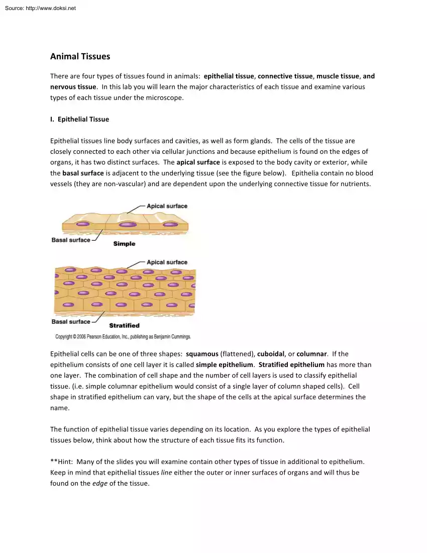
Source: http://www.doksinet Animal Tissues There are four types of tissues found in animals: epithelial tissue, connective tissue, muscle tissue, and nervous tissue. In this lab you will learn the major characteristics of each tissue and examine various types of each tissue under the microscope. I. Epithelial Tissue Epithelial tissues line body surfaces and cavities, as well as form glands. The cells of the tissue are closely connected to each other via cellular junctions and because epithelium is found on the edges of organs, it has two distinct surfaces. The apical surface is
exposed to the body cavity or exterior, while the basal surface is adjacent to the underlying tissue (see the figure below). Epithelia contain no blood vessels (they are non-‐vascular) and are dependent upon the underlying connective tissue for nutrients. Epithelial cells can be one of three shapes: squamous (flattened), cuboidal, or columnar. If the epithelium consists of one cell layer it is called simple epithelium. Stratified epithelium has more than one layer. The combination of cell shape and the number of cell layers is used to classify epithelial tissue. (ie
simple columnar epithelium would consist of a single layer of column shaped cells) Cell shape in stratified epithelium can vary, but the shape of the cells at the apical surface determines the name. The function of epithelial tissue varies depending on its location. As you explore the types of epithelial tissues below, think about how the structure of each tissue fits its function. *Hint: Many of the slides you will examine contain other types of tissue in additional to epithelium. Keep in mind that epithelial tissues line either the outer or inner surfaces of organs
and will thus be found on the edge of the tissue. Source: http://www.doksinet Simple squamous epithelium Blood vessels (artery, vein, and nerve slide): Simple squamous epithelium comprises the inner lining of blood vessels, where it provides a smooth surface that reduces friction as blood travels through the vessels. The blood vessel slide shows a cross section of an artery and a vein The wavy lining of the vessel lumen (interior) is simple squamous epithelium. Lung slide: The walls of lung air sacs (alveoli) are also composed of simple squamous epithelium.
Air sacs are the location of gas exchange between the air and blood. How does the structure of this epithelial type allow for efficient gas exchange? (hint: the gases have to travel through the epithelial cells to move between the air and the blood) Simple cuboidal epithelium Simple cuboidal epithelium (Kidney) slide: The tubules of the kidney are composed of a single layer of cuboidal cells. The kidney slide shows cross sections of many tubules, all of which are lined with simple cuboidal epithelium. These cells are active
in absorption and secretion of various substances from or into the kidney filtrate (which ultimately becomes urine). Note the shape of the epithelial cells and the centrally located nuclei. Source: http://www.doksinet Simple columnar epithelium Duodenum/Small intestine slide: The intestinal lining is a simple columnar epithelium. The primary function of these cells is absorption of nutrients. As you examine the slide, note the large, oval shaped nuclei that are positioned near the basal edge of the cells. Also note the large, clear goblet cells that are interspersed in the epithelial
layer. These are glandular cells that secrete mucus that helps protect the underlying tissues. Stratified squamous epithelium Esophagus/stomach slide: Stratified squamous epithelium consists of multiple layers, with squamous cells at the apical surface. The primary function of this type of epithelium is protection Areas subject to abrasion, like the mouth, esophagus, and skin, have stratified epithelium. Cells at the apical surface can be scraped away (for instance, by food particles traveling down the esophagus), but the layered nature of the epithelium ensures that the underlying tissues are
protected. Note the thick layer of epithelium on the esophagus slide. (*This slide also contains stomach tissue, which has a simple columnar epithelium) Source: http://www.doksinet Keratinized stratified squamous epithelium Palmar Skin (Human skin corpuscle) slide: The epidermis (most superficial layer) of the skin is composed of stratified squamous epithelial cells that contain large quantities of the protein keratin. Keratin is a tough fibrous protein that offers protection from abrasion and water loss. New cells are produced at the basal surface of the epithelium and are gradually pushed
towards the apical surface. As they move upwards, they become filled with keratin and eventually die, forming a layer of dead, keratin filled cells on the apical surface of the epidermis. Examine the palmer skin slide, noting the entire epidermis and the layer of dead cells at the apical surface. The dermis, which lies deep to the epidermis, is composed of connective tissue. Compare the skin and esophagus slide. How are they similar? How are they different? II. Connective Tissue Connective tissues vary widely in their form and function, but
they are all characterized by the presence of extracellular matrix. The extracellular matrix is nonliving material composed of protein fibers and ground substance. The protein fibers are composed of collagen (which gives strength) or elastin (which Source: http://www.doksinet gives flexibility). The number and type of fibers differs between the various types of connective tissue The ground substance fills the spaces between the cells and the fibers. It contains interstitial fluid (tissue fluid) and large polysaccharide molecules. The consistency of the ground substance can vary from liquid to
gel-‐like to a solid. *Hint: Because connective tissue consists largely of extracellular matrix, the cells that are present will be scattered among the matrix components. For most of these slides (adipose tissue is an exception), you will not see cells directly adjacent to other cells as they are in epithelial tissue. Dense connective tissue Palmar Skin (Human skin corpuscle) slide: The layer of skin that lies deep to the epidermis is called the dermis and is composed of dense connective tissue. This tissue contains densely packed bundles of irregularly arranged collagen fibers.
It is found in areas of the body that are subject to tension from many different directions. Note the thick layer of dense connective tissue that lies deep to the epithelium on the skin slide. Nuclei of the connective tissue cells are scattered throughout the collagen fibers. Adipose tissue slide: Adipose tissue consists of adipocytes, or fat storage cells. It functions in energy storage, insulation, and cushioning. Small pockets of adipose tissue can be found all over the body, but accumulates under the skin (subcutaneous fat) and around certain organs, such as the kidneys.
Unlike other connective tissues, it has very little matrix and the cells are closely packed together. Each cell contains a large fat droplet, which pushes the nucleus to the side. Note the clear cytoplasm and the peripherally located nuclei of the fat cells in the slide. Source: http://www.doksinet Hyaline cartilage slide: Hyaline cartilage is the most abundant type of cartilage in the body and is found in the rib cage, the nose, the trachea, and the ends of long bones. It provides structural support (but is more flexible than bone) and has cushioning properties. Hyaline
cartilage has a firm matrix with abundant collagen fibers, but the individual fibers cannot be seen under the microscope. When viewed under the microscope the matrix an amorphous quality (no discernable structures). The cells, which are known as chondrocytes, reside in small cavities within the matrix called lacunae. Bone tissue slide: Bone tissue forms the skeletal system. It functions in structural support, protection, and mineral (calcium) storage. The extracellular matrix of bone tissue contains abundant collagen fibers as well as a hard, calcified ground substance. Mature bone cells,
called osteocytes, reside in cavities within the matrix called lacunae. As bone tissue is formed, channels remain in the hardened matrix that provide passageways for blood vessels and nerves. The larger channels are called central canals (Haversian canals). Bone tissue forms in rings (lamellae) around these canals, creating a structure called an osteon. Examine the bone tissue slide, noting the osteons with their lamellae and bulls-‐eye like central canals. The lacunae, which contain the bone cells, are visible as small dark patches in the lamellae. Source: http://www.doksinet III.
Muscle Tissue Muscle tissue is specialized for contraction. The cells are elongated, and are also known as muscle fibers. They contain the contractile proteins actin and myosin, which interact to shorten and elongate the cells. There are three different types of muscle tissue: skeletal, cardiac, and smooth Examine each type of tissue using the muscle composite slide. (*The skeletal and smooth muscle are shown as part of organs, so they are not the only tissue present) Skeletal muscle (muscle composite slide) Skeletal muscles are attached to bones, and contraction of these
muscles generates body movements (limb movement, jaw movement, breathing, etc.) The skeletal muscle fibers are long and cylindrical, with multiple peripherally located nuclei. The cells have striations, alternating light and dark bands that result from the ordered arrangement of actin and myosin within the cell. Cardiac muscle (muscle composite slide) Cardiac muscle is present in the heart. Cells are striated, but the striations are much less obvious than in skeletal muscle tissue. The cells are shorter than skeletal muscle fibers, have a single nucleus and are often branched.
Individual cells are connected via gap junctions and desmosomes These cellular connections are visible under the microscope as dark bands called intercalated disks. These cellular communication junctions are necessary for the coordinated beating of the heart. Source: http://www.doksinet Smooth muscle (muscle composite slide & artery/vein/nerve slide) Smooth muscle tissue is found in the walls of hollow organs, such as the gastrointestinal tract, blood vessels, and the urinary bladder. Contractions of these muscles propel fluid or materials through the organs (i.e food through the GI tract, blood through
blood vessels, urine pushed out of bladder) Smooth muscle cells are not striated (hence the name “smooth” muscle); they have a single nucleus, and have tapered ends. Examine the smooth muscle on the muscle composite slide as well as the blood vessel slide. In blood vessels there is a layer of smooth muscle deep to the epithelial layer It is thicker on the artery than on the vein, but can be seen in both. IV. Nervous Tissue Nervous tissue is specialized for communication and composes the brain, spinal cord, and peripheral nerves. The tissue
consists of two major cell types: neurons and glial cells Neurons communicate with each other via electrical and chemical signals. They have nucleated cell bodies and two types of elongated cellular processes: dendrites – which receive signals, and axons – which send signals. Glial cells are the support cells of nervous tissue. There are several different types with various functions, including maintaining proper ion concentrations in the fluid surrounding neurons, generating myelin (an insulating material that surrounds some axons), and cleaning up debris. Examine the slide of
nervous tissue (giant multipolar neuron slide). Note the large neurons with their elongated cellular processes and the smaller, more numerous glial cells.


