Értékelések
Nincs még értékelés. Legyél Te az első!
Tartalmi kivonat
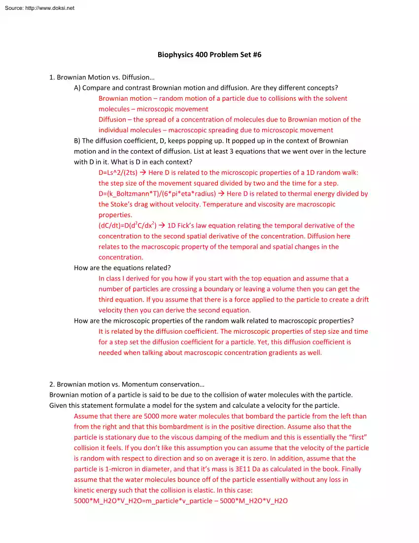
Source: http://www.doksinet Biophysics 400 Problem Set #6 1. Brownian Motion vs Diffusion A) Compare and contrast Brownian motion and diffusion. Are they different concepts? Brownian motion – random motion of a particle due to collisions with the solvent molecules – microscopic movement Diffusion – the spread of a concentration of molecules due to Brownian motion of the individual molecules – macroscopic spreading due to microscopic movement B) The diffusion coefficient, D, keeps popping up. It popped up in the context of Brownian motion and in the context of diffusion. List at least 3 equations that we went over in the lecture with D in it. What is D in each context? D=Ls^2/(2ts) Here D is related to the microscopic properties of a 1D random walk: the step size of the movement squared divided by two and the time for a step. D=(k Boltzmann*T)/(6pietaradius) Here D is related to thermal energy divided by the Stoke’s drag without velocity. Temperature and viscosity are
macroscopic properties. (dC/dt)=D(d2C/dx2) 1D Fick’s law equation relating the temporal derivative of the concentration to the second spatial derivative of the concentration. Diffusion here relates to the macroscopic property of the temporal and spatial changes in the concentration. How are the equations related? In class I derived for you how if you start with the top equation and assume that a number of particles are crossing a boundary or leaving a volume then you can get the third equation. If you assume that there is a force applied to the particle to create a drift velocity then you can derive the second equation. How are the microscopic properties of the random walk related to macroscopic properties? It is related by the diffusion coefficient. The microscopic properties of step size and time for a step set the diffusion coefficient for a particle. Yet, this diffusion coefficient is needed when talking about macroscopic concentration gradients as well. 2. Brownian motion
vs Momentum conservation Brownian motion of a particle is said to be due to the collision of water molecules with the particle. Given this statement formulate a model for the system and calculate a velocity for the particle. Assume that there are 5000 more water molecules that bombard the particle from the left than from the right and that this bombardment is in the positive direction. Assume also that the particle is stationary due to the viscous damping of the medium and this is essentially the “first” collision it feels. If you don’t like this assumption you can assume that the velocity of the particle is random with respect to direction and so on average it is zero. In addition, assume that the particle is 1-micron in diameter, and that it’s mass is 3E11 Da as calculated in the book. Finally assume that the water molecules bounce off of the particle essentially without any loss in kinetic energy such that the collision is elastic. In this case: 5000*M H2OV H2O=m particlev
particle – 5000M H2OV H2O Source: http://www.doksinet v particle = 10000*M H2OV H2O/m particle v particle=1E5*(18 Da)(500 m/s)/(3E11 Da)(0.3 nm/1 ps) – speed of liquid water is 300 m/s which is slightly less than rms speed of steam given by ideal gas law v particle= 180 microns/sec How does this velocity relate to what we know about the particle’s random walk? Is your model a good one? Let’s estimate that the mean free path or distance that the particle goes before hitting another “shell” of water molecules is 0.3 nm This is also our step distance The time for a step is just this distance divided by the velocity we calculated above, which equals 1.7 microseconds The diffusion coefficient is then Ls^2/(2ts) = 2.6E-14 m^2/s The actual diffusion coefficient is 44E13 m^2/s, which is off by one order of magnitude So I guess it depends on what we mean by good! I leave it to you to tweak the model to get the right order of magnitude. 3. The mean of the squared displacement for
a one-dimensional random walk is equal to 2Dt, for two dimensions it is 4Dt, and for three dimensions it is 6Dt. Why? For 1D <x^2>=2Dt For 2D <r^2>=<x^2+y^2>=<x^2>+<y^2>=2Dt+2Dt=4Dt For 3D <r^2>=<x^2+y^2+z^2>=<x^2>+<y^2>+<z^2>=2Dt+2Dt+2Dt=6Dt 4. Assume that a protein undergoes a two-dimensional random walk in x and y What is the function for the probability density in this case? 2D Gaussian distribution Plot the probability density at three different time points all on the same graph. (Mathematica is your friend!) Plots should show that the probability density spreads out as the number of steps or time increases. The probability at the origin decreases and the width of the Gaussian increases How does the probability at the initial position change with time? Select a different position and relate how the probability changes with time at that position. The initial position in this case is at the origin. The
probability to be at the initial position decreases with time. At a position that is not the origin the probability should be 0 before any steps are taken. Then as time progresses there is a nonzero probability for being at the position, indicating that the probability has increased! Finally, as time continues to go on the probability function keeps spreading out and the probability for being at that position decreases. What does this say about the protein’s diffusion? (Make sure to pick reasonable numbers such that your plots make sense.) If you assume that the protein is small (radius=1 nm) then D=2.2E-10 m^2/s If you also assume isotropic diffusion then the standard deviation should be 940 nm after 1 ms. That is you should be able to pull out that <r^2>=4Dt from your graph. 5. 1312 (from Solution manual) Source: http://www.doksinet 6. Design an experiment to measure the diffusion coefficient This is an open-ended question so you should be as specific as possible. (Bonus:
Calculate the error for your experiment Will it give you a reasonable answer?) There are several ways to do this and I went over them in class. As you should have it in your lecture notes I will not go into detail and instead will just mention them here. Method 1 D=(k b*T)(v drift)/(F x) so apply a force in x, F x, to a particle and measure the particle’s drift velocity, v drift. Method 2 Use video microscopy or some other method to track the Brownian motion of a particle. This should give you r (distance) vs t (time) Then square the distance and find the Source: http://www.doksinet mean. Plot this as a function of time You should be able to pull out D from the slope of the graph. Method 3 Use Fick’s law and measure changes in concentration over time and space to pull out D. In the notes I told you about one case where you use a micropipette to release fluorescent dye in solution and you track the concentration over time and space. Fit this to the Gaussian distribution
I gave you and you can pull out D. 7. Brownian Motion vs Lipid Rafts Read the article: “Dynamic clustered distribution of hemagglutinin resolved at 40 nm in living cell membranes discriminates between raft theories” by Hess et al., PNAS 2007 A pdf is available on the website. You will find several movies in the supplementary information that can be downloaded from the PNAS website through a school computer or by using a VPN client. Let’s see what diffusion has to say about lipid rafts. A) Protein molecules are nanometer-sized; how were the researchers able to visualize them using a light microscope? The article: “Review PALM and STORM: Unlocking Live-Cell SuperResolution” by Henriques et al. Biopolymers 2011, should be helpful in answering this question You can localize fluorophores to less than the diffraction limit of a microscope. (Diffraction limit is ~250nm and localization is in nanometers.) There are only three requirements – you need enough time to average position,
you need enough photons from fluorophore to see it, and you need the fluorophores to be sparse enough so that photons from one fluorophore don’t overlap another. The problem is that in cells typically many molecules are fluorescently tagged and so you see all of them at once. In FPALM (technique used in paper) the experimentalist photobleaches the region with one laser. Then shines another laser on the region, this is the activation laser If everything is still photobleached you would see nothing – but actually what happens is that some fluorophores will respond and you can track them very precisely. Then you can photobleach again and shine the activation laser again and track new fluorophores. You keep doing this and you can reconstruct the positions of multiple fluorophores. If you want more fluorophores then you have to repeat the process again and again and your image time increases. B) Summarize the findings of the article. Do lipid rafts exist? The authors find that HA
protein is localized in particular regions on the membrane and diffuses locally within that region. This is consistent with a lipid raft model See my possible objections below. C) Do you have any scientific objections to the author’s argument? -HA is known to form elongated clusters. These clusters may be forming independently of a lipid raft. -Diffusion coefficient of HA is 9E-14 m^2/s, which is the same as the diffusion coefficient for a 10 micron particle and is incredibly slow. Perhaps the proteins are being inserted at the locations they are clumped at and diffusion has not equilibrated the concentrations. -They assume a 1D diffusion when the membrane is 2D. They do say that diffusion occurs along a particular path, which would be 1D. However, I think that if you want to Source: http://www.doksinet look at the existence of lipid rafts you need a protein that is more promiscuous in its diffusion, but that is just me. -Definitely see diffusion in a confined region for HA
molecules, whether or not this is caused by a lipid raft or something else remains to be tested. D) It is always irksome that the techniques of PALM and STORM are called super-resolution techniques. This is NOT true Why? The author’s are not resolving the molecules, they are localizing the molecules to a specific point. There is not a resolution limit on localization for a microscope Indeed micron-sized beads can be localized to 1 nm. So calling them super-resolution techniques is just annoying


