A doksi online olvasásához kérlek jelentkezz be!

A doksi online olvasásához kérlek jelentkezz be!
Nincs még értékelés. Legyél Te az első!
Tartalmi kivonat
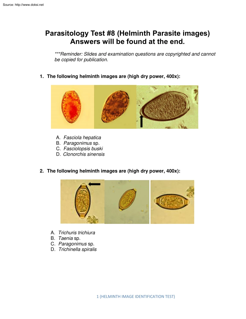
Source: http://www.doksinet Parasitology Test #8 (Helminth Parasite images) Answers will be found at the end. *Reminder: Slides and examination questions are copyrighted and cannot be copied for publication. 1. The following helminth images are (high dry power, 400x): A. Fasciola hepatica B. Paragonimus sp C. Fasciolopsis buski D. Clonorchis sinensis 2. The following helminth images are (high dry power, 400x): A. Trichuris trichiura B. Taenia sp C. Paragonimus sp D. Trichinella spiralis 1 (HELMINTH IMAGE IDENTIFICATION TEST) Source: http://www.doksinet 3. The following helminth images are (high dry power, 400x): A. B. C. D. Diphyllobothrium latum Paragonimus sp. Fasciolopsis buski Clonorchis sinensis 4. The following helminth images are (high dry power, 400x): A. Hookworm B. Ascaris lumbricoides C. Trichuris trichiura D. Opisthorchis sp 5. The following helminth images are (adult worm; GI tract image): 2 (HELMINTH IMAGE IDENTIFICATION TEST) Source:
http://www.doksinet A. Enterobius vermicularis B. Trichuris trichiura C. Onchocerca volvulus D. Ascaris lumbricoides 6. The following helminth images are (egg packet 400x, proglottid, 10x): A. Taenia saginata B. Diphyllobothrium latum C. Dipylidium caninum D. None of the above 7. The following helminth images are (high dry power, egg, adult worm, low power) A. Enterobius vermicularis B. Ascaris lumbricoides C. Trichuris trichiura D. Diphyllobothrium latum 3 (HELMINTH IMAGE IDENTIFICATION TEST) Source: http://www.doksinet 8. The following helminth images are (high dry power, 400x): A. Toxocara canis B. Toxocara cati C. Hookworm D. Ascaris lumbricoides 9. The following helminth images are (high dry power, 400x): A. Hymenolepis nana B. Hymenolepis diminuta C Taenia sp. D. Dipylidium caninum 10. The following helminth image is (high dry power, 400x) 4 (HELMINTH IMAGE IDENTIFICATION TEST) Source: http://www.doksinet A. Schistosoma mansoni B. Schistosoma haematobium C.
Schistosoma mekongi D. Schistosoma japonicum 11. The following helminth image is (low power, 10x): A. Taenia solium B. Taenia saginata C. Dipylidium caninum D. Echinococcus sp 12. The following helminth image is (low power, 10x): A. Echinococcus sp B. Taenia solium C. Taenia saginata D. Diphyllobothrium latum 5 (HELMINTH IMAGE IDENTIFICATION TEST) Source: http://www.doksinet 13. The following helminth images are (high dry power, 400x) A. Schistosoma japonicum B. Schistosoma haematobium C. Schistosoma mekongi D. Schistosoma mansoni 14. The following helminth images are (high dry power, 400x) A. Taenia sp B. Taenia saginata C. Hymenolepis diminuta D. Hymenolepis nana 15. The following helminth images are (high dry power, 400x): 6 (HELMINTH IMAGE IDENTIFICATION TEST) Source: http://www.doksinet A. Echinococcus sp B. Dipylidium caninum C. Taenia sp D. Hymenolepis nana 16. The following helminth images are (gross specimen, 400x, 1000x): A. Taenia solium B. Echinococcus
granulosus C. Echinococcus multilocularis D. Taenia saginata 17. The following helminth images are (high dry power, 400x) and measure approximately 30 microns: A. Fasciolopsis buski B. Fasciola hepatica C. Paragonimus sp D. Clonorchis sinensis 18. The following helminth images are (high dry power, 400x): 7 (HELMINTH IMAGE IDENTIFICATION TEST) Source: http://www.doksinet A. Taenia sp B. Ascaris lumbricoides C. Hookworm D. Artifacts 19. The following helminth image is (low power, 100x): A. Diphyllobothrium latum B. Taenia solium C. Taenia saginata D. Hymenolepis diminuta 20. The following helminth image is (low power, 100x): 8 (HELMINTH IMAGE IDENTIFICATION TEST) Source: http://www.doksinet A. Diphyllobothrium latum B. Taenia solium C. Taenia saginata D. Hymenolepis diminuta 21. The following helminth images are (high dry power, 400x): A. Strongyloides stercoralis rhabditiform larvae B. Strongyloides stercoralis filariform larvae C. Hookworm larvae D. Trichinella spiralis
larvae 22. The following helminth images are (low power 40x and high dry power, 400x): A. Trichinella spiralis B. Trichuris trichiura C. Ascaris lumbricoides D. Hookworm 23. The following helminth images are (low power, 40x and high dry power 400x): 9 (HELMINTH IMAGE IDENTIFICATION TEST) Source: http://www.doksinet A. Taenia sp B. Diphyllobothrium latum C. Hymenolepis nana D. None of the above 24. The following helminth images are (high dry power, 400x; right image is cellophane tape at a low magnification): A. Heterophyes heterophyes B. Toxocara sp C. Hookworm D. Enterobius vermicularis 25. The following helminth images are (high dry power, 400x): A. Hookworm unfertilized egg B. Ascaris lumbricoides unfertilized egg C. Ascaris lumbricoides fertilized egg D. Pollen grains 10 (HELMINTH IMAGE IDENTIFICATION TEST) Source: http://www.doksinet ANSWERS: ANSWER. 1 B These images represent Paragonimus sp eggs Note the typical opercular shoulders and the thickened abopercular end
(arrow). All of these morphological details are very typical for these eggs. This report should read: “Paragonimus sp. eggs seen” ANSWER. 2 A These organisms are Trichuris trichiura eggs (whipworm) Note the two clear polar plugs, one at each end of the egg (arrow). The egg morphology is very typical, and it is not difficult to identify the correct nematode species. ANSWER 3. A The correct response is Diphyllobothrium latum eggs (broad fish tapeworm). The eggs are very typical with no opercular shoulders (smooth outline of the shell) and a small knob at the abopercular end (see oval). In the image on the right you can see eggs with the opercula popped open – this can often be accomplished by taping on the coverslip of a wet preparation with a pencil or pen. ANSWER. 4 B These organisms are Ascaris lumbricoides fertilized eggs Note the eggs have a tuberculated/bumpy shell. Occasionally fully mature larvae can be seen within the egg shells; these eggs are not easily preserved, even
with 10% formalin. ANSWER. 5 D These organisms are adult Ascaris lumbricoides roundworms Note that the image on the left is an adult male (see curved tail within the oval). The image on the right shows two adult ascarids visible during visualization of the GI tract. This configuration is often called “railroad tracks” – a very revealing image. ANSWER. 6 C The correct identification is Dipylidium caninum, the dog tapeworm. The image on the left shows a typical egg packet; the individual eggs resemble Taenia sp. eggs (see egg in the oval) The image on the right is a gravid proglottid filled with egg packets (arrow). The proglottids look like cucumber seeds when fresh and rice grains when dry. ANSWER. 7 A The correct identification is Enterobius vermicularis (pinworm), probably one of the most common nematode infections throughout the world. The eggs mature within about 24 h, so occasionally eggs containing larvae may be seen (see left image). The right image shows the adult female
worms, about 3/8” and white to light tan in color. Occasionally the adult worms may be seen on the surface of the stool; the test of choice is the scotch tape preparation. However, many physicians treat on the basis of symptoms alone. ANSWER. 8 C These images are of hookworm eggs The egg morphology is very typical with rounded ends, a clear space between the egg shell and the 11 (HELMINTH IMAGE IDENTIFICATION TEST) Source: http://www.doksinet developing embryo, and development of the embryo to the 8-16 ball stage. Occasionally, the eggs mature and may contain a fully developed larva as seen in the right image. ANSWER. 9 B These images are consistent with Hymenolepis diminuta, the rat tapeworm. The egg is very typical Note the six-hooked embryo (oncosphere) and the lack of polar filaments that lie between the oncosphere and the shell (seen in Hymenolepis nana, but not H. diminuta) (see the area within the oval – no polar filaments). ANSWER 10. A This image is the trematode egg of
Schistosoma mansoni (blood fluke). Note the presence of the miracidium larva within the egg shell and the large lateral spine. ANSWER. 11 B The image is a Taenia saginata (beef tapeworm) gravid proglottid that has been injected with India ink. Note the uterine branches are visible once they are filled with ink. The branches should be counted on one side only where they come off of the main stem; usually T. saginata is more than 12 and Taenia solium is less than 12/around 8). Counting the branches confirms that this gravid proglottid is T. saginata ANSWER 12. B The image is a Taenia solium (pork tapeworm) gravid proglottid that has been injected with India ink. Note the uterine branches are visible once they are filled with ink. The branches should be counted on one side only where they come off of the main stem; usually T. saginata is more than 12 and Taenia solium is less than 12/around 8). Counting the branches confirms that this gravid proglottid is T. solium ANSWER. 13 B These
image are the trematode egg of Schistosoma haematobium (blood fluke). Note the presence of the miracidium larva within the egg shell and the large terminal spine. The image on the right represents a bladder biopsy containing S. haematobium eggs (again, note the terminal spine) ANSWER. 14 D These images are consistent with Hymenolepis nana, the dwarf tapeworm. The egg is very typical Note the six-hooked embryo (oncosphere) and the polar filaments that lie between the oncosphere and the shell (seen in Hymenolepis nana, but not H. diminuta) (see the area within the oval –polar filaments). ANSWER 15. C These images represent Taenia spp eggs; identification of T saginata vs. T solium cannot be made from the eggs without using special stains. Note the presence of the six-hooked embryo (oncosphere) and the striated egg shell. The presence of these eggs in a human clinical specimen should be reported as: “Taenia spp. eggs seen; unable to identify to the species level without special
testing.” 12 (HELMINTH IMAGE IDENTIFICATION TEST) Source: http://www.doksinet ANSWER. 16 B These structures are from cases of hydatid disease with Echinococcus granulosus. The left image is a cyst containing a daughter cyst; the middle image contains protoscolices that are contained within the cyst, the immature helminth stages (the black line represents the hooklets contained in each protoscolex). The group is called hydatid sand The image on the right is a high magnification of the hooklets from the protoscolices. ANSWER. 17 D These structures are trematode eggs, probably Clonorchis sinensis, (Chinese liver fluke). However, the morphology of this egg closely resembles eggs within the genera Heterophyes, Metagonimus, and Opisthorchis. Note the opercular shoulders (within oval) and the small knob at the abopercular end; these eggs are the smallest within the trematode group of human parasites. ANSWER. 18 D These structures are most likely pollen grains, artifacts that often mimic
nematode eggs such as Ascaris and/or Taenia. They often tend to be pleomorphic in shape and do not exhibit typical internal morphology present in actual helminth eggs. ANSWER. 19 C This image is a Taenia saginata (beef tapeworm) scolex; note the four suckers and no hooklets. Without extensive examination of post-therapy fecal specimens, the tapeworm scolex is rarely recovered or seen. ANSWER. 20 B This image is a Taenia solium (pork tapeworm) scolex; note the four suckers and the presence of hooklets. Without extensive examination of post-therapy fecal specimens, the tapeworm scolex is rarely recovered or seen. ANSWER. 21 A The correct response is Strongyloides stercoralis rhabditiform larvae. The image on the left (oval) reveals the packet of genital primordial cells, a structure that is not visible in the rhabditiform larvae of hookworm. The same structure is seen in the oval (right image). Another characteristic of these larvae is a short mouth opening, which is not visible in
either image. The mouth openings (buccal capsule) of the hookworm larvae are more elongated. ANSWER. 22 B The image on the left is an adult Trichuris trichiura (whipworm) nematode. The head end (thin section/whip portion) embeds itself in the intestinal mucosa, while the thicker/handle end is free in the lumen. The image on the right is the typical Trichuris egg; note the two polar plugs (not opercula), one at each end of the egg shell. ANSWER. 23 B The image on the left is a group of three Diphyllobothrium latum (broad fish tapeworm) proglottids. Note the reproductive structures are found in the center of the proglottid (rosette appearance). The adult tapeworm can be as long as 30ft, and large sections of the worm can be passed in the fecal specimen. The egg seen in the right image is very typical with no opercular 13 (HELMINTH IMAGE IDENTIFICATION TEST) Source: http://www.doksinet shoulders (smooth egg shell all the way around) and a small abopercular bump at the opposite end of
the egg. ANSWER. 24 D These images are Enterobius vermicularis (pinworm) The eggs mature within about 24 h, so occasionally eggs containing larvae may be seen (see left image). The right image shows the scotch tape preparation with many of the typical eggs (football shaped with one flattened side). Because it requires six consecutive negative tapes to rule out an infection, many physicians treat on the basis of symptoms alone. ANSWER. 25 B These images represent Ascaris lumbricoides unfertilized eggs. Note these eggs are more elongate than the more common rounder fertilized eggs. Also, the bumpy (tuberculated) egg shell is more pronounced than that of the fertilized egg. The presence or absence of unfertilized eggs in the clinical specimen merely indicates the presence or absence of the adult male worm. The eggs should merely be reported as Ascaris lumbricoides without specifying fertilized or unfertilized (not clinically relevant). REFERENCES 1. Garcia, LS 2016 Diagnostic Medical
Parasitology, 6th Ed, ASM Press, Washington, D.C 14 (HELMINTH IMAGE IDENTIFICATION TEST)


