Please log in to read this in our online viewer!
Please log in to read this in our online viewer!
No comments yet. You can be the first!
What did others read after this?
Content extract
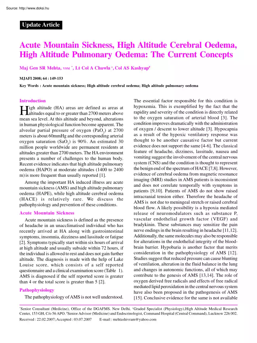
Update Article Acute Mountain Sickness, High Altitude Cerebral Oedema, High Altitude Pulmonary Oedema: The Current Concepts Maj Gen SR Mehta, VSM *, Lt Col A Chawla +, Col AS Kashyap# MJAFI 2008; 64 : 149-153 Key Words : Acute mountain sickness; High altitude cerebral oedema; High altitude pulmonary oedema Introduction igh altitude (HA) areas are defined as areas at altitudes equal to or greater than 2700 meters above mean sea level. At this altitude and beyond, alterations in human physiological function become apparent. The alveolar partial pressure of oxygen (PaO2) at 2700 meters is about 60mmHg and the corresponding arterial oxygen saturation (SaO2) is 90%. An estimated 30 million people worldwide are permanent residents at altitudes greater than 2700 meters. The HA environment presents a number of challenges to the human body. Recent evidence indicates that high altitude pulmonary oedema (HAPO) at moderate altitudes (1400 to 2400 m) is more frequent than usually reported [1].
Among the important HA induced illness are acute mountain sickness (AMS) and high altitude pulmonary oedema (HAPE), while high altitude cerebral oedema (HACE) is relatively rare. We discuss the pathophysiology and prevention of these conditions. H Acute Mountain Sickness Acute mountain sickness is defined as the presence of headache in an unacclimatised individual who has recently arrived at HA along with gastrointestinal symptoms, insomnia, dizziness and lassitude or fatigue [2]. Symptoms typically start within six hours of arrival at high altitude and usually subside within 72 hours, if the individual is allowed to rest and does not gain further altitude. The diagnosis is made with the help of Lake Louise score, which consists of a self reported questionnaire and a clinical examination score (Table 1). AMS is diagnosed if the self reported score is greater than 4 or the total score is greater than 5 [2]. Pathophysiology The pathophysiology of AMS is not well understood. The
essential factor responsible for this condition is hypoxemia. This is exemplified by the fact that the rapidity and severity of the condition is directly related to the oxygen saturation of arterial blood [3]. The condition improves dramatically with the administration of oxygen / descent to lower altitude [3]. Hypocapnia as a result of the hypoxic ventilatory response was thought to be another causative factor but current evidence does not support the same [4-6]. The classical feature of headache, dizziness, lassitude, nausea and vomiting suggest the involvement of the central nervous system (CNS) and the condition is thought to represent the benign end of the spectrum of HACE [7,8]. However, evidence of cerebral oedema from magnetic resonance imaging (MRI) studies in AMS patients is inconsistent and does not correlate temporally with symptoms in patients [9,10]. Patients of AMS do not show raised intracranial tension either. Therefore the headache of AMS is not due to meningeal
stretch or raised cerebral blood flow. A likely possibility is a hypoxia mediated release of neuromodulators such as substance P, vascular endothelial growth factor (VEGF) and bradykinin. These substances may sensitize the pain nerve endings in the brain resulting in headache [11,12]. Additionally, the same molecules may also be responsible for alterations in the endothelial integrity of the bloodbrain barrier. Hypobaria is another factor that merits consideration in the pathophysiology of AMS [12]. Studies suggest that reduced pressure can cause blunting of ventilation, alteration in the fluid balance in the lung and changes in autonomic functions, all of which may contribute to the genesis of AMS [13,14]. The role of oxygen derived free radicals and effects of free radical mediated lipid peroxidation in the central nervous system have also been proposed in the pathogenesis of AMS [15]. Conclusive evidence for the same is not available * Senior Consultant (Medicine), Office of the
DGAFMS. New Delhi +Graded Specialist (Physiology),High Altitude Medical Research Center, 153 GH, C/o 56 APO. #Senior Advisor (Medicine) and Endocrinologist, Command Hospital (Central Command), Lucknow 226 002 Received : 22.022007; Accepted : 03072007 E-mail : mehtashivram@yahoo.com 150 Mehta, Chawla and Kashyap Table 1 Lake Louise scoring system for diagnosis of AMS Self Reported Symptoms Headache None at all Mild headache Moderate headache Severe incapacitating headache Gastrointestinal Symptoms Good appetite Poor appetite or nausea Moderate nausea or vomiting Severe incapacitating nausea and vomiting Fatigue and/or Weakness Not tired or weak Mild fatigue/weakness Moderate fatigue/weakness Severe incapacitating nausea and vomiting Dizziness/lightheadedness None Mild Moderate Severe incapacitating Difficulty in Sleeping Slept as well as usual Did not sleep as well as usual Woke many times, poor night’s sleep Could not sleep at all Physical Examination Signs Change in Mental
Status None Lethargy, lassitude Disoriented, confused Stupor, semiconscious Coma Ataxia in Heel to Toe Walking None Balancing manoeuvres Steps off line Falls down Cannot stand Peripheral Oedema None At one location At two or more locations 0 1 2 3 0 1 2 3 0 1 2 3 0 1 2 3 0 1 2 3 0 1 2 3 4 0 1 2 3 4 0 1 2 at present. Recent reports suggest that fluid retention, of AMS, is not an essential requirement/accompaniment of the condition [16]. Management Patients of AMS require rest, prevention of further gain in altitude and correction of the hypoxemia. The latter can be achieved by supplemental oxygen or descent to lower altitudes, if symptoms persist despite oxygen therapy. Tablet acetazolamide 250 mg twice a day should be administered to the patient. This drug results in a mild bicarbonate diuresis, thus correcting the hyperventilation induced respiratory alkalosis.In addition, it improves tissue oxygen delivery, reduces the formation of cerebro-spinal fluid (CSF) and improves sleep.
In severe cases or in patients sensitive to sulphonamides, dexamethasone 4 mg six hourly for 48 hours may be given [17]. The headache usually responds to common analgesics such as aspirin, ibuprofen and paracetamol. High Altitude Cerebral Oedema High altitude cerebral oedema is a potentially fatal condition that can develop in an individual suffering from AMS or HAPE. It can even occur in the absence of the above conditions. The diagnosis is made in the setting of a recent gain in altitude with either, a change in mental status and/or ataxia in a person with AMS, or the presence of both mental status changes and ataxia in a person without AMS. Associated findings may include papilledema, retinal haemorrhages and cranial nerve palsy [17]. MRI may reveal increased signal intensity in the region of the corpus callosum and splenium [18]. Pathophysiology The pathophysiology of HACE centres around three main factors ie. hypoxia mediated cerebral vasodilatation coupled with a possible
impairment of the autoregulation of cerebral blood flow [19,20], disruption of the integrity of the blood brain barrier possibly by hypoxia mediated release of certain neuromodulators such as VEGF and calcitonin gene related peptide (CGRP) [21,22] and higher ratio of brain mass to CSF volume resulting in impaired ability to buffer a rise in intracranial pressure (ICP) [7]. The generalized increase in sympathetic nervous activity at HA may aggravate the condition by increasing the levels of the anti diuretic hormone (ADH) and aldosterone thus resulting in salt and water retention in the body. All the above possibly result in vasogenic oedema. One attractive unifying hypothesis is that hypoxia leads to overperfusion of microvascular beds, endothelial leakage and hence oedema. Precise mediators are likely to be activation of VGEF by hypoxiainducible factor and possibly nitric oxide. Management All cases of cerebral oedema must be evacuated to a lower altitude as an emergency. In case
where the same is not possible, a simulated descent to lower altitude by use of pressurized chambers must be attempted if facilities exist. Portable recompression chambers which are capable of generating up to 130 mmHg pressure, which is equivalent to a reduction of altitude of 1800m to 2400m may be life saving at remote posts. The patient should be kept inside the chamber till evacuation to lower altitude is possible. The patient must be reassessed every 90-120 minutes by decompressing the chamber. At MJAFI, Vol. 64, No 2, 2008 Acute Mountain Sickness, High Altitude Cerebral Oedema and High Altitude Pulmonary Oedema locations where permanent recompression chambers are available, the patient should be pressurized to 760 mmHg i.e one atmospheric absolute (1 ATA) or in severe HACE to 1.2 ATA Clinical assessment is carried out every six hours and the patient is kept within the chamber till symptoms regress or evacuation to lower altitudes is possible. Hypoxemia should be corrected
using supplemental oxygen. Dexamethasone should be administered 8mg initially orally/IM/IV and followed by 4 mg every six hours. Acetazolamide 250 mg twice a day can also be added if descent to lower altitude is delayed. In severe cases, raised ICP should be reduced using mannitol. High Altitude Pulmonary Oedema High altitude pulmonary oedema usually occurs within the first four days of ascent to high altitude. Most cases present on the second or third day of arrival at high altitude. The condition must be suspected in any individual who has recently arrived at high altitude and develops dry cough and decreased physical performance. Classical pink frothy sputum and respiratory distress usually occur late in the illness, while orthopnea and frank haemoptysis are uncommon. Resting tachycardia and tachypnea becomes pronounced as the illness progresses [23]. The diagnosis is made in the setting of a recent gain in altitude with any two of the symptoms of dyspnea at rest; cough,
weakness/decreased exercise performance and chest tightness or congestion and any two signs of crackles/wheeze in at least one lung field, central cyanosis, tachypnea or tachycardia. Unlike AMS and HACE,electrocardiogram usually shows sinus tachycardia, with right ventricular strain, right axis deviation, RBBB and P wave abnormalities. Chest radiograph shows a normal size heart, full pulmonary arteries and patchy infiltrates generally confined to the right middle and lower lobes in mild cases and involve both lungs in severe cases. Arterial blood gas analysis shows severe hypoxemia and respiratory alkalosis. Pathophysiology Elevated pulmonary artery pressures (PAP) consequent to the phenomenon of hypoxic pulmonary vasoconstriction are central to the pathogenesis of HAPE [24]. The heterogeneous nature of the vasoconstriction results from the variation of the alveolar partial pressure of oxygen in different segments of the lungs. Recent studies using single photon emission computed
tomography (SPECT) suggest that HAPE susceptible individuals show greater heterogeneity in pulmonary blood flow as compared to HAPE resistant subjects [25,26]. HAPE susceptibles possibly have blunted ventilatory responses to hypoxia, exaggerated hypoxic pulmonary vasoconstriction [27], aided possibly by MJAFI, Vol. 64, No 2, 2008 151 reduced endogenous production of vasodilators such as nitric oxide and sympathetic overactivity [28]. The HAPE fluid contains a very high protein concentration suggesting disruption of the alveolar endothelial barrier and endothelial dysfunction. Current evidence indicates that inflammation does not trigger the fluid leak in HAPE but is secondary to the exposure of the basement membrane to high protein levels and resultant chemotaxis of inflammatory infiltrates [29]. Pre-existing inflammation may prime the endothelium making it more prone to disruption. This is suggested by the presence of pre-existing upper respiratory infection in a significant
percentage of HAPE patients, especially children. Dysfunction of coagulation, as suggested by the postmortem findings of thrombi in the lungs, is also not a primary event in the pathogenesis of HAPE and occurs later in the illness. Recent studies have examined the role of alveolar fluid clearance mechanism in the lung in HAPE patients. Preliminary reports indicate that reduced activity of the apical sodium pump in the alveolar epithelial cells may contribute to the fluid accumulation in the alveoli [30]. Preliminary studies addressing the issue of genetic susceptibility to HAPE suggest an association between certain HLA subtypes i.e HLA DR6 and HLA DQ4 [31]. Management The management of HAPE requires the correction of hypoxemia and interventions to reduce the elevated pulmonary arterial pressure. The former can be achieved by lowering the altitude and/or using supplemental oxygen. Elevated pulmonary artery pressures usually reduce with the correction of hypoxemia. In certain cases
inhaled nitric oxide may be administered to the patient to achieve the same. Drugs that enhance the endogenous production of nitric oxide or those that increase intracellular cyclic guanosyl monophosphate (cGMP) levels, thereby resulting in vascular smooth muscle relaxation, have also been assessed for treating HAPE. Trials with sildenafil and tadalafil have shown promising results that await validation [32]. Inhaled beta agonists such as salbutamol and salmetrol have also been reported to be useful in treating HAPE by reducing PAP [33]. Salmetrol has the added effect of improving the alveolar fluid clearance mechanism by potentiating the action of the alveolar epithelial sodium pump. Nifedipine, a calcium channel blocker has also been used successfully in lowering elevated PAP in HAPE patients and must be considered especially in situations where supplemental oxygen and/or reduction of altitude is not possible [17,34]. For the treatment of HAPE, nifedipine is usually administered 10
mg orally initially followed by 20-30 mg of extended release formulation every 12 hours. 152 Diagnosis of HA Illness Many clinical conditions may mimic the HA illnesses. Table 2 lists some of the common conditions considered in the differential diagnosis. The diagnosis of AMS and HACE must be re-considered if the onset of symptoms is after three days of arrival at HA, headache is absent, there is a rapid response to fluids or rest and there is absence of a response to descent to lower altitude, oxygen, acetazolamide or dexamethasone. Prevention The occurrence of HA illness is largely determined by the rate of ascent, final altitude reached, sleeping altitude, previous history of HA illness, pre-existing cardio-pulmonary conditions, improper acclimatization and individual susceptibility. The most appropriate strategy to prevent HA illness is a gradual ascent to altitude. A commonly followed thumb rule is that once above an altitude of 2500 meters, the altitude at which one sleeps
should not increase by more than 400 - 600 meters in 24 hours [17,35]. For an increase in altitude between 600 to 1200 meters, an extra day should be added for acclimatization. The process of acclimatization to HA provides the body time to execute appropriate changes in physiological functions and processes, thus enabling it to function optimally in the new environment. The current acclimatization schedule being practiced in our scenario of six days for altitudes between 2700 to 3600 meters, four days for altitudes between 3600 to 4500 meters and a further four days for altitudes above 4500 meters is time-tested and recommended. An Table 2 Differential diagnosis of high altitude illnesses AMS and HACE Carbon monoxide poisoning Dehydration Central nervous system infections Acute psychosis Exhaustion Hangover Hypoglycaemia Hypothermia Migraine Seizures Stroke Transient ischaemic attacks Brain tumour HAPE Asthma Bronchitis Congestive cardiac failure Hyperventilation syndrome Myocardial
infarction Pneumonia Pulmonary embolism Mehta, Chawla and Kashyap absence from HA of more than 28 days requires the entire schedule to be followed once again on induction to HA. Acetazolamide and dexamethasone are effective in preventing AMS. Acetazolamide is preferred to dexamethasone because it does not interfere with the acclimatization process, has no rebound effect on stopping the drug and has no significant side effects [36]. Dexamethasone is also effective in preventing AMS but it does not allow acclimatization to occur. It may also lead to neurocognitive impairment and other side effects of steroid therapy such as gastritis. Dexamethasone may be considered in individuals who cannot be offered acetazolamide such as individuals with sulphonamide sensitivity or individuals with chronic obstructed pulmonary disease with carbon dioxide retention. The recommended dosage of acetazolamide for the prophylaxis of AMS is a debated subject. While the standard dosage of 250 mg twice a day
is considered appropriate, it has been suggested that low dose therapy with 125 mg twice a day is effective to prevent AMS without any accompanying side effects. A recent metaanalysis however concluded that any dose lower than 750 mg per day is ineffective in preventing AMS [37]. Nifedipine in a dose of 20-30 mg extended release formulation twice a day has been found effective in preventing HAPE in susceptible individuals [17]. To conclude, adherence to acclimatization procedure, avoiding rapid ascent to altitude, maintaining a high degree of clinical suspicion especially in individuals with preexisting upper respiratory tract infection and previous history of HA illness will not only enable reduction in the incidence of HA illnesses but also decrease the morbidity and mortality due to these disorders. Conflicts of Interest None identified References 1. Gabry AL, Ledoux X, Mozziconacci M, Martin C HighAltitude pulmonary edema at moderate altitude (2,400 m;7870 feet) A series of 52
patients. Chest 2003; 123:49-53 2. Roach RC, Bärtsch P, Oelz O, Hackett PH The Lake Louise acute mountain sickness scoring system. In: Hypoxia and molecular medicine Sutton JR, Houston CS, Coates G, editors. Queen City Printers, Burlington, VT: 1993:272-4. 3. Bartch P, Vock P, Maggiorini M et al Respiratory symptoms, radiographic and physiologic correlations at high altitude. In: Sutton JR, Cotes G, Remmers JE, eds. Hypoxia: The adaptations. Philadelphia: BC Decker, Inc, 1990:241-5 4. Bartch P, Baumgartner RW, Waber U, Maggiorini M, Olez O Comparison of carbon dioxide-enriched, oxygen enriched and normal air treatment of acute mountain sickness. Lancet 1990; 336:772-5. 5. Hohenhaus E, Paul A, Bartch P Hypoxic ventilatory response and gas exchange in acute mountain sickness. Eur Respir J MJAFI, Vol. 64, No 2, 2008 Acute Mountain Sickness, High Altitude Cerebral Oedema and High Altitude Pulmonary Oedema 1994; 7 : 200. 6. Krasney JA A neurogenic basis for acute altitude illness Med
Sci Sports Exerc 1994; 26:195-208. 7. Hackett PH High altitude cerebral edema and acute mountain sickness: a pathophysiology update. In: Roach RC, Wagner PD, Hackett PH. editors Hypoxia: into the next millennium Vol. 474 of Advances in experimental medicine and biology New York: Kluwer Academic/Plenum, 1999:23-45. 8. Kilgore D, Loeppky J, Sanders J, Caprihan A, Icenogle M, Roach RC. Corpus callosum (CC) MRI: early altitude exposure In: Roach RC, Wagner PD, Hackett PH, editors. Hypoxia: into the next millennium. Vol 474 of Advances in experimental medicine and biology. New York: Kluwer Academic/Plenum, 1999:396-7. 9. Muza SR, Lyons TP, Rock PB, et al Effects of altitude exposure on brain volume and development of AMS (abstr). In: Roach RC, Hackett PH, Wagner PD, editors. Hypoxia: Into the next millennium. New York: Plenum/Kluwer Academic Publishing 1999; 414. 10. Icenogle M, Kilgore D, Sanders J, Caprihan A, Roach RC Cranial CSF volume is reduced by altitude exposure but is not related
to early AMS (abstr). In: Roach RC, Hackett PH, Wagner PD, editors. Hypoxia: Into the next millennium New York: Plenum/Kluwer Academic Publishing 1999; 392. 11. Moskowitz MA Basic mechanisms in vascular headache Neurology 1990; 8:801-15. 12. Bartch P, Roach R Acute mountain sickness and high altitude cerebral edema. In: Hornbein TF, Schoene RB, eds High altitude: An exploration of human adaptation. New York: Marcel Dekker Inc., 2001; 752 13. Grover RF, Tucker A, Reeves JT Hypobaria: an etiologic factor in acute mountain sickness. In: Loeppky JA, Riedesel ML, eds. Oxygen transport to human tissue New York: Elsevier/ North Holland, 1982:223-30. 14. Roach RC, Loeppky JA, Robergs R Fluid balance in humans at HA: Does hypobaria play a role. FASEB J 1994; 8:A553 15. Bartch P, Bailey DM, Berger M M, Knauth M, Baumgartner R. Acute mountain sickness: controversies and advances High Alt Med Biol 2004; 5:110-24. 16. Hildebrandt W, Buclin T, Swenson E, Bartch P, Biollaz J Development of acute
mountain sickness without sodium and fluid retention (abstr). Int J Sports Med 1998; 19:S16 17. Hackett PH, Roach RC High altitude illness N Engl J Med 2001; 345:107-14. 18. Hackett PH, Yarnell PR, Hill R, Reynard K, Heit J, McCormick J. High-altitude cerebral edema evaluated with magnetic resonance imaging: clinical correlation and pathophysiology. JAMA 1998; 280:1920-5. 19. Jensen JB, Sperling B, Severinghaus JW, Lassen NA Augmented hypoxic cerebral vasodilation in men during 5 days at 3,810 m altitude. J Appl Physiol 1996; 80:1214-8 20. Levine BD, Zhang R, Roach RC Dynamic cerebral autoregulation at high altitude. In: Roach RC, Wagner PD, Hackett PH, eds. Hypoxia: into the next millennium Vol 474 of Advances in experimental medicine and biology. New York: Kluwer Academic/Plenum, 1999:319-22. MJAFI, Vol. 64, No 2, 2008 153 21. Severinghaus JW Hypothetical roles of angiogenesis, osmotic swelling, and ischemia in high-altitude cerebral edema. J Appl Physiol 1995; 79:375-9. 22.
Schilling L, Wahl M Mediators of cerebral edema In: Roach RC, Wagner PD, Hackett PH, editors. Hypoxia: into the next millennium. Vol 474 of Advances in experimental medicine and biology. New York: Kluwer Academic/Plenum, 1999:123-41 23. Hultgren HN High-altitude pulmonary edema: current concepts. Annu Rev Med 1996; 47:267-84 24. Maggiorini M, Melot C, Pierre S, et al High altitude pulmonary edema is initially caused by an increase in capillary pressure. Circulation 2001; 103:2078-83. 25. Elser H, Swenson E, Hildebrandt J, et al Regional distribution of pulmonary perfusion after five hours of normobaric hypoxia in subjects susceptible to high altitude pulmonary edema. Eur Respir J 1998; 12:A2328. 26. Schoene RB, Swenson ER, Hultgren HN High altitude pulmonary edema. In: Hornbein TF, Schoene RB, eds High altitude: An exploration of human adaptation. New York: Marcel Dekker Inc., 2001; 793-4 27. Hohenhaus E, Paul A, McCullough RE, Kucherer H, Bartsch P Ventilatory and pulmonary vascular
response to hypoxia and susceptibility to high altitude pulmonary edema. Eur Respir J 1995; 8:1825-33. 28. Duplain H, Vollenweider L, Delabays A, Nicod P, Bartsch P, et al. Augmented sympathetic activation during short-term hypoxia and high-altitude exposure in subjects susceptible to high-altitude pulmonary edema. Circulation 1999; 99:1713-8 29. Kubo K, Hanaoka M, Hayano T, et al Inflammatory cytokines in BAL fluid and pulmonary hemodynamics in high-altitude pulmonary edema. Respir Physiol 1998; 111:301-10 30. Scherrer U, Sartori C, Lepori M, et al High-altitude pulmonary edema: from exaggerated pulmonary hypertension to a defect in transepithelial sodium transport. In: Roach RC, Wagner PD, Hackett PH, editors. Hypoxia: into the next millennium Vol 474 of Advances in experimental medicine and biology. New York: Kluwer Academic/Plenum, 1999:93-107. 31. Hanaoka M, Kubo K, Yamazaki Y, et al Association of high altitude pulmonary edema with major histocompatibility complex. Circulation
1998; 97:1124-8 32. Maggiorini M, Brunner-La Roccca HP, Peth S, et al Both tadalafil and dexamethasone may reduce the incidence of high altitude pulmonary edema. A randomized trial Ann Int Med 2006; 145:497-506. 33. Sartori C, Lipp E, Duplain H, et al Prevention of HAPE by beta-adrenergic stimulation of the alveolar transepithelial sodium transport. Am J Crit Care Med 2000; 161: Suppl: A415 34. Hackett PH, Roach RC, Hartig GS, Greene ER, Levine BD The effect of vasodilators on pulmonary hemodynamics in high altitude pulmonary edema: a comparison. Int J Sports Med 1992; 13: Suppl 1:S68-S71. 35. West JB The Physiological Basis of High-Altitude Diseases Ann Intern Med 2004;141 :789-800. 36. Barry JW, Pollard AJ Altitude illness BMJ 2003; 326:915-9 37. Dumont L, Mardirosoff C, Tramer MR Efficacy and harm of pharmacological prevention of acute mountain sickness: quantitative systemic review. BMJ 2000; 321:267-72

 When reading, most of us just let a story wash over us, getting lost in the world of the book rather than paying attention to the individual elements of the plot or writing. However, in English class, our teachers ask us to look at the mechanics of the writing.