Please log in to read this in our online viewer!

Please log in to read this in our online viewer!
No comments yet. You can be the first!
Content extract
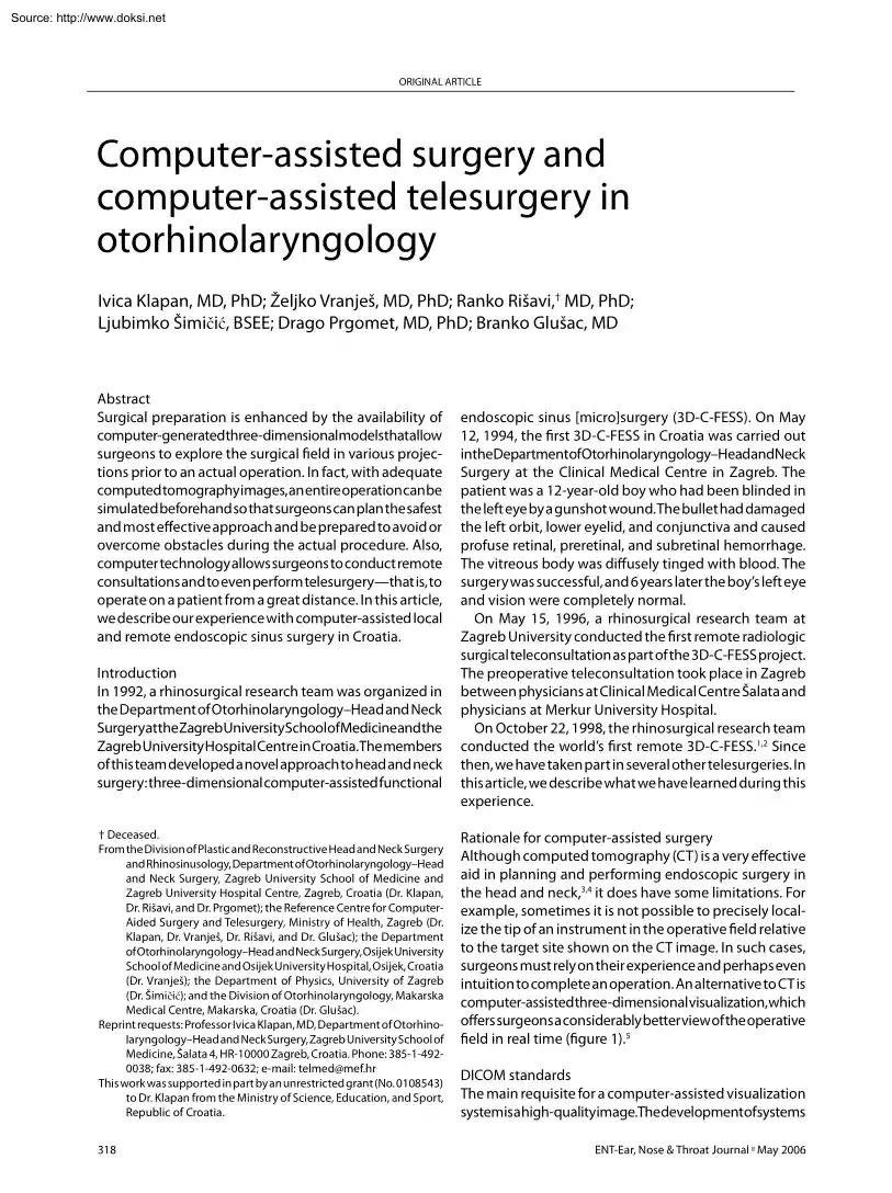
Source: http://www.doksinet ORIGINAL KLAPAN, VRANJEŠ, RIŠAVI,ARTICLE ŠIMIČIĆ, PRGOMET, GLUŠAC Computer-assisted surgery and computer-assisted telesurgery in otorhinolaryngology Ivica Klapan, MD, PhD; Željko Vranješ, MD, PhD; Ranko Rišavi,† MD, PhD; Ljubimko Šimičić, BSEE; Drago Prgomet, MD, PhD; Branko Glušac, MD Abstract Surgical preparation is enhanced by the availability of computer-generatedthree-dimensionalmodelsthatallow surgeons to explore the surgical field in various projections prior to an actual operation. In fact, with adequate computed tomography images, an entire operation can be simulated beforehand so that surgeons can plan the safest and most effective approach and be prepared to avoid or overcome obstacles during the actual procedure. Also, computer technology allows surgeons to conduct remote consultations and to even perform telesurgerythat is, to operate on a patient from a great distance. In this article, we describe our experience with
computer-assisted local and remote endoscopic sinus surgery in Croatia. Introduction In 1992, a rhinosurgical research team was organized in the Department of Otorhinolaryngology–Head and Neck SurgeryattheZagrebUniversitySchoolofMedicineandthe Zagreb University Hospital Centre in Croatia.The members of this team developed a novel approach to head and neck surgery: three-dimensional computer-assisted functional † Deceased. From the Division of Plastic and Reconstructive Head and Neck Surgery and Rhinosinusology, Department of Otorhinolaryngology–Head and Neck Surgery, Zagreb University School of Medicine and Zagreb University Hospital Centre, Zagreb, Croatia (Dr. Klapan, Dr. Rišavi, and Dr Prgomet); the Reference Centre for ComputerAided Surgery and Telesurgery, Ministry of Health, Zagreb (Dr Klapan, Dr. Vranješ, Dr Rišavi, and Dr Glušac); the Department of Otorhinolaryngology–Head and Neck Surgery, Osijek University School of Medicine and Osijek University Hospital, Osijek,
Croatia (Dr. Vranješ); the Department of Physics, University of Zagreb (Dr. Šimičić); and the Division of Otorhinolaryngology, Makarska Medical Centre, Makarska, Croatia (Dr. Glušac) Reprint requests: Professor Ivica Klapan, MD, Department of Otorhinolaryngology–Head and Neck Surgery, Zagreb University School of Medicine, Šalata 4, HR-10000 Zagreb, Croatia. Phone: 385-1-4920038; fax: 385-1-492-0632; e-mail: telmed@mefhr This work was supported in part by an unrestricted grant (No. 0108543) to Dr. Klapan from the Ministry of Science, Education, and Sport, Republic of Croatia. 318 endoscopic sinus [micro]surgery (3D-C-FESS). On May 12, 1994, the first 3D-C-FESS in Croatia was carried out intheDepartmentofOtorhinolaryngology–HeadandNeck Surgery at the Clinical Medical Centre in Zagreb. The patient was a 12-year-old boy who had been blinded in the left eye by a gunshot wound. The bullet had damaged the left orbit, lower eyelid, and conjunctiva and caused profuse retinal,
preretinal, and subretinal hemorrhage. The vitreous body was diffusely tinged with blood. The surgery was successful, and 6 years later the boy’s left eye and vision were completely normal. On May 15, 1996, a rhinosurgical research team at Zagreb University conducted the first remote radiologic surgical teleconsultation as part of the 3D-C-FESS project. The preoperative teleconsultation took place in Zagreb between physicians at Clinical Medical Centre Šalata and physicians at Merkur University Hospital. On October 22, 1998, the rhinosurgical research team conducted the world’s first remote 3D-C-FESS.1,2 Since then, we have taken part in several other telesurgeries. In this article, we describe what we have learned during this experience. Rationale for computer-assisted surgery Although computed tomography (CT) is a very effective aid in planning and performing endoscopic surgery in the head and neck,3,4 it does have some limitations. For example, sometimes it is not possible to
precisely localize the tip of an instrument in the operative field relative to the target site shown on the CT image. In such cases, surgeons must rely on their experience and perhaps even intuition to complete an operation. An alternative to CT is computer-assistedthree-dimensionalvisualization,which offers surgeons a considerably better view of the operative field in real time (figure 1).5 DICOM standards The main requisite for a computer-assisted visualization systemisahigh-qualityimage.Thedevelopmentofsystems ENT-Ear, Nose & Throat Journal May 2006 Source: http://www.doksinet COMPUTER-ASSISTED SURGERY AND COMPUTER-ASSISTED TELESURGERY IN OTORHINOLARYNGOLOGY Figure 1. Computer-assisted visualization is a good alternative to CT for data exchange between multiple diagnostic instruments and computer networks led to the establishment of DICOM standards, which codify the forms and modes of data exchange. DICOM is an acronym for Digital Imaging and Communications in
Medicine.6 Before DICOM standards became widely accepted, image recordings were stored on film. But even under ideal conditions, film could record only 16 different image levels at most. Also, the process of transferring film images onto computer storage disks resulted in the loss of some anatomic information and the probable introduction of unwanted artifact. Moreover, the level setting and window width of the images could not be changed. Because video images seen on diagnostic device monitors are considerably better than film images, they are used for storage in computer media. These video images are capable of containing as many as 256 image levels, and it is possible to subsequently modify the level setting and window width once they have been stored in the computer system. When images are transferred to computer systems in accordance with DICOM protocols, they are stored in the same form that was generated by the diagnostic device without data loss. This is particularly important
when images are retrieved for use during complex examinations andduringpreoperativepreparation,asaprecisedemarcation is needed to distinguish diseased from healthy tissue. DICOM images can be visualized from different aspects and used to develop three-dimensional models. Volume 85, Number 5 Computer-assisted preoperative preparation Surgical preparation is enhanced by the availability of three-dimensionalmodelsthatallowsurgeonstoexplorethe surgical field in various projections and to simultaneously view multiple model sections (figure 2). With programs such as Virtual Endoscopy and Virtual Surgery, an entire operation can be simulated prior to the actual surgery Figure 2. A three-dimensional model shows the cranial anatomy 319 Source: http://www.doksinet KLAPAN, VRANJEŠ, RIŠAVI, ŠIMIČIĆ, PRGOMET, GLUŠAC Figure 3. With programs such as Virtual Endoscopy, entire operations can be planned and simulated prior to the actual surgery. (figure 3).7,8 As a result, surgeons can plan
the safest and most effective approach and be prepared to avoid or overcome obstacles during the actual procedure. Also, these models can be entered into a variety of software programs and transmitted to distant radiologic and surgical sites for preoperative consultation (Tele-Virtual Endoscopy).1,9 The development of our 3D-C-FESS system involved the use of a variety of computer programs and systems. The initial modeling was done with VolVis, VolPack/ vprender, and GL Ware programs on a DECstation 3100 computer. As programs were upgraded and refined, we subsequently used 3D Viewnix V1.0 and V11 software, the AnalyzeAVW system, the T-Vox system, and the OmniPro 2 system on Silicon Graphics O2, Origin200, and Origin2000 computers. Computer-assisted surgery The use of a computer during surgery/telesurgery requires highlyreliable,stable,andfastcomputersystems.Computer work stations with UNIX-compatible operating systems are most commonly used. Because a surgeon’s hands are engaged in
performing surgery, he or she cannot operate the computer, and the presence of a computer system expert in the operating theater is necessary. However, a surgeon can operate some computer systems by voice. Model movements on the monitor and various projections and sections can be viewed by issuing simple, short voice instructions during surgery. During our initial computer-assisted procedures, spatial orientationwithintheoperativefieldofathree-dimensional computer model and transfer of a particular point to the real operative field within the patient were performed by arbitrary approximation of the known reference points of the operative field anatomy.10 In this way, the given enti320 Figure 4. The major problem with computer-assisted surgery is transmission of the actual operative-field coordinate system to the coordinate system of the three-dimensional spatial model. ties were simultaneously recognized on the model and in the real operative field.11 This method facilitated access
to the operative field, but it could not guarantee absolute safety at critical points. The use of a three-dimensional spatial model of the operative field during surgery has pointed to the need for simulating the position of the tip of the instrument (e.g, endoscope and forceps) within the computer model. The major problem is transmission of the actual operative field coordinate system to the coordinate system of the three-dimensional spatial model that has been previously designed from a series of CT images during preoperative preparation (figure 4).10,12 Several modes of instrument localization within the operative field are usedelectromagnetic, optic, and mechanical: • The electromagnetic method is very sensitive to environmental electromagnetic fields (e.g, those generated by electrical devices) and to large amounts of metal (e.g, cabinets, tables, and instruments). Therefore, the basic default precision of localization within the field is inadequate for surgery. • Optic
locators have proved to be suitable, but they are relatively expensive and less precise than mechanical locators. ENT-Ear, Nose & Throat Journal May 2006 Source: http://www.doksinet COMPUTER-ASSISTED SURGERY AND COMPUTER-ASSISTED TELESURGERY IN OTORHINOLARYNGOLOGY Computer networks Once a local DICOM-compliant computer-assisted surgery system is established, the next step is to create an interactive network of such programs among appropriate institutions. With such a network, physicians at participating institutions can consult almost instantaneously with each other and transmit textual, image, and other data. Of course, the consultation itself can be recorded and stored for further use. Figure 5. Computer-assisted telesurgery can be performed at a distance with the assistance of video and audio transmission and sophisticated endoscopic cameras. •
�������������������������������������������������� The primary shortcoming of mechanical locators is their inability to reach deep areas within the operative field. This problem might be solved by redesigning instruments so that the tips are thinner and longer. Teleconsultation and telesurgery Computer-assisted consultation and surgery can be performed at a distance with the assistance of video and audio transmission and sophisticated endoscopic cameras (figure 5). Preoperatively, a consulting surgeon can receive CT images from a remote location, examine the images, develop a three-dimensional spatial model, and transfer all this information back to the remote location.2 Intraoperatively, staff and consultants both near to and far from the actual operating table can view the operation “live” via the endoscopic camera images, and they can
followtheprogressofthesurgeryonthethree-dimensional computer models.1,2 In most cases, a network can be set up so that intraoperative consultations can be obtained from multiple locations. The underlying principle behind telesurgery is that it is often better to move the data than to move the patient. Postoperative analysis All relevant pre- and intraoperative data (e.g, CT images, test results, three-dimensional models, and video of the surgery) can be stored on a CD-ROM disk and reviewed for postoperative analysis.7 An analysis and critique of a computer-assisted surgical procedure may identify shortcomings and areas that need improvement. This may be especially valuable when reviewing the particularly critical points of an operation. Such a record is also useful as a teaching tool and as permanent documentation in case a medicolegal issue arises. Volume 85, Number 5 Acknowledgment The authors gratefully acknowledge the support of the
DepartmentofOtorhinolaryngology–HeadandNeckSurgery at the Zagreb University Hospital Centre; the Merkur University Hospital, Zagreb; T-Com Croatia, Zagreb; InfoNET Projekt, Zagreb; and SiliconMaster, Zagreb. References 1. Klapan I, Simicic Lj, Pasaric K, et al Realtime transfer of live video images in parallel with three-dimensional modeling of the surgical field in computer-assisted telesurgery. J Telemed Telecare 2002;8:125-30. 2. Klapan I, Simicic L, Risavi R, et al Tele-3-dimensional computerassisted functional endoscopic sinus surgery: New dimension in the surgery of the nose and paranasal sinuses. Otolaryngol Head Neck Surg 2002;127:549-57. 3. Mladina R, Hat J, Klapan I, Heinzel B An endoscopic approach to metallic foreign bodies of the nose and paranasal sinuses. Am J Otolaryngol 1995;16:276-9. 4. Risavi R, Klapan I, Handzic-Cuk J, Barcan T Our experience with FESS in children. Int J Pediatr Otorhinolaryngol 1998;43:271-5 5. Hassfeld S, Muhling J Navigation in maxillofacial
and craniofacial surgery. Comput Aided Surg 1998;3:183-7 6. Levine BA, Cleary KR, Norton GS, Mun SK Experience implementing a DICOM 30 multivendor teleradiology networkTelemed J 1998;4:167-75. 7. Holtel MR, Burgess LP, Jones SB Virtual reality and technologic solutions in otolaryngology [abstract]. Otolaryngol Head Neck Surg 1999;121:181. 8. Klapan I, Barbir A, Simicic L, et al Dynamic 3D computer-assisted reconstruction of a metallic retrobulbar foreign body for diagnostic and surgical purposes. Case report of orbital injury with ethmoid bone involvement. Orbit 2001;20:35-49 9. Klapan I, Simicic L, Besenski N, et al Application of 3D computer-assistedtechniquestosinonasalpathologyCasereport:War wounds of paranasal sinuses caused by metallic foreign bodies. Am J Otolaryngol 2002;23:27-34. 10. Klimek L, Mosges R, Schlondorff G, Mann W Development of computer-aided surgery for otorhinolaryngology. Comput Aided Surg 1998;3:194-201. 11. Mann W, Klimek L Indications for computer-assisted
surgery in otorhinolaryngology. Comput Aided Surg 1998;3:202-4 12. Anon J Computer-aided endoscopic sinus surgery Laryngoscope 1998;108:949-61. 321


 When reading, most of us just let a story wash over us, getting lost in the world of the book rather than paying attention to the individual elements of the plot or writing. However, in English class, our teachers ask us to look at the mechanics of the writing.