Comments
No comments yet. You can be the first!
Most popular documents in this category


Content extract
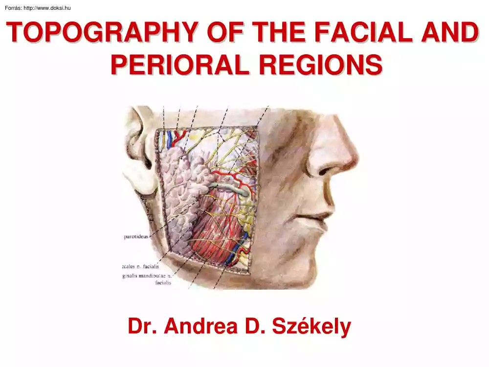
TOPOGRAPHY OF THE FACIAL AND PERIORAL REGIONS Dr. Andrea D Székely ANATOMICAL RELATIONS DEVELOPMENT OF THE FACE 5-week embryo 6-week embryo The nasal prominences are gradually separated from the maxillary prominence by deep furrows. C. Scanning electron micrograph of a mouse embryo at a stage similar to that of B. 7-week embryo. 10-week embryo Maxillary prominences have fused with the medial nasal prominences. C Scanning electron micrograph of a human embryo at a stage similar to that of A. COMPONENTS OF THE FACIAL SKELETON Upper portion - frontal bone (partially forms neurocranium too) Middle portion - nasal bone, bone lacrimal bone, maxilla, zygomatic bone ethmoidal bone, palatine bone, vomer, inferior nasal concha Lower portion - mandible SUPERFICIAL AND DEEP FACIAL VESSELS INNERVATION OF THE FACE FRONTAL AND TEMPORAL REGIONS INNERVATION OF THE FACE PERIORAL TOPOGRAPHY - CROSS SECTION PAROTID NEST AND DEEP FACIAL STRUCTURES COMPONENTS
OF THE FACIAL REGIONS SALIVARY GLANDS CROSS SECTIONS CROSS SECTIONS CROSS SECTIONS CROSS SECTIONS ANATOMICAL REGIONS Anterior facial region Borders Layers infraorbital margin, zygomatic bone, piriform aperture, masseteric m., mandible - Skin and CT - muscles of facial expression - Bichat fat pad - buccinator, levator anguli oris Structures Structures facial a.+v (anastomosis with ophtalmic vein), facial n, infraorbital a+v+n, mental a+v+n Parotideomasseteric region Borders Layers mandible, zygomatic bone and arch, masseteric muscle - Skin and CT - parotid fascia - parotid gland and nerves passing through it (facial+ auriculotemporal), parotid duct - masseteric muscle - mandibular ramus Structures facial nerve, superficial temporal a.+v, transverse facial a, auriculotemporal n Retromandibular fossa Borders Layers mandible (head and ramus), mastoid proc, external acoustic meatus (cartilege) - Skin and CT (with the superfic lamina of the cervical fascia) -
lateral wall and cavity of parotid nest: masseteric m, mandible, med pterygoid m, mastoid process+ muscles (SCM and digastric posterior belly), CONTENT: parotid gland and lymph nodes -medial wall of parotid nest: styloid proc + muscles, carotid sheath+ content Structures facial n, int. jugular v, ext carotid a, auriculotemporal n ANATOMICAL REGIONS Infratemporal region Borders mandible (head and ramus), mastoid proc, tuber of maxilla, greater wing of sphenoid, zygomatic Layers - space 1. between ramus and pterygoid muscles - space 2. below lat pterygoid (with foramen ovale) Structures - in space 1.: maxillary a, inferior alveolar n, lingual n, buccal n - in space 2.: med meningeal a, auriculotemporal n, tympanal chord, venous plexus Sublingual region Borders Layers mandible (body), root of the tongue - mucosa - floor of the mouth (muscles)- lateral sulcus, medial sulcus Structures LS: lingual n., submandibular duct, hypoglossal n, (then more anteriorly the sublingual gland) MS:
lingual a. Ramus region Borders 3rd lower molar tooth, ramus (mandible), medial pterygoid m. Structures lingual n., inferior alveolar n MOUTH IN GENERAL