A doksi online olvasásához kérlek jelentkezz be!
A doksi online olvasásához kérlek jelentkezz be!
Nincs még értékelés. Legyél Te az első!
Mit olvastak a többiek, ha ezzel végeztek?
Tartalmi kivonat
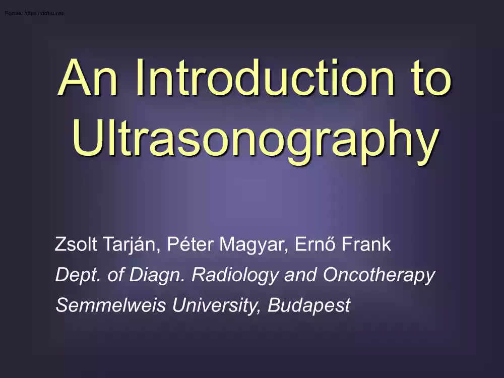
An Introduction to Ultrasonography Zsolt Tarján, Péter Magyar, Ernő Frank Dept. of Diagn Radiology and Oncotherapy Semmelweis University, Budapest Advantages? – Limitations? Ultrasonography 1. Physics 2. The US examination 3. Future Ultrasonography 1. Physics 2. The US examination 3. Future 1. Physics a) Physical features of US b) Production of US c) Interaction between US and tissue d) Image construction e) Transducers 1. Physics a) Physical features of US b) Production of US c) Interaction between US and tissue d) Image construction e) Transducers Physical features of US • Sound – Mechanical wave = propagating mechanical vibration Longitudinal waves – Longitudinal ↔ waves on water: transversal – Requires medium ↔ x-ray Transversal waves 1. Physics a) Physical features of US b) Production of US c) Interaction between US and tissue d) Image construction e) Transducers Producing US • 1st generation compound scanner TRANSDUCER
=PROBE Ultrasound transducer. Piezoelectric effects Electrode Connection Protective layer Piezoelements ~ EMISSION US transducer (Shung et al. 1992, pg 106, Figure 82) Lead zirconate titanate or polyvinyldene fluoride based piezodiscs or piezosegments Ultrasound transducer. Piezoelectric effects Electrode Connection Protective layer Piezoelements ~ RECEPTION US transducer (Shung et al. 1992, pg 106, Figure 82) 1. Physics a) Physical features of US b) Production of US c) Interaction between US and tissue d) Image construction e) Transducers a) b) c) d) (+ Interaction between US & tissue Reflection Refraction Absorption Scattering Divergation ATTENUATION) a) b) c) d) (+ Interaction between US & tissue Reflection Refraction Absorption Scattering Divergation ATTENUATION) Reflection • Imaging is based on it • Echos coming from edge surfaces = different acoustic impedances (Z) Acoustic impedance Tissue Z [g cm-2 sec-1] Air
0.0004 Bone 4–7.5 Fat 1.33 Blood 1.61–166 Liver 1.65 Muscle 1.7 Water 36 °C 1.53 Reflection coefficient for edge surfaces: intensity reflected α refl = intensity immitted Z1 − Z2 α refl = Z1 + Z2 2 – this is what pixel brightness is proportional to (B mode) a) b) c) d) (+ Interaction between US & tissue Reflection Refraction Absorption Scattering Divergation ATTENUATION) Refraction • Significance: e.g puncturing a small cyst a) b) c) d) (+ Interaction between US & tissue Reflection Refraction Absorption Scattering Divergation ATTENUATION) Absorption • A part of US penetrating a medium is being gradually absorbed • Significance: – Also limits imaging depth – Hazards (later) a) b) c) d) (+ Interaction between US & tissue Reflection Refraction Absorption Scattering Divergation ATTENUATION) a) b) c) d) (+ Interaction between US & tissue Reflection Refraction
Absorption Scattering Divergation ATTENUATION) a) b) c) d) (+ Interaction between US & tissue Reflection Refraction Absorption Scattering Divergation ATTENUATION) Attenuation (Obesity is an important limiting condition) a) b) c) d) (+ Interaction between US & tissue Reflection Refraction Absorption Scattering Divergation ATTENUATION) Reflectivity mapping: US. Attenuation mapping: x-ray. 1. Physics a) Physical features of US b) Production of US c) Interaction between US and tissue d) Image construction e) Transducers How do you get images? How do you get images? How do you get images? How do you get images? Image composition by 1st generation compound scanner How do you get images? Why not to build several transducer elements in a single unit? B mode Imaging by waves. Resolution • Spatial resolution limit for imaging by wave/radiation: – λ / 2 (for the 1st step) c λ f Imaging by waves. Resolution • Audible
sound – 20 Hz–20 kHz – In air: c = 330 m/sec – λ = c / f (16.5 m– 1.65 cm) Imaging by waves. Resolution • Ultrasound – 2 MHz–15 MHz – In water: c = 1540 m/sec – λ = c / f (0.77–01 mm) Ways of imaging • B mode (brightness) – gray-scale • M mode (motion) • (A mode [amplitude=intensity of US reflected]) • Doppler modes B (brightness) mode • • • • A row of US beams in a single plane (2D) One crystal (in movement) or a crystal array (256 lines) Gray-scale for echo intensity 2D section (slice = tomogram) Tissue depth Width M (motion) mode • Keeping on scanning in a single line (1D) • Shifting section image to the right ~ ECG Series of 1D sections in time • Exact measuring, echocardiography B Wall Cusp No. 1 Wall Cusp No. 2 Normal mitral valve Tissue depth M Time A (amplitude) mode - HISTORY • Curve, not image • Single US beam • Amplitude (intensity) on ordinate, tissue depth on absciss Echo amplitude
Tissue depth Doppler modes. Doppler effect • A moving object emits/reflects waves or radiation • Frequency perceived: • Approaching object: f ↑ (λ ↓) • Object moving away: f ↓ (λ ↑) Δf can be displayed acoustically or visually Source of waves moving Doppler modes 1. Continuous Wave Doppler (CWI) 2. Pulsed wave Doppler: • Doppler flow curve • Color Doppler • Power Doppler Continuous Wave Doppler (CWI) • ∆f is „displayed” as (audible) sound • Encoding: Pitch level ~ Δf during reflection ~ Flow velocity Loudness ~ Energy of frequencyshifted beams ~ Flow intensity Color Doppler • ∆f is displayed in colors • Encoding: Position on color scale ~ Δf during reflection in a certain voxel ~ Flow velocity Doppler flow curve (B-mode + Doppler flow curve = duplex Doppler) Power Doppler • Flow intensity is displayed in colors • Encoding: Position on color scale ~ Energy of frequencyshifted beams from a
certain voxel ~ Flow intensity, regardless of direction 1. Physics a) Physical features of US b) Production of US c) Interaction between US and tissue d) Image construction e) Transducers Depth vs. resolution • Higher frequency (7.5 MHz λ ≈ 02 mm Shallow penetration High resolution if v = 1530 m/sec) • Lower frequency (3.5 MHz λ ≈ 044 mm) Deep penetration Low resolution • Tricks to visualize deep structures: – Acoustic windows (e.g full urinary bladder) – Compression – Endoscopic transducers Biopsy transducers Ultrasonography 1. Physics 2. The US examination 3. Future? 2. The US examination a) Terminology b) Diagnostic value c) Hazards d) Preparations for US exam e) Consultation 2. The US examination a) Terminology b) Diagnostic value c) Hazards d) Preparations for US exam e) Consultation B mode – terminology • Reflectivity of a voxel • Shape – Anechoic – Hypoechoic – Isoechoic – Hyperechoic • Borders
• Transmission – (Relative) inforcement – Acoustic shadowing B mode – patterns – pathology? 2. The US examination a) Terminology b) Diagnostic value c) Hazards d) Preparations for US exam e) Consultation Diagnostic value of sonography • Possible diagnoses – Simple cyst – Hemangioma – Lipoma – Metastasis – Echinococcal cyst – Cystadenoma – • Diagnosis based on – Cystic or solid appearance – Other patterns – Vascularisation Doppler modes – Patient history – Experience, intuition Diagnostic value of sonography • Experience, intuition • Accuracy ~ 70–90%? 2. The US examination a) Terminology b) Diagnostic value c) Hazards d) Preparations for US exam e) Consultation Admission – Hazards? Based on US absorption Heating Cavitation Reactive oxygen species Cell membrane damage? Mutagenity, teratogenity? Admission – Hazards? • Intrauterine damage? • No correlation found to various disease • Left-handedness
– possible correlation but not evidence-based Hazards of US. Σ • OUTPUT POWER MAXIMALIZED in diagnostic US equipments: – Average < 0.01 W/cm2 – Peak < 0.2 W/cm2 • ALARA principle = „As Low As Reasonably Achievable” SONOGRAPHY IS SECURE! 2. The US examination a) Terminology b) Diagnostic value c) Hazards d) Preparations for US exam e) Consultation Preparation for exam Abdominal US: Transvaginal / transrectal US: • Empty stomach & bowels • Empty urinary bladder Gas-fluid surfaces; full gallbladder • Patient informing! • Adult: fasting 5–6 hours • Presence of one more person • Covering transducer • Child: feasting 3–4 hours • Not after gastroscopy • Pancreas: fluid intake before exam Pelvic US: • Full urinary bladder (acoustic window) • Reaching uterine fundus • Intake of fluid without bubbles before exam • Filling by catheter? 2. The US examination a) Terminology b) Diagnostic value c)
Hazards d) Preparations for US exam e) Consultation Admission to US exam Consultation Ultrasonography 1. Physics 2. The US examination 3. Future Novel strategies • Contrast enhanced US • US elastography • 3D / volume rendering • Panoramic imaging • THI, CHI • EUS – Transrectal – Transvaginal – Transesophageal – Laparoscopic – Intraoperative – Intravascular Contrast-enhanced US • Microbubbles < 10 μm • Echo enhancing • Penetrate capillary wall • Safe • B mode, Doppler • Application e.g: • Echocardiography • Liver nodules CONTRAST AGENTS • More details • Better contrast • Depiction of movement E.g circulation kinetics Contrast-enhanced US After contrast injection: liver masses clearly visible Before contrast injection Contrast-enhanced US • Cirrhosis, ascites • Bulging hyperechoic mass (8th segment) • Regenerative nodule / hepatocell. cc? • Contrast enhancement in early arterial phase
• Later isoechoic Probably HCC Elastographic imaging Breast, B mode: some hypoechogenicity US elastogram: bifocal tumor Panoramic imaging 2nd generation compound scanner Panoramic imaging Kidney Panoramic imaging ? 3D multiplanar rendering Ultrasonography. Σ • • • • • • • Reflected mechanical waves Secure Sophisticated diagnostic tool Skills & experience needed CONSULTATION Some preparations Often 1st choice (by Terry Pratchett)
 Ahogy közeledik a történelem érettségi, sokan döbbennek rá, hogy nem készültek fel eléggé az esszéírás feladatra. Módszertani útmutatónkban kitérünk a történet térbeli és időbeli elhelyezésére, a források elemzésére és az eseményeket alakító tényezőkre is.